
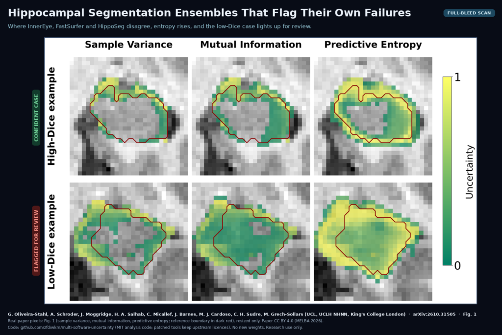
Every hippocampal volume report has the same weak spot. The segmentation tool returns a mask, the platform turns it into millilitres and a percentile, and nothing tells the reader when the mask is wrong. A bad case looks exactly as confident as a good one. Gabriel Oliveira-Stahl, Anna Schroder, James Moggridge, Hamza A. Salhab, Caroline Micallef, Josephine Barnes, M. Jorge Cardoso, Carole H. Sudre and Matthew Grech-Sollars (University College London, the National Hospital for Neurology and Neurosurgery at UCLH, and King’s College London) tackle that gap in a paper published in the Journal of Machine Learning for Biomedical Imaging (MELBA) and posted as arXiv:2610.31505 on 22 October 2026. They run several hippocampus segmentation tools that were built in different ways, average their probability maps, and use the places where they disagree as an uncertainty signal that can send a case to a human.
Why a viewer or platform team should care
Hippocampal volume is an established MRI biomarker for Alzheimer’s disease and epilepsy, and volumetry is a common feature request for neuro platforms. Most products ship one segmentation engine. This paper is useful because it does not ask you to train anything new. It takes tools that exist, some of which you may already run, and shows how to combine them so the output carries a confidence map and a per-hemisphere review flag. With that flag, a reporting workflow can hold a volume back before it lands in a report.
The tools in the ensemble
The authors picked five hippocampus segmentation methods on purpose so they would disagree in useful ways. Two are deep learning models, InnerEye and FastSurfer. Three are atlas-based, HippoSeg and ASHS (both multi-atlas) and FreeSurfer (probabilistic atlas). Their training data or atlases also differ: some come from healthy controls and mild cognitive impairment, some include Alzheimer’s disease, and HippoSeg’s atlas includes epilepsy patients. The reasoning is that a deep ensemble of the same network trained five times tends to make the same mistakes, while tools with different methods and different training cohorts fail in different places.
They benchmarked all five tools, every combination of three, and the full five-tool ensemble on 30 ADNI subjects with manual reference masks. A triad of InnerEye, FastSurfer and HippoSeg matched the full ensemble’s accuracy while dropping the two slowest tools, ASHS and FreeSurfer. Inference emissions for the 30 subjects fell from 4.65 kg to 0.85 kg CO2 equivalent, about 82% less. The rest of the paper uses that triad.
How the uncertainty is computed
None of the three tools outputs probabilities by default, so the authors patched each one. FastSurfer was changed to save the raw hippocampus outputs before the argmax, InnerEye to save its per-class posteriors, and HippoSeg to average the votes of its atlas templates into a probability map. With three probability maps per hemisphere, they computed three voxel-wise uncertainty measures: sample variance, mutual information and predictive entropy. They then tried three ways to combine the masks (plain averaging, confidence weighting and a winner-takes-all rule). Plain averaging of the probabilities gave the highest Dice on all three test sets and the best calibrated confidence of anything tested, including each single tool.
For quality control, the voxel maps are reduced to one number per hippocampus by averaging the uncertainty over the predicted region plus a one-voxel margin. A threshold on that number decides whether the case goes to review. Left and right hippocampi are handled separately, because epilepsy often affects one side.
What it hangs on a scan
For each T1-weighted brain MRI, the output is an averaged hippocampus mask per hemisphere, three uncertainty maps that can be overlaid on the slices, and one aggregated score per hippocampus. Fig. 1 of the paper shows the overlays on a good case and a failed case. In the good case, uncertainty follows the boundary in a band a few voxels wide. In the failed case, high uncertainty forms a wide band that reaches well past the reference outline, and the interior is uncertain too. That difference is what a reviewer would see in a viewer, and it is also where they should look when correcting the mask.
What the evaluation shows
The data are small but varied. Beyond the 30 ADNI subjects, the authors tested on two held-out local cohorts scanned on a Siemens Prisma 3T at UCLH: 21 patients from a dementia clinic and 8 patients with temporal lobe epilepsy. A neuroradiology-trained clinician drew the reference masks, confirmed by a consultant neuroradiologist. The local data were processed on a MacBook Pro with an Intel Core i5 and about 8 GB of memory, because the ethics approval did not allow the data to leave the hospital. So the triad runs on hardware a hospital already has.
For failure detection, the authors labelled a segmentation as failed when it fell below 0.82 Dice or above 2.5 mm HD95, then tuned an uncertainty threshold on 30% of the cases and tested on the rest. Predictive entropy worked best. It caught about 92% of the failed segmentations, but fewer than half of the flagged cases were real failures. The authors translate this into workload. In a cohort of 100 scans, about 35 would be sent for review, roughly 15 of them true failures and 20 false alarms.
On the public MR-ART dataset, in which healthy volunteers were scanned still and while nodding their heads, uncertainty rose with motion artefact severity, and clinically unusable scans had significantly higher uncertainty than medium-quality ones. Good and medium scans could not be told apart. Adding the uncertainty values to hippocampal volume gave a modest improvement for separating cognitively normal subjects from Alzheimer’s disease, and no improvement for mild cognitive impairment.
What it does not show
The cohorts are small, with only 8 epilepsy patients. The triad was chosen on the same ADNI data used to report its ADNI accuracy, so that part is optimistic. The local cohorts are the fairer test. The review threshold was tuned per dataset, which means a deployment needs its own calibration set and will not get a universal cut-off from the paper. FreeSurfer was run as version 6.0.0 to fit the laptop’s memory, so its ranking may not reflect current releases. The authors did not compare against temperature scaling, conformal prediction or probabilistic segmentation networks, and they make no claim about which kind of uncertainty the disagreement measures. The study covers adult T1-weighted MRI only. Children, other sequences, post-surgical brains and scanners other than those in ADNI and the local Prisma data are untested. The flag rate has a cost too: sending roughly a third of cases to review may be more than a busy service will accept.
Code, data and license
The analysis code is public at github.com/zfdiwkm/multi-software-uncertainty under the MIT license. It includes the patched files for FastSurfer, InnerEye and HippoSeg that save probability maps, plus scripts for the calibration, quality control, motion and classification experiments. The patched tool files stay under their upstream licenses, which the repository’s NOTICE file lists as Apache 2.0 for FastSurfer, MIT for InnerEye, and HippoSeg’s original academic license. There are no new model weights; you use each tool’s own released models, and each tool needs its own environment or container. No imaging data ships with the repository. ADNI is available through its own application process, MR-ART is public, and the two UCLH cohorts are not shared. The paper itself is open access under CC BY 4.0. This is a research evaluation, not a cleared medical device.
How we would use it
We build DICOM viewers and imaging platforms, and this pattern needs no new model from anyone. For a hippocampal volumetry feature, we would run the three tools as separate containers behind one job, average their probability maps, and store the entropy map as an overlay series next to the mask. The report would show the volume only when the per-hemisphere score is under a threshold set on the site’s own reviewed cases, and otherwise mark the side as “needs review” with the uncertainty overlay open in the viewer. The HippoSeg academic license is the first thing we would check before any commercial deployment, since it decides whether the triad can ship as is or needs a substitute. For related reading, see our posts on uncertainty-guided tumour segmentation in 3D Slicer, on flagging quiet segmentation failures, and on cortical surfaces from any brain MRI.
Sources
- Oliveira-Stahl, G., Schroder, A., Moggridge, J., Salhab, H. A., Micallef, C., Barnes, J., Cardoso, M. J., Sudre, C. H., Grech-Sollars, M. Evaluation of Uncertainty-Aware Multi-Software Ensembles for Hippocampal Segmentation. Machine Learning for Biomedical Imaging, 2026. https://doi.org/10.59275/j.melba.2026-e77b. arXiv:2610.31505: https://arxiv.org/abs/2610.31505. PDF: https://arxiv.org/pdf/2610.31505.
- Code (MIT for analysis code, upstream licenses for the patched tools): https://github.com/zfdiwkm/multi-software-uncertainty.
- FastSurfer: https://github.com/Deep-MI/FastSurfer. InnerEye Deep Learning toolbox: https://github.com/microsoft/InnerEye-DeepLearning.
- Featured image: Fig. 1 of arXiv:2610.31505 (sample variance, mutual information and predictive entropy maps for a high-Dice and a low-Dice example, reference boundary in dark red), real paper pixels, cropped and resized only. The paper is licensed CC BY 4.0.