
If you build 3D tools for surgeons, you have probably been asked whether a foundation model can draw a cranial implant. It is a fair question. After a decompressive craniectomy or a tumour resection, the patient is left with a hole in the skull vault, and the plate that closes it is still designed by hand in CAD by a specialist, case by case. A skull with a defect looks like a partial shape, and shape completion models, including the first ones sold as foundation models, promise to fill in partial shapes with no task-specific training. Marek Wodzinski, Kamil Kwarciak, Daria Hemmerling and Mateusz Daniol of AGH University of Krakow and the Sano Centre for Computational Medicine tested that promise in arXiv:2610.30035, posted on 21 October 2026 and accepted at the TrustFMI workshop at ACCV 2026. Their answer, for the checkpoints they could obtain, is no.
Why an imaging or planning team should care
It is easy to demo a completion model on a skull. You load the defective skull, run the checkpoint, and the hole is covered by something that looks like bone. The usual completion metrics score how closely that output resembles a reference skull. They do not say whether the model added anything the patient’s own bone did not already supply, or whether the result could be placed in that patient. If your product turns a segmented skull into a printable implant, a model output that intersects the patient’s remaining bone is a plate that will not seat in the operating room, no matter how good the render looks.
What the audit does
The authors took three publicly released point-cloud completion checkpoints and ran them zero-shot, as released:
- MSN-FM, which describes itself as the first multi-modal foundation model for medical point cloud completion. It was trained on MedShapeNet and accepts text prompts.
- MGPC, a more recent model trained at foundation scale, with text and image branches.
- AdaPoinTr, a strong generalist trained on ShapeNet-55, whose 55 everyday object classes include no skull. It serves as a non-medical control.
The test set is 239 skulls: 210 synthetic defects carved from the SkullBreak and SkullFix benchmarks and 29 real post-craniectomy cases from MUG500+, where the reference is the implant an expert designed for that patient. Each model received the surface points of the remaining skull and returned a completed point cloud.
Two checks carry the study:
- A do-nothing baseline. Every output is scored against handing back the patient’s own remaining bone unchanged. The removed bone is a thin shell over the vault, so bone near the edge of the defect already “covers” part of it. A model only earns credit for material it adds beyond that.
- A physical acceptance test. An output counts as implantable only if it does not cut into the retained bone by more than 1 mm and also covers the defect at least as well as the do-nothing baseline. The authors call it a necessary geometric screen, not a clinical approval.
For scale, they also scored a task-specific volumetric model they trained themselves on the SkullBreak and SkullFix training splits, a 3D convolutional encoder-decoder that segments the missing bone. They are explicit that this reference was selected on the same test cases, so it shows the criteria can be met, not how well such a model would do in general.
What they found
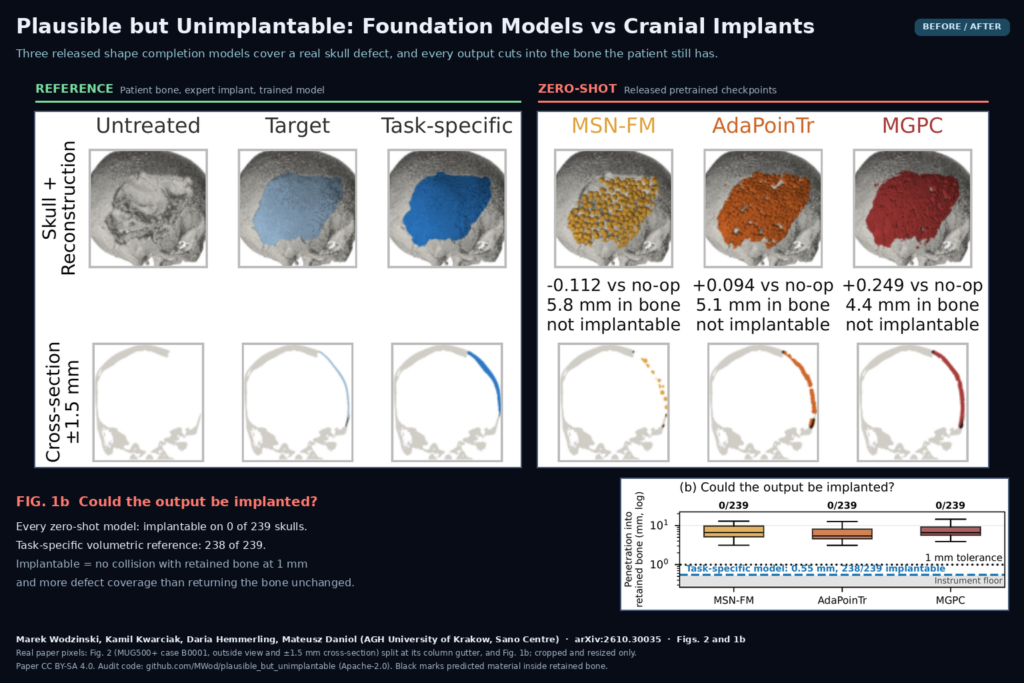
None of the three models was implantable on any of the 239 skulls, while the task-specific reference passed on 238 of 239. On the carved defects, all three placed less useful material near the missing bone than the do-nothing baseline. On the 29 real cases, MGPC did beat the baseline on most skulls, yet every one of its outputs still ran into bone the patient has. Medical pretraining did not help: MSN-FM, the model trained on medical shapes, scored worst of the three on the baseline comparison.
Fig. 2 of the paper, reproduced in the featured image above, shows one real MUG500+ case. All three outputs cover the hole. In cross-section, each one pushes material several millimetres into bone the patient still has.
The authors spent the second half of the study trying to rescue the models. They swept six protocol settings, tried the training-time alignment, and handed each model the complete skull as a positive control. Nothing produced an implantable row.
Why thickness is the deeper problem
These models emit a few thousand surface points. At 6,144 points, the mean spacing between neighbouring points on a skull is about 2.56 mm, which is too coarse to tell the inner and outer face of a 3 mm plate apart. Thickness is lost before the model is asked to recover it, and more than a third of the real MUG500+ implants are thinner than 3 mm. Any pipeline that wants a printable plate from these outputs would need to surface the cloud, trim it against the bone and thicken it, with thresholds set per patient. That is the manual work the model was supposed to remove.
The audit also tested the text prompts both foundation models advertise. In MGPC, changing the prompt moved the geometry far less than swapping in a second blank image. In MSN-FM, almost all of the text code is exactly zero after the text branch’s final ReLU, so most prompt changes did less than running the model twice with a new seed.
What this means for a product roadmap
- Keep task-specific volumetric or implicit models in the implant pipeline. The paper’s reference shows that a model trained on the defect task can pass the same screen.
- Add the two checks from this paper to your own validation: a per-case comparison against returning the patient’s bone unchanged, and a collision test against retained bone at a tolerance your measurement can resolve.
- Check that the output representation can carry thickness. The authors note that a surface point cloud needs point spacing below half the plate thickness, or the output should be volumetric or implicit.
- Score outputs with the collision and baseline checks before anyone reviews renders. In the paper, AdaPoinTr’s output looked the most like a skull and was no more placeable than the others.
Where it falls short
- Released checkpoints only. The models were used as they come. What fine-tuning on cranial data would buy is not tested.
- Geometric screen, no clinician. Failing the test rules an output out, but passing it would not make an implant acceptable, and no surgeon or designer reviewed the outputs.
- 29 real defects. Enough to show that carved benchmarks flatter these models, not enough to settle the clinical question alone.
- No task-specific point-cloud method. The study does not separate the effect of the point-cloud output from the effect of the pretraining.
- Partial release. The audit code is public under Apache-2.0 at MWod/plausible_but_unimplantable, with scripts that fetch and pin the three upstream checkpoints. The task-specific reference model, the stored result tables and the predicted point clouds are not released, and the skull datasets come from their own sources under their own terms.
Related reading on PYCAD
- DART: One Shape Model That Repairs Broken Segmentation Masks
- MedBOE: Rebuilding the Full Body Surface From Truncated PET/CT
- Partial Intraop Ultrasound In, a Complete Lumbar Spine Mesh Out
Sources
- Marek Wodzinski, Kamil Kwarciak, Daria Hemmerling, Mateusz Daniol. Plausible but Unimplantable: A Trustworthiness Audit of Shape Completion Foundation Models for Cranial Defect Reconstruction. arXiv:2610.30035, 2026. Abstract and PDF. Licensed CC BY-SA 4.0.
- Code: github.com/MWod/plausible_but_unimplantable (Apache-2.0).
- Featured image: Figs. 2 and 1b of the paper, cropped and resized only.