
Spine surgeons often plan from two studies. The MRI shows discs, cord, nerve roots and tumours. A separate CT shows the bone they will put screws into. If the MRI could also give a usable picture of the vertebrae, some patients would skip a CT and its radiation dose. Eduardo Nieto, Ibai Genua, Davide Scorza and Karen López-Linares describe a three-stage pipeline for that in arXiv:2610.37134. Nieto and López-Linares are at Fundación Vicomtech (Basque Research and Technology Alliance) in San Sebastián, Genua and Scorza at the R&D department of Cyber Surgery in the same city, and López-Linares is also at the Biogipuzkoa Health Research Institute.
The real problem is pairing
MRI-to-CT translation is an old idea. Most supervised methods need the same patient scanned in both modalities and registered, and full-spine MRI and CT pairs are rare. Unpaired methods such as CycleGAN or SynDiff can learn from two separate piles of scans. Even random slices from the same spinal region still differ in which vertebrae they show and where, so the model sees loose correspondences. The authors’ answer is to build pseudo-pairs: for each MRI slice, find a CT slice from a different patient whose vertebrae look the same, align it, and train on that.
Stage 1: vertebra-by-vertebra matching
Everything starts from vertebral masks. Some datasets ship them, the rest were generated with TotalSegmentator. Scans are reoriented, resampled to 1 x 1 x 2.5 mm, cut into 288 x 288 subvolumes along the spine, and sliced sagittally. Slices with fewer than four vertebrae are dropped.
Matching runs in two passes.
- Cheap filter. Each slice mask becomes a 25-value descriptor: the share of pixels belonging to each vertebra from C1 down to the sacrum. Cosine distance between MRI and CT descriptors gives the 50 closest CT candidates, restricted to slices that differ by at most one vertebra.
- Registration and scoring. Each candidate CT mask is rigidly registered to the MRI mask using the centroids of the vertebral bodies and spinous processes, then scored with a Dice weighted by vertebra size. The best candidate wins, the same rigid transform is applied to the CT image, the pair is cropped to the central 192 x 192 pixels, and that CT slice is removed from the pool.
The 50-candidate limit came from an elbow plot of matching quality against run time. The result is 11,319 training pairs, 3,555 for validation and 1,911 for test.
Stage 2: adversarial diffusion on the pairs
The translator is SynDiff, an existing unsupervised adversarial diffusion model from Özbey, Çukur and colleagues, with its original settings: 256 x 256 input, 1,000 forward diffusion steps and four denoising steps. The authors did not modify the network. The comparison in the paper isolates the effect of the training data: SynDiff trained on slices from the same spinal region with no matching, against SynDiff trained on the matched pseudo-pairs.
Stage 3: a 3D clean-up pass
SynDiff works on 2D sagittal slices. Stack them and the axial and coronal views show steps and seams between neighbouring slices. The third stage is a 3D GAN (residual encoder-decoder generator, volumetric PatchGAN discriminator) trained to remove those seams. For training data, the team took 670 real CT subvolumes and rebuilt each one from look-alike sagittal slices of other CT scans, using the same matching code. Each rebuilt volume has realistic stacking artefacts, and the original CT is the target. The loss mixes adversarial, L1, SSIM, perceptual and an inter-slice term, with weights tuned by Optuna.
What comes out on a scan
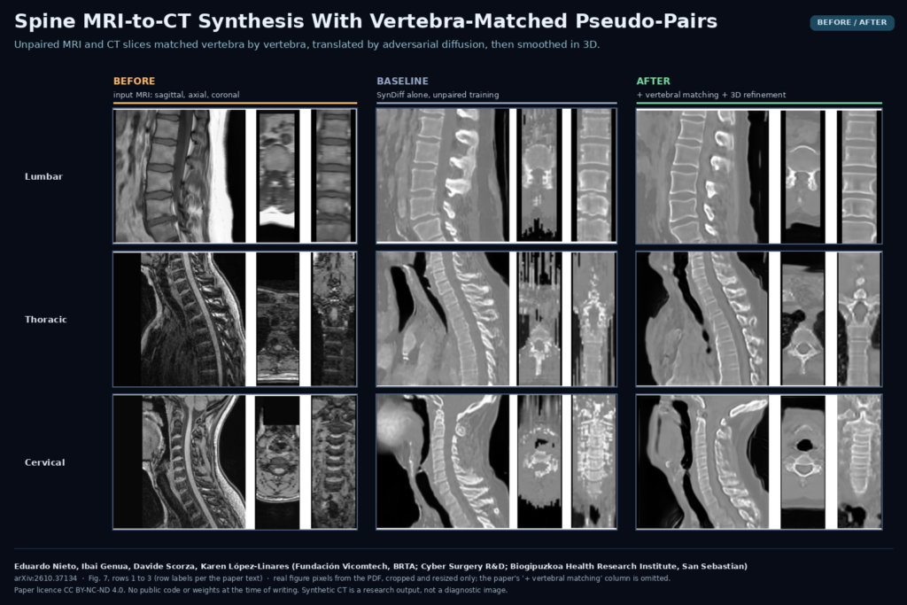
Feed in a sagittal spine MRI (the authors chose T1-weighted series where available) and you get a CT-like volume: bright cortical bone, vertebral bodies with clear endplates, posterior elements, and grey soft tissue. In Fig. 7 of the paper, the matched model draws spinous processes as separate, regular shapes where the baseline tends to inflate them and merge them across levels. After the 3D pass, axial and coronal reformats look like a continuous volume rather than a stack of strips.
There is no paired ground truth, so the authors cannot compute voxel-wise error. Their main anatomical check segments the vertebrae on the input MRI and on the synthetic CT with TotalSegmentator and measures overlap. Treating all vertebrae as one class, the 3D Dice went from 0.666 for SynDiff alone to 0.739 with pseudo-pairing, on 83 test MRI subvolumes covering all spinal regions. The 3D pass kept that level (0.732) while improving per-vertebra overlap a little.
Why a DICOM viewer or planning shop would care
Spine planning tools are built around CT: bone thresholds, pedicle measurements, screw trajectory planning, registration to fluoroscopy and navigation. A CT-like volume derived from MRI could feed those tools without new code. A plausible workflow in a viewer looks like this.
- Receive the sagittal spine MRI series from PACS.
- Run vertebral segmentation, then the translator slice by slice and the 3D refinement on the stack.
- Store the result as a separate derived series, clearly labelled synthetic, linked to the source MRI.
- Show MRI and synthetic CT side by side or fused, with the vertebral masks from both overlaid so the reader can see where they disagree.
The paper’s own evaluation compares masks from MRI and synthetic CT. The same comparison works as a live QC signal in step 4. If the synthetic vertebrae do not overlap the MRI vertebrae at a level, the reader should not trust that level.
Where it fails
The authors list the weak spots themselves.
- Sacrum. Both models sometimes merge the sacrum with L5, and the sacrum has the worst intensity agreement with real CT. The 3D pass makes it worse there.
- Upper cervical spine. The baseline sometimes fuses C1 with C2. Matching helps, but this is a region with high variability between patients.
- Lower thoracic spine. T7 to T12 show higher intensity error than the other regions, with T7 the hardest. Public sagittal thoracic MRI is scarce, and the training set reflects it.
- Smoothing. The 3D refinement removes seams but blurs soft tissue and shifts vertebral intensities. Full-image FID rose from 80.88 to 87.41 after it, so the cleaner 3D volume is a less realistic CT slice by slice.
- Excluded cases. Scans with metal implants, very low resolution or heavy noise were removed by visual inspection. Post-operative spines with hardware, a large share of real spine surgery work, are untested.
- Proxy evaluation. Every anatomical number relies on TotalSegmentator on both sides. A segmenter that is forgiving on synthetic images would flatter the result.
Code, data and licence
All training data comes from ten open-access datasets, including SPIDER, Spine Generic, the TotalSegmentator MRI and CT datasets, CTSpine1K and Spine-Mets-CT-Seg. I could not find released code or weights for this pipeline. SynDiff and TotalSegmentator are public, so the matching step and the 3D refinement are the parts you would have to rebuild from the paper. The preprint is licensed CC BY-NC-ND 4.0. The work was partly funded by the Basque Government HAZITEK 2024 programme, and two authors work for Cyber Surgery. Synthetic CT here is a research output and has no regulatory clearance for planning.
My take
The pseudo-pairing step is what I would reuse. It costs a segmentation model and a rigid registration, it works with any unpaired translator, and in this paper it moved vertebral overlap more than the 3D network did. I would not plan screws on these images yet. I would try the matching trick on our own unpaired MRI and CT archives, and I would test it on instrumented spines and sacral anatomy before showing a synthetic CT to a surgeon.