
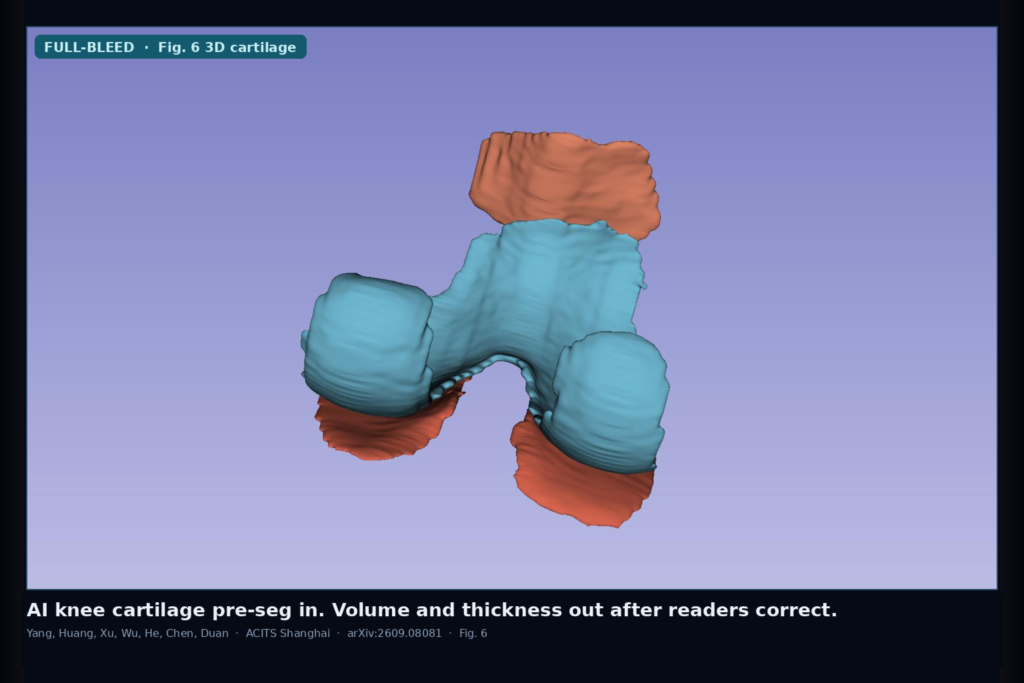
Hang a three-dimensional knee MRI from a multicenter osteoarthritis trial and ask for cartilage volume, mean thickness, and an estimate of where the surface is thinner than 1.5 mm. Manual slice-by-slice delineation is slow. Vendor and field-strength differences make boundaries drift. Deep learning can propose an initial mask, but a structure-modifying drug trial still needs masks that readers can correct, adjudicate, and turn into metrics that stay stable across visits. Binbin Yang, Rui Huang, Yuanjing Xu, Jingshu Wu, Chengzhang He, Yinan Chen, and Qi Duan (Academy for Clinical Innovation and Translation of Shanghai, ACITS) posted arXiv:2609.08081 on 8 September 2026. The paper is Development, Evaluation, and Multicenter Clinical-Trial Application of an Artificial Intelligence-Assisted MRI Method for Quantitative Knee Cartilage Morphometry.
Their workflow keeps AI in the assist lane. nnU-Net pre-segmentation proposes femoral, tibial, and patellar cartilage. Two blinded readers correct the mask. A third reader adjudicates. A partitioning model splits medial and lateral compartments. Then volume, 3D ray-tracing thickness (3D-RT), and a ray-based estimate of surface area below 1.5 mm (3D-RBA) run on the frozen gold-standard mask. They ran that stack on 1,189 phase III trial MRIs and used the same rules on 1,188 examinations from 416 participants after unblinding.
What the AI actually does in the reading room
Pre-segmentation uses the 3D full-resolution nnU-Net configuration with a combined Dice and cross-entropy loss. DICOM goes to NIfTI, gets resampled and Z-score normalized, and the mask is mapped back to the original physical space. Version 1.0 ran separate femorotibial and patellar models (OAI-ZIB, SKI10, and early Shenkang labels). Version 2.0 switched to one three-class model trained on 428 gold-standard Shenkang exams after the facility had enough adjudicated labels. In deployment, V1.0 covered 384 examinations and V2.0 covered 805. Overall Dice against the adjudicated mask on the full 1,189 set was 0.964 ± 0.030 (median 0.970), with 78.7% of exams at Dice ≥ 0.95. Use it as a deployment check against the adjudicated mask, not as a stand-alone product claim.
For a viewer or trial shop, the human loop that follows is the product. Two readers independently edit the AI mask slice by slice without seeing each other. The adjudicator picks the better match to the source series, or sends both back. Inter-reader ICCs for cartilage volume sat between 0.959 and 0.995 depending on compartment and whether the adjudicator was included. Intra-reader ICCs were similarly high except for one patellar sample. AI never writes the final morphometry input. The frozen adjudicated mask does.
From adjudicated mask to volume, thickness, and defect area
Medial-lateral partitioning uses OAI-ZIB plus the CLAIR-Knee-103R atlas so femoral and tibial cartilage split into MFC, LFC, MTC, and LTC, with patellar cartilage kept as its own class. Volume is computed in physical coordinates. Mean thickness uses three-dimensional ray tracing from the inner surface. For protocol-style defect area, 3D-RBA counts surface facets where local thickness falls below 1.5 mm, borrowing the depth idea behind ICRS-style grading and earlier 1.5 mm threshold work. A second area method (3D-PMA) and three alternate thickness definitions (3D-NN, 3D-MN, 3D-SDT) ride along as consistency checks, not as replacements for the prespecified 3D-RT and 3D-RBA trial measures.
On 20 synthetic thinning models, 3D-RBA showed MAPE 5.73%, CCC 0.822, and spatial Dice 0.956 against geometric ground truth. In 69 participants with strictly rising total volume across V0, V6, and V8 (207 exams), mean total volume rose from about 14,184 mm³ to 15,359 mm³, 3D-RBA fell by 4.70%, and all four thickness methods peaked at V8. After unblinding on the full 1,188-exam database, the treatment group moved +3.45% volume, +2.46% mean thickness, and −4.54% 3D-RBA from V0 to V8, while the control group did not show that coordinated pattern.
Where it still fails
The preprint still has placeholders for trial registration, ethics IDs, center count, and dosing, so treat efficacy language as directional imaging evidence, not as a finished regulatory package. V1.0 and V2.0 hit different image subsets at different project stages, so the paper correctly refuses a head-to-head model bake-off. Thickness methods disagree in absolute millimeters even when they agree on direction, which means a viewer should hang the chosen method name next to the number. 3D-RBA is an MRI estimate of thin-surface area, not an arthroscopic or histologic defect map. Outliers remain in the Dice tail, so a fail-closed path still needs human review when the initial mask is fragmented or when medial-lateral partitioning looks anatomically wrong. Multicenter 1.5 T and 3.0 T FLASH/SPGR-style cartilage sequences help realism, but the workflow assumes same-scanner longitudinal follow-up for a given participant.
For a viewer or clinic AI shop
Treat this as a cartilage morphometry rail on 3D knee MRI: series in, AI pre-seg overlay out, two-reader correction and adjudication before any volume or thickness leaves the workstation, then medial-lateral labels plus 3D-RT thickness and 3D-RBA thin-area maps a reader can audit. Hang the AI mask beside the source slices with an edit history, hang the adjudicated mask as the only input to metrics, and show visit-to-visit volume, thickness, and thin-area together so a coordinated change is visible. Fail closed when the pre-seg Dice against a quick QC contour is poor, when partitioning flips medial and lateral, when the series is not a dedicated 3D cartilage sequence, when visits jump scanners mid-trial, or when someone wants to publish morphometry straight from the raw nnU-Net output without adjudication. If your DICOM viewer already supports SEG overlays and structured measurement export, this paper is a concrete blueprint for keeping AI assist and trial-grade numbers on the same case without letting the network own the endpoint.
Rebuild from arXiv:2609.08081. As of 15 September 2026 the abstract and PDF respond. Public code and weights are not linked in the preprint.
Sources
- Yang, B., Huang, R., Xu, Y., Wu, J., He, C., Chen, Y., Duan, Q. Development, Evaluation, and Multicenter Clinical-Trial Application of an Artificial Intelligence-Assisted MRI Method for Quantitative Knee Cartilage Morphometry. arXiv:2609.08081, posted 8 September 2026. https://arxiv.org/abs/2609.08081. PDF: https://arxiv.org/pdf/2609.08081.
- Isensee et al. nnU-Net (cited segmentation backbone).
- OAI-ZIB and SKI10 knee MRI resources; CLAIR-Knee-103R atlas (cited for partitioning labels).
- OARSI guidance on structural MRI assessment in KOA trials (cited clinical context).