
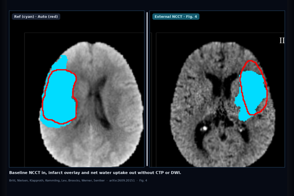
Hang an infarct overlay on baseline non-contrast CT alone, then read net water uptake from that mask, without waiting on CT perfusion or DWI to draw the lesion. That is the clinic ask Britt, Nielsen, Klapproth, Kemmling, Lev, Broocks, Werner, and Sentker take on in arXiv:2609.20151, posted around mid September 2026. The paper is Ischemic Stroke Segmentation and Net Water Uptake Quantification on Multicenter Non-Contrast CT Using Supervised Target-Domain Adaptation. Sentker is corresponding author at University Medical Center Hamburg-Eppendorf. Code is planned at github.com/IPMI-ICNS-UKE/ncct-stroke-seg after acceptance.
Early ischemic change on NCCT is subtle. Today many shops still draw the hypodensity by hand, often with CTP or DWI open as a guide, before they trust an NWU number. The authors train an nnU-Net on NCCT only, then fine-tune a small labeled slice when the scanner site shifts. On held-out Boston cases, automated NWU sits within about 1.37 percentage points mean absolute error of the expert reference. Larger lesions are where the overlay looks most usable.
What hangs on the viewer
Upstream is a baseline NCCT volume. Downstream you get a binary infarct mask on that same NCCT space, plus an NWU percentage computed from mean Hounsfield units inside the lesion and its contralateral mirror. No CTP map and no DWI are required at inference for the delineation step. For a DICOM viewer or stroke AI rail, that means you can hang the overlay beside the admission head CT and surface NWU as a derived measurement once the mask is non-empty.
NWU is only reported for cohorts imaged within six hours of known onset (UKE and Boston). AISD and ISLES were kept out of the NWU analysis because of longer or missing onset windows and, for ISLES, a post-treatment final-infarct endpoint that is the wrong target for baseline edema.
How the domain adapt works in plain words
Preprocessing is atlas registration, skull strip with TotalSegmentator, and HU clipping to [0, 80], then resample to 1.0 × 1.0 × 3.0 mm. The network is nnU-Net 3d_fullres with a Residual Encoder U-Net preset. Input is dual-channel: the native NCCT plus a left-right mirror, so the model can lean on hemispheric asymmetry the way a reader does.
Baseline training uses UKE (n=198, CTP-guided references) and AISD (n=397, DWI-guided references) with five-fold patient-level cross-validation. When the site changes, each fold model is fine-tuned on a small target set with the encoder frozen and only the decoder updated: 11 Boston cases (10 epochs) or 75 ISLES cases (30 epochs). A probability threshold is picked on that fine-tune set (0.05 Boston, 0.03 ISLES) and locked before held-out testing. At inference the five fine-tuned folds are ensembled, then thresholded. Across 801 patients, 595 go to development and internal testing, 86 to fine-tuning, and 120 to held-out external tests.
What the numbers say
On Boston acute lesion segmentation after adaptation, median Dice is 0.54 (IQR 0.30–0.70) across all held-out cases, with lesion detection rate 1.00. For lesions ≥30 mL, median Dice rises to 0.68 on Boston (n=26) and 0.56 on ISLES final-infarct prediction (n=23). Small and multifocal lesions pull the overall score down hard. ISLES is a harder endpoint: preinterventional NCCT predicting DWI final infarct 2–9 days after reperfusion, median Dice 0.20 overall.
The product-facing number is NWU agreement. On Boston, automated NWU mean absolute error is 1.37 percentage points (SD 1.61), with Lin’s concordance correlation 0.877. UKE internal NWU MAE is 1.10 percentage points. Absolute NWU error did not track lesion volume on Boston. Ablations on Boston show mirrored input plus fine-tuning plus thresholding together lift median Dice from a native single-channel baseline of 0.27 up to 0.54.
Where it fails and what not to trust
You still need a labeled fine-tune set at each new site (11 cases at Boston, 75 at ISLES). That is a real deployment cost. Small lesions under about 15 mL, multifocal infarcts, and hyperacute cases with almost no hypodensity remain weak. Thick-slice NCCT resampled to 3 mm cannot invent missing detail. ISLES-style final-infarct prediction from baseline NCCT alone is not ready for patient-level use at mean Dice 0.26. Reference standards also differ across cohorts (CTP vs DWI, pretreatment vs post-treatment), so Dice is not one universal score.
Treat the overlay as a decision-support hang for larger, clearer hypodensities and for NWU when onset time is early and known. Fail closed on tiny lesions, on sites with no local fine-tune labels, and when governance still requires CTP or DWI guidance for the mask. Rebuild and measure on your own scanners before quoting clinic numbers.
For a viewer or clinic AI shop
Wire baseline NCCT in, infarct mask and NWU out, with a supervised target-domain fine-tune step when acquisition shifts. Keep CTP and DWI out of the inference path for delineation if your product promise is admission-CT-only. Prefer hanging the rail on larger MCA-territory hypodensities first. Audit small-lesion miss rate and NWU limits of agreement on your own hold-outs. Prospective outcome work is still needed before NWU from auto masks drives treatment choice.
Rebuild from arXiv:2609.20151. As of 20 September 2026 the abstract and PDF respond. Planned code: https://github.com/IPMI-ICNS-UKE/ncct-stroke-seg after acceptance.
Sources
- Britt, L., Nielsen, M., Klapproth, S., Kemmling, A., Lev, M.H., Broocks, G., Werner, R., Sentker, T. Ischemic Stroke Segmentation and Net Water Uptake Quantification on Multicenter Non-Contrast CT Using Supervised Target-Domain Adaptation. arXiv:2609.20151, 2026. https://arxiv.org/abs/2609.20151. PDF: https://arxiv.org/pdf/2609.20151.
- Datasets: UKE (institutional); AISD (public); Boston / MGH (institutional); ISLES 2024 (public training set). Planned code: github.com/IPMI-ICNS-UKE/ncct-stroke-seg.
- Related prior on the same Boston test set: Sentker et al. classical NWU / NCCT delineation (cited as [9] in the paper).