
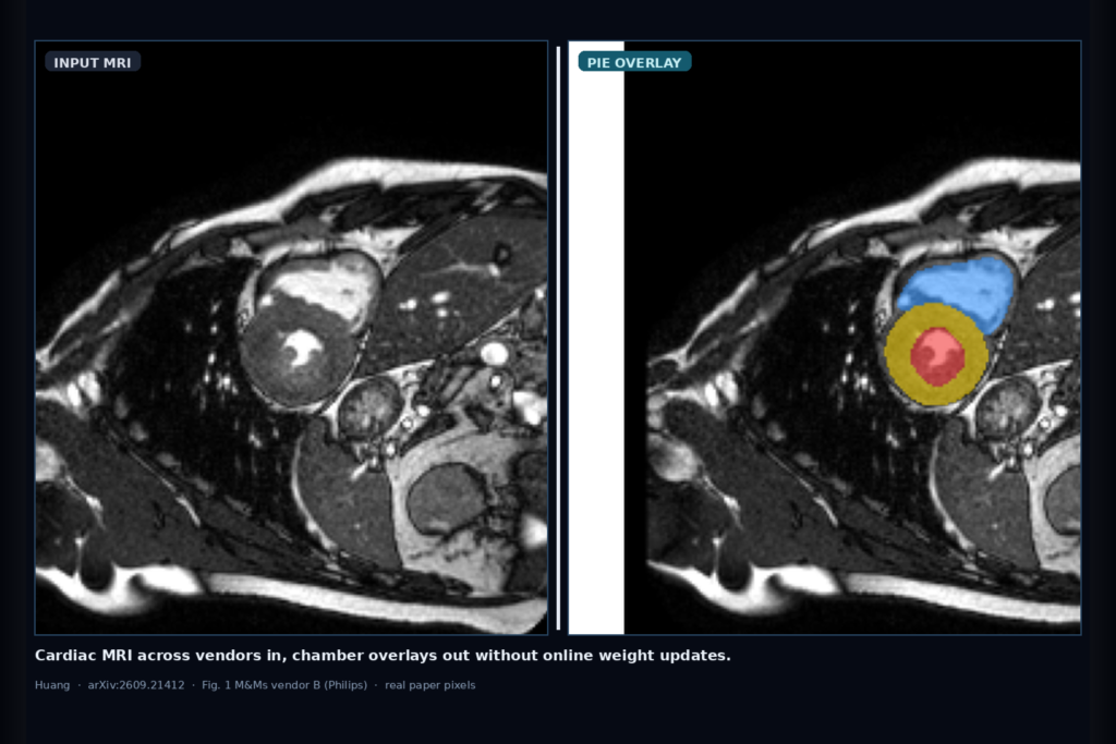
Hang LV, myocardium, and RV chamber overlays on cardiac MRI that arrived from a different scanner vendor than the one that trained your segmenter, without letting online weight updates rewrite the model on the fly. That is the clinic ask Huang takes on in arXiv:2609.21412, posted around mid September 2026. The paper is When Online Adaptation Hurts: Parameter-Frozen Test-Time Ensembling for Continual Medical Image Segmentation. The method is nicknamed PIE (parameter-frozen inference enhancement). A demo Space is at huggingface.co/spaces/RyanJoyice/medseg-ctta.
Continual test-time adaptation (CTTA) tries to fix vendor and protocol shift with unlabeled target data. On a non-stationary stream those updates can accumulate bad pseudo-labels and make normalization stats unreliable on small clinical batches. Huang asks how much you can get from the source segmenter if you never touch its weights or running statistics, and answers with a frozen scale-and-flip ensemble that maps each view back to native coordinates and averages probabilities.
What hangs on the viewer
Upstream is a short-axis cardiac MRI slice (here resized to 224 × 224) from the M&Ms multi-vendor stream. Downstream you get a four-class overlay: background, left-ventricular cavity (LV), myocardium (MYO), and right-ventricular cavity (RV). For a DICOM viewer or cardiac AI rail, that means chamber masks that stay tied to the source-trained model while still averaging complementary geometric views of the same frame. The quantitative protocol trains on vendor A (Siemens), then runs B (Philips) → C (GE) → D (Canon) in order. Qualitative examples also cover REFUGE2 fundus disc/cup nesting; those are illustrative only, not scored in the main table.
How it works in plain words
PIE keeps θ_t = θ_0 for every time step, including frozen batch-norm statistics. At inference it builds a set G of invertible spatial transforms: seven scales from 0.7 to 1.3 and four flip states (identity, horizontal, vertical, both), for 28 views. Each view is segmented independently, resized predictions are restored to H × W, probabilities are averaged uniformly, then argmax gives the label map. There is no target loss, optimizer, replay buffer, pseudo-label threshold, or source-restoration hyperparameter. Cost is 28 forward passes that can be batched.
The project also ships a lightweight anatomy-prior network for visualization. Reported PIE outputs come from the frozen ensemble, not from that prior. Confidence weighting, class-prior reweighting, largest-component filtering, morphological refinement, probability sharpening, and 3D inter-slice smoothing were tried and dropped when they were neutral or harmful.
What the numbers say
On the M&Ms B→C→D stream, PIE mean Dice is 0.7786 versus 0.7680 for source-only inference. Five online baselines land lower: SAR 0.7416, EATA 0.7409, TENT 0.7406, CoTTA 0.7388, BN-Adapt 0.6744. Per-vendor PIE Dice is 0.8025 (B), 0.7584 (C), 0.7395 (D). Per-class means are LV 0.8439, MYO 0.7575, RV 0.7343. Source-only beats every update-based method in aggregate on this stream, which is the paper’s warning shot about unsupervised online updates.
Ablations saturate at 28 views (mean Dice about 0.7816 in the multi-scale TTA core). Extending to 36 views adds cost without a gain. Class-prior reweighting and 3D slice smoothing show the largest negative transfer (−0.0125 and −0.0096 vs the full multi-scale core). Ninety-degree rotations help MYO but can hurt RV, so the retained PIE config stays scale plus flip. The manuscript reports point estimates without patient-level intervals or multi-seed runs, and flags itself as a structured research draft rather than finished clinical validation.
Where it fails and what not to trust
PIE cannot learn a genuinely new target representation; it only averages invertible views of the frozen source function. Cost scales with view count. Residual errors on cardiac MRI still sit near class boundaries. Largest-component filters can delete valid disconnected regions; morphology can nibble thin walls; slice smoothing can erase abrupt anatomy. Prostate MRI and fundus tasks appear only qualitatively. Rebuild on your own vendor mix, measure latency on the hardware you ship, and treat the Hugging Face Space as a visualization prototype, not a medical device.
For a viewer or clinic AI shop
Wire short-axis cardiac MRI in, LV/MYO/RV overlay out, with a frozen multi-view ensemble at inference instead of an online adapter on the live stream. Prefer hanging overlays when the source model already covers the anatomy you care about, and treat CTTA weight updates as optional extras you have to justify against a strong frozen baseline. Surface vendor tags next to the overlay so readers know which scanner the frame came from. Audit against source-only and against any adapter you still want to ship, on a hold-out that matches your acquisition mix.
Rebuild from arXiv:2609.21412. Demo: https://huggingface.co/spaces/RyanJoyice/medseg-ctta.
Sources
- Huang, R. When Online Adaptation Hurts: Parameter-Frozen Test-Time Ensembling for Continual Medical Image Segmentation. arXiv:2609.21412, 2026. https://arxiv.org/abs/2609.21412. PDF: https://arxiv.org/pdf/2609.21412.
- Demo Space: huggingface.co/spaces/RyanJoyice/medseg-ctta (prototype, not a medical device).
- Dataset: M&Ms multi-centre, multi-vendor cardiac MRI challenge (Campello et al., IEEE TMI 2021). Related CTTA baselines: TENT, EATA, SAR, CoTTA, BN-Adapt.