
Congenital heart disease work often hangs on a patient-specific 3D model. Chambers and great vessels get painted on CT or CMR, then the mesh drives surgical planning or catheter paths. That paint job is still mostly manual when anatomy is weird. Deep models promise to fill the masks, but most papers only test in-distribution: same dataset, same scanners, same labeling style. Aniketh Vijesh, Shrisharanyan Vasu, Abhijit Ramesh, Clare Pomeroy-Ward, Harikrishnan Anil Maya, Sarin Xavier, Mahesh Kappanayil, and Gilad Gressel (Amrita Vishwa Vidyapeetham with Amrita Institute of Medical Sciences, Kochi) posted arXiv:2609.17068 on 15 September 2026. The paper is Beyond In-Distribution Metrics: A Systematic Out-of-Distribution Evaluation of Congenital Heart Disease Segmentation, accepted at STACOM 2026 with MICCAI.
Their punchline is blunt for anyone shipping cardiac overlays. The model that looks best on the home validation split can be the one that falls apart on a held-out public CHD CT cohort. Architecture choice and a handful of target-site labels matter more than polishing the in-distribution Dice board.
What hangs on the viewer
Input is a cardiac CT or CMR volume from a CHD study. Output is multi-structure overlays for left and right ventricles, left and right atria, aorta, and pulmonary artery. In a DICOM viewer that is six colored masks on the axial cine or MPR, plus the mesh you export for planning. The authors train and compare nnU-Net, Zhu-Net, CardiacSeg, and SwinUNETR (from scratch and with MAE or JEPA pretraining) under combined CT+CMR training, CT-only training, and progressive fine-tuning on labeled ImageCHD cases.
ImageCHD is the held-out OOD target: public contrast-enhanced CT with septal and other CHD phenotypes never used as the training home. Training draws from a private 3D-Labs CT cohort and HVSMR-2.0 CMR. The viewer-facing question is simple. If you hang these overlays in a second hospital, do the chambers still look like chambers, or do you get structure-level nonsense that a surgeon should never trust?
In-distribution scores hide the site break
On the authors’ in-distribution validation, nnU-Net leads with about 0.77 Dice. On ImageCHD that same setup drops to about 0.51. SwinUNETR trained from scratch lands around 0.67 on ImageCHD with HD95 near 42.7 mm. MAE and JEPA pretraining nudge Swin only modestly (about 0.68 and 0.67). So the headline for a clinic shop is not “transformers always win.” It is that leaderboard order on the home split is a poor proxy for cross-cohort transfer in CHD.
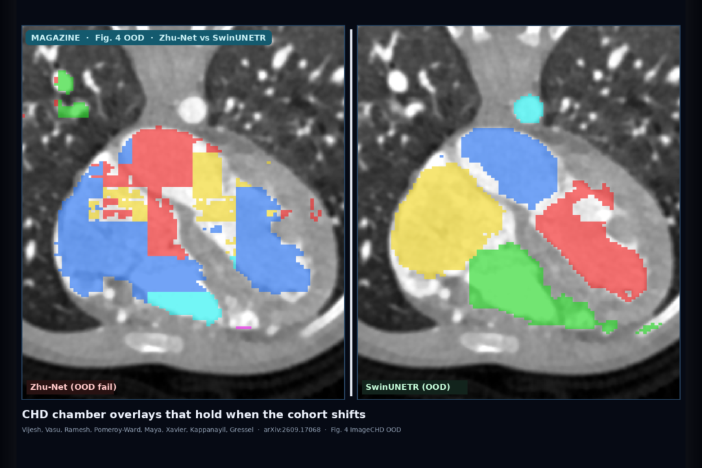
Qualitative Fig. 4 matches that story. On HVSMR and 3D-Labs, most methods look usable. On the ImageCHD OOD row, Zhu-Net, nnU-Net, and CardiacSeg show broken chamber topology, while SwinUNETR variants keep coherent LV/RV/LA/RA and vessel blobs. That is the failure mode a viewer QA gate should catch: not a 2-point Dice dip, but a mask that no longer respects chamber boundaries.
Why multi-modal training and a few target labels help
When they drop HVSMR-2.0 and train CT-only on 3D-Labs, ImageCHD performance falls further. nnU-Net drops from about 0.51 to 0.36. SwinUNETR slips from about 0.67 to 0.64. Joint CT/CMR training is not a magic domain adapter, but it buys a bit of anatomy prior that survives the next CT site better than CT-only specialization.
The practical lever is target-domain adaptation. Adding labeled ImageCHD cases in steps of 11, all SwinUNETR variants clear about 0.76 Dice after the first tranche on a fixed 22-case holdout. Zhu-Net and nnU-Net jump after the first labels too, but they do not consistently match Swin in the low-label regime. For a hospital that can spare a small annotated pilot set, that is the difference between hanging a home-site model and hanging one that has seen eleven local CHD CTs.
Where it still fails
CHD is long-tailed. Rare combinations, unusual vessel routing, and scanner-specific contrast timing still break overlays even when average Dice looks fine. Structure-wise tables in the paper show uneven OOD behavior across chambers and vessels. MAE and JEPA help only a little here, so do not treat self-supervised pretraining as a substitute for an OOD holdout or a local adaptation set. The study is an evaluation protocol, not a cleared planning device. Labels, ROI crops, and intensity normalization still have to match how your site builds ground truth.
Fail closed when the overlay topology looks wrong on a quick radiologist glance, when the study modality or age band sits outside the training mix, or when someone wants silent auto-meshes for surgical planning without a human review of the six masks.
For a viewer or clinic AI shop
Hang CHD segmentation as a gated overlay rail: volume in, multi-chamber and vessel masks out, human edit in, mesh export only after QC. Prefer SwinUNETR-class backbones when you already know the next site will not match your training cohort. Budget for an ImageCHD-style held-out test before go-live, and for about a dozen labeled local cases if the first OOD check fails. Keep nnU-Net in the bake-off for in-distribution speed, but do not ship it on OOD Dice alone.
Rebuild the comparison from arXiv:2609.17068. As of 19 September 2026 the abstract and PDF respond. The authors frame this as the first systematic OOD evaluation for CHD segmentation and argue that cross-dataset testing should sit next to in-distribution metrics whenever the overlay feeds planning.
Sources
- Vijesh, A., Vasu, S., Ramesh, A., Pomeroy-Ward, C., Maya, H. A., Xavier, S., Kappanayil, M., Gressel, G. Beyond In-Distribution Metrics: A Systematic Out-of-Distribution Evaluation of Congenital Heart Disease Segmentation. arXiv:2609.17068, posted 15 September 2026 (STACOM 2026 / MICCAI). https://arxiv.org/abs/2609.17068. PDF: https://arxiv.org/pdf/2609.17068.
- ImageCHD public CT cohort; HVSMR-2.0 CMR; private 3D-Labs CT (as described in the paper).
- nnU-Net, SwinUNETR, CardiacSeg, Zhu-Net (cited baselines and architectures).