
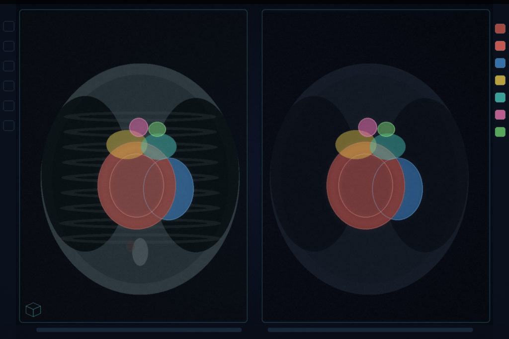
Hang a cardiac CT or MRI and you want the same seven overlays: left-ventricular myocardium, left atrium, left ventricle, right atrium, right ventricle, ascending aorta, pulmonary artery. LISynSeg keeps a 3D full-resolution nnU-Net and mixes real CARE volumes with images synthesized from those seven-class label maps. Jiacheng Wang and Ipek Oguz (Vanderbilt University), with Ivana Isgum (Mayo Clinic), posted arXiv:2608.31073 on 31 August 2026. The paper is LISynSeg: Data-Centric Label-to-Image Synthesis for Cross-Modality Whole-Heart Segmentation. The claimed code URL github.com/MedICL-VU/Care26_LISynSeg returned 404 when checked on 1 September 2026.
Inference takes one preprocessed image channel. There is no modality flag and no synthesis step at test time. The same 3D configuration is used for CT and MRI. CARE-WHS labels still vary by site: myocardium thickness at the Myo-LV interface, how far the AO and PA annotations run, and whether the scan is a tight cardiac CT or a wider thoracoabdominal MRI.
Ten percent synthetic, ninety percent real
After nnU-Net spatial and intensity augmentation, each training example is replaced with a label-to-image synthesis with probability 0.10. The synthesizer paints Gaussian intensities on the seven cardiac labels plus background, then adds partial volume, a bias field, gamma variation, noise, and a resolution profile sampled from native-to-target spacing ratios in the current fold (capped at a factor of three). Extra affine or elastic warp inside the synthesizer is skipped because nnU-Net already warped the pair.
The seven labels do not describe lung, fat, bone, or other thoracic tissue, so a volume synthesized from labels alone is missing that context. Training on synthetic images only drops MRI Dice by 5.87 percentage points versus the real-image nnU-Net. Mixing real and synthetic keeps the thorax that the labels omit, and adds contrast and acquisition variation that MRI needs more than CT.
Wall thickness on the synthetic branch, distal vessels ignored in the loss
Myocardial thickness is a site-level difference that global elastic warp barely controls. On synthetic examples only, LISynSeg exchanges a smooth random subset of the Myo-LV interface: thicken by moving LV voxels into myocardium, or thin the other way, with equal probability. Combined Myo-plus-LV extent stays fixed. Volume ratios are clamped (Myo in [0.72, 1.35], LV in [0.80, 1.20]). If a patch has no Myo or LV, the map is left alone.
AO and PA endpoints in the released CARE labels do not sit on a consistent anatomical landmark. Some AO maps stop in the ascending segment; others continue through the arch. Some PA maps stop before the bifurcation; others continue past it. The CARE protocol wants AO from the aortic valve to the superior atrial level, and PA from the pulmonary valve to the bifurcation. The loss still sees every voxel as a hard target. LISynSeg finds the proximal vessel from AO-LV and PA-RV contact, keeps the closest 90 percent of annotated vessel voxels in the Dice-plus-cross-entropy loss, and ignores the distal remainder plus nearby background. Masking is skipped if the distal set would exceed 40 percent of the vessel, or if the patch has no ventricular contact. It runs on every training example, real or synthetic, and is not used at validation or inference.
CARE-WHS split, and what actually moved
Released CARE-WHS training is 106 labeled volumes from five site groups: 60 CT and 46 MRI. Models train on both modalities together. The development split used for the controlled tables is 85 train and 21 val (12 CT, 9 MRI), 200 epochs, largest-component postprocessing per label. Real-only nnU-Net already sits high: overall Dice 0.9203. Full LISynSeg is 0.9219. The MRI column moves more than CT. Myo-LV exchange alone, without vessel masking, hurts MRI. The authors point to qualitative cases rather than that leaderboard delta: a CT Site-B Myo-LV interface that was under-segmenting LV, an MRI AO that was running too far, an MRI PA that was stopping short.
A separate official-validation ensemble (real-only, real-plus-synthetic, and a DA5 setting) ranked in the top five on the combined CARE 2026 track at the time of writing. That ranking is the ensemble, not the isolated data recipe in Tables 1 and 2.
How this lands in a viewer
If you already hang cardiac CT and MRI in a DICOM viewer, treat this as one seven-class overlay rail on a mixed-modality worklist. Load the volume, run the same 3D nnU-Net, hang myocardium, chambers, AO, and PA. Do not expect the overlay to resolve a site’s preferred AO or PA endpoint; distal extent was ignored in the loss on purpose. Fail closed when the FOV misses the ventricles, so proximal AO-LV or PA-RV contact cannot be established, or when weights are not yet public. Do not treat a 0.16-point overall Dice move on 21 validation cases as a device claim.
Cine aorta contours keep ascending and descending aorta overlays through systole. SLIP is interactive 3D segmentation with latency and undo. SAT3D is tumour plus uncertainty in Slicer. This paper is cardiac CT or MRI in, seven whole-heart overlays out, no modality flag.
Rebuild from arXiv:2608.31073 when code and weights land. As of 1 September 2026 the GitHub URL in the abstract still 404s. The preprint is the source of record until a camera-ready version exists.
Sources
- Wang, J., Isgum, I., Oguz, I. LISynSeg: Data-Centric Label-to-Image Synthesis for Cross-Modality Whole-Heart Segmentation. arXiv:2608.31073, posted 31 August 2026. https://arxiv.org/abs/2608.31073. Claimed code: github.com/MedICL-VU/Care26_LISynSeg (HTTP 404 on 1 September 2026).
- CARE 2026 Organizers. CARE 2026 Whole Heart Segmentation Track. https://www.zmic.org.cn/care_2026/track_wholeheart/. Accessed 25 June 2026 in the paper.
- Billot, B., et al. SynthSeg: Segmentation of brain MRI scans of any contrast and resolution without retraining. Med. Image Anal. 2023. DOI: 10.1016/j.media.2023.102789
- Zhuang, X., et al. Evaluation of algorithms for multi-modality whole heart segmentation: An open-access grand challenge. Med. Image Anal. 2019. DOI: 10.1016/j.media.2019.101537
- Isensee, F., et al. nnU-Net: a self-configuring method for deep learning-based biomedical image segmentation. Nat. Methods 2021. DOI: 10.1038/s41592-020-01008-z
- Zhuang, X. Multivariate mixture model for myocardial segmentation combining multi-source images. IEEE Trans. Pattern Anal. Mach. Intell. 2019. DOI: 10.1109/TPAMI.2018.2869576
- Zhuang, X., Shen, J. Multi-scale patch and multi-modality atlases for whole heart segmentation of MRI. Med. Image Anal. 2016. DOI: 10.1016/j.media.2016.02.006
- Hatamizadeh, A., et al. UNETR: Transformers for 3D medical image segmentation. WACV 2022.
- Çiçek, Ö., et al. 3D U-Net: Learning dense volumetric segmentation from sparse annotation. MICCAI 2016. DOI: 10.1007/978-3-319-46723-8_49