
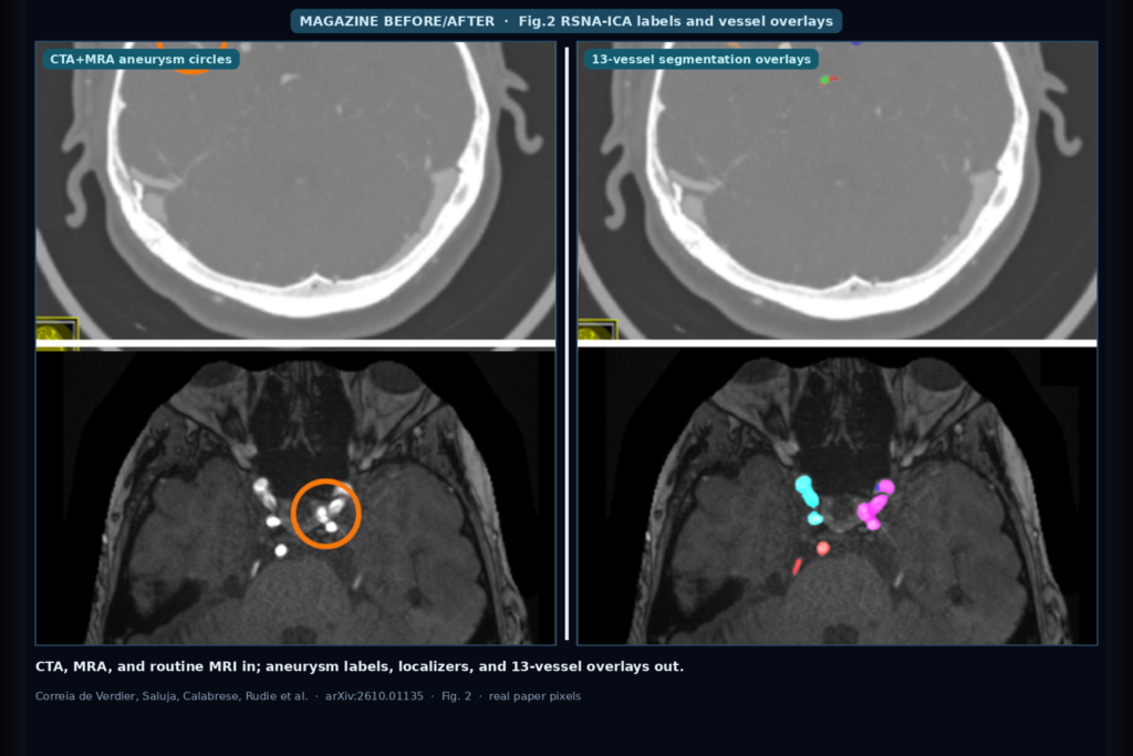
Hang a head CTA, TOF MRA, or even a routine T2 / T1-post brain MRI into a DICOM viewer and the shop question is not another single-site MRA CSV. It is whether aneurysm presence and the 13 challenge-defined vascular locations come with the series, whether a crosshair localizer lands on the matching SOPInstanceUID slice, and whether a subset of cases also ships NIfTI overlays for those vessels so a Circle-of-Willis hang is more than a yellow bounding box. Maria Correia de Verdier, Rachit Saluja, Evan Calabrese, Jeffrey D. Rudie, and co-authors take that hang seriously in arXiv:2610.01135. They release the RSNA Intracranial Aneurysm (RSNA-ICA) Dataset: 7202 CTA / MRA / MRI series from 4278 adult patients across 21 institutions, 12 countries, and five continents, built with ASNR, SNIS, and ESNR for the 2025 RSNA Intracranial Aneurysm Detection Challenge.
What hangs on the viewer
Upstream inputs are DICOM series organized by SeriesInstanceUID, with SOPInstanceUID-named frames that order by position in the series. Downstream hangables for a viewer or clinic AI shop include series-level aneurysm presence and location labels (13 challenge-defined vascular locations), per-aneurysm crosshair localizers keyed to the DICOM frame, and, for 178 series, 3D NIfTI segmentations of those same 13 arterial segments. Modality mix is deliberate: 2566 CTA, 2166 MRA, and 2470 MRI, including routine non-angiographic brain MRI (T2-weighted and post-contrast T1) where vessels show as flow voids or enhancement. That last piece is the opportunistic screening angle: aneurysms can appear on exams ordered for other indications, not only on dedicated CTA or MRA.
Public release is through MIRA at https://mira.rsna.org/dataset/7: 4346 challenge training series after quality exclusions, plus a 695-series post-challenge out-of-distribution cohort from three sites that never entered the original challenge. The original challenge training set also remains on Kaggle. Labels arrive as CSV (dataset.csv / dataset_localizers.csv on MIRA; train.csv / train_localizers.csv on Kaggle). PatientGroupID and StudyGroupID link series across modalities for the same patient.
How it works in plain words
Sites contributed series with initial presence and location labels from clinical reports, using the 13 locations: infraclinoid ICA left/right, supraclinoid ICA left/right, MCA left/right, ACOM, ACA left/right, PCOM left/right, basilar tip, and other posterior circulation. Sixty-five attending radiologists from 20 countries then independently annotated presence and location and placed a crosshair at each aneurysm center on a web platform, after a scored training module. Discrepancies between site labels and independent reads went to high-performing adjudicators who saw both label sets plus the images. Indeterminate cases after adjudication were marked negative to cut false positives.
The 178-series vessel subset used MONAI Label with a TopCoW-pretrained model, iterative refinement, and neuroradiologist review from the organizing committee. When an aneurysm was visible on the segmented series, it was included inside the parent-vessel label. That gives a viewer shop a real overlay path for Circle-of-Willis anatomy on a curated subset, while the bulk of the dataset stays at presence, location, and crosshair localizer density that scales to thousands of series.
What the numbers say
Across the full collection: 7202 series, 4278 adults, 21 institutions, 12 countries, five continents. Aneurysms: 4099 lesions in 2261 patients, about 1.8 per positive patient, split across training (2252), public test (397), private test (1093), and the post-challenge OOD set (357). Challenge splits assigned all exams and modalities for one patient to the same fold. After post-challenge QC, two artifact-heavy series left the training release, leaving 4346 training series on MIRA. Field of view always covers the Circle of Willis, with variable neck and distal coverage that mirrors real protocol choices. Acquisition and reconstruction parameters stay heterogeneous on purpose so models see clinic-like DICOM, not a single scanner cookie-cutter.
Where it fails and what not to trust
There are no rupture or subarachnoid-hemorrhage annotations. Enrichment for rarer locations and a higher hemorrhage prevalence in the aneurysm-positive group can bias detectors toward predicting aneurysm when blood is visible. Small aneurysms under 3 mm remain hard to label, especially versus arterial infundibula; conservative adjudication may leave some tiny true positives as negatives. Location grouping is coarse by design: clinoid / ophthalmic / supraclinoid ICA segments collapse into the intradural-vs-extradural split the challenge needed, and non-basilar-tip posterior circulation collapses into one label. Border aneurysms (for example at the carotid terminus) are ambiguous. Annotations were done on a web platform without clinical PACS monitors, multiplanar thin stacks, priors, or full clinical context. Noncommercial research use only. Re-validate localizer-to-SOP mapping and NIfTI overlays on your own viewer before you treat this as production ground truth.
For a viewer or clinic AI shop
Wire CTA, MRA, and routine brain MRI DICOM in; hang series-level presence and 13-location labels, crosshair localizers on the matching frames, and optional 13-vessel NIfTI overlays on the 178-series subset out. Prefer this dataset when you need multi-modality aneurysm detection that includes opportunistic MRI, when you want public multi-continent DICOM rather than a few hundred same-site MRAs, and when a challenge-grade CSV plus localizer path is enough to stand up a hangable demo before you invest in full voxel masks everywhere.
If you integrate, keep patient-level grouping so CTA and MRI from the same adult do not leak across train and test. Do not invent rupture risk from this release. Start from MIRA https://mira.rsna.org/dataset/7 or the Kaggle challenge mirror, and rebuild from arXiv:2610.01135. PDF: https://arxiv.org/pdf/2610.01135.
Sources
- Correia de Verdier, M., Saluja, R., Calabrese, E., Rudie, J.D., et al. The RSNA Intracranial Aneurysm (RSNA-ICA) Dataset. arXiv:2610.01135, 2026. https://arxiv.org/abs/2610.01135. PDF: https://arxiv.org/pdf/2610.01135.
- Counts: 7202 series (2566 CTA, 2166 MRA, 2470 MRI) from 4278 adults; 21 institutions, 12 countries, 5 continents; 4099 aneurysms in 2261 patients (~1.8/patient); 178 series with 13-location NIfTI segs.
- Public: MIRA https://mira.rsna.org/dataset/7 (4346 training + 695 OOD); Kaggle challenge training mirror. Built for 2025 RSNA Intracranial Aneurysm Detection Challenge with ASNR/SNIS/ESNR. Noncommercial research use.