Viewing Medical Images Made Easy
The Digital Imaging and Communications in Medicine (DICOM) standard has transformed medical imaging, enabling seamless handling of X-rays, CT scans, MRIs, and ultrasounds. Before DICOM emerged, healthcare professionals struggled with incompatible systems using proprietary formats and physical media for image exchange. Developed in the late 1980s through collaboration between the American College of Radiology (ACR) and the National Electrical Manufacturers Association (NEMA), DICOM created a common language for medical imaging. This breakthrough enabled true interoperability and digital advancement in radiology. Standardization proved essential for improving patient care, research capabilities, and medical education. A good DICOM viewer delivers smooth access, manipulation, and analysis of these images regardless of their origin. The most helpful viewers offer intuitive navigation, powerful visualization tools like 3D rendering and multiplanar reconstruction, plus comprehensive measurement and annotation capabilities. For any organization working with medical imaging data, understanding DICOM viewer software is essential. Whether you’re developing medical devices, building healthcare tech platforms, conducting research, or managing hospital IT systems, choosing the right one directly impacts your workflow efficiency and diagnostic accuracy. In this article, we’ll examine seven outstanding DICOM viewer options, exploring their features, benefits, and best use cases. By the end, you’ll have the information needed to select the perfect solution for your specific requirements and budget constraints.1. OsiriX MD
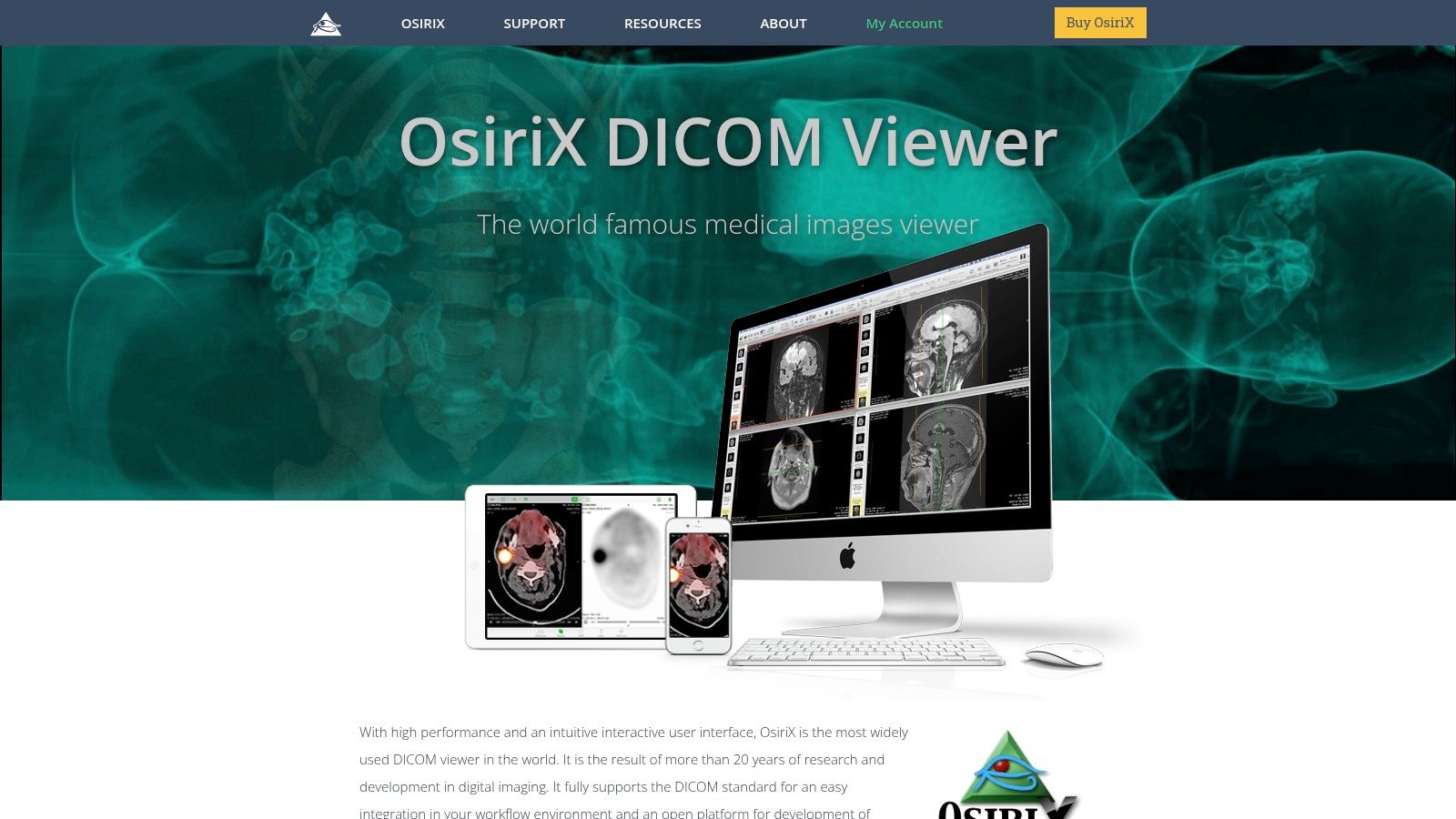 OsiriX MD stands out as a powerful DICOM viewer designed specifically for the macOS ecosystem. Its comprehensive toolset addresses the needs of medical professionals, researchers, and healthcare institutions working with medical imaging. While it comes with a premium price tag, its advanced capabilities and Mac-friendly interface make it an excellent choice for those who prioritize performance and seamless integration with Apple devices.
More than just a basic viewer, OsiriX MD functions as a complete PACS workstation, allowing users to view, manage, process, and analyze DICOM images. The software offers advanced 3D/4D visualization tools, including MPR, MIP, volume rendering, and surface rendering, giving clinicians detailed anatomical insights for more accurate diagnosis and treatment planning. Researchers can use these same tools for in-depth image analysis and data extraction. Its built-in DICOM PACS server functionality with query/retrieve capabilities makes image management and sharing straightforward in clinical or research environments.
Key Features and Benefits:
OsiriX MD stands out as a powerful DICOM viewer designed specifically for the macOS ecosystem. Its comprehensive toolset addresses the needs of medical professionals, researchers, and healthcare institutions working with medical imaging. While it comes with a premium price tag, its advanced capabilities and Mac-friendly interface make it an excellent choice for those who prioritize performance and seamless integration with Apple devices.
More than just a basic viewer, OsiriX MD functions as a complete PACS workstation, allowing users to view, manage, process, and analyze DICOM images. The software offers advanced 3D/4D visualization tools, including MPR, MIP, volume rendering, and surface rendering, giving clinicians detailed anatomical insights for more accurate diagnosis and treatment planning. Researchers can use these same tools for in-depth image analysis and data extraction. Its built-in DICOM PACS server functionality with query/retrieve capabilities makes image management and sharing straightforward in clinical or research environments.
Key Features and Benefits:
- Advanced 3D/4D Post-Processing: Provides detailed anatomical visualization for accurate diagnostics and surgical planning
- DICOM PACS Server Functionality: Makes image management simpler and enables smooth data exchange
- Support for All DICOM Modalities: Works with various imaging data types including CT, MR, US, PET, and SPECT
- Powerful Image Fusion and Registration: Combines images from different modalities for a more complete view of patient anatomy
- AI-Assisted Annotation and Segmentation: Boosts workflow efficiency and accuracy during image analysis
- Intuitive Mac-centric Interface: Delivers a smooth, user-friendly experience for Apple users
- Powerful Rendering Capabilities: Offers high-quality image rendering comparable to specialized workstations
- Excellent Performance with Large Datasets: Handles complex and large image files without slowdowns
- Regular Updates with New Features: Keeps the software current with the latest medical imaging advancements
- macOS Exclusivity: Only available for Apple devices
- High Cost: Requires significant investment compared to free alternatives
- Steep Learning Curve for Advanced Features: Needs dedicated training to fully use all capabilities
- Limited Free Version (Lite): The free version has restricted functionality, making the full version necessary for professional use
- Download OsiriX MD from the official website and follow the installation guide
- Review the user manual and tutorials to learn the software’s interface and functions
- Begin with smaller datasets to practice navigation and image manipulation
- Consider attending training sessions to master advanced features and optimize your workflow
2. Horos
 Horos is a powerful, free, and open-source designed specifically for macOS users. Developed as an offshoot of the respected OsiriX platform, it delivers an impressive range of capabilities that rival paid solutions, making it an excellent choice for various medical imaging applications outside primary diagnosis.
This makes Horos particularly valuable in settings with budget constraints, such as research laboratories, educational institutions, and early-stage medtech companies. Many physicians also find it useful as a secondary review tool during consultations, case discussions, and teaching sessions.
One of Horos’s standout features is its advanced visualization toolkit. Users can access 3D and 4D rendering capabilities, including Multiplanar Reformatting (MPR), Maximum Intensity Projection (MIP), and volume rendering. These tools allow detailed manipulation and exploration of image datasets, helping to reveal subtle anatomical structures and enhance understanding of complex pathologies.
Medical device manufacturers and healthcare technology developers often use Horos to evaluate new imaging technologies and algorithms. For researchers, it provides robust capabilities for image analysis and processing that can support various study designs.
Horos includes a built-in DICOM database with effective search functionality that streamlines management of large datasets. It also offers basic PACS server capabilities, enabling image sharing within local networks—especially beneficial for smaller clinics or research teams seeking cost-effective solutions for image storage and distribution.
The software’s plugin architecture supports customization and expansion to meet specific requirements. Additionally, Horos is compatible with all common DICOM modalities, ensuring it works with a wide range of medical imaging equipment.
Key Features:
Horos is a powerful, free, and open-source designed specifically for macOS users. Developed as an offshoot of the respected OsiriX platform, it delivers an impressive range of capabilities that rival paid solutions, making it an excellent choice for various medical imaging applications outside primary diagnosis.
This makes Horos particularly valuable in settings with budget constraints, such as research laboratories, educational institutions, and early-stage medtech companies. Many physicians also find it useful as a secondary review tool during consultations, case discussions, and teaching sessions.
One of Horos’s standout features is its advanced visualization toolkit. Users can access 3D and 4D rendering capabilities, including Multiplanar Reformatting (MPR), Maximum Intensity Projection (MIP), and volume rendering. These tools allow detailed manipulation and exploration of image datasets, helping to reveal subtle anatomical structures and enhance understanding of complex pathologies.
Medical device manufacturers and healthcare technology developers often use Horos to evaluate new imaging technologies and algorithms. For researchers, it provides robust capabilities for image analysis and processing that can support various study designs.
Horos includes a built-in DICOM database with effective search functionality that streamlines management of large datasets. It also offers basic PACS server capabilities, enabling image sharing within local networks—especially beneficial for smaller clinics or research teams seeking cost-effective solutions for image storage and distribution.
The software’s plugin architecture supports customization and expansion to meet specific requirements. Additionally, Horos is compatible with all common DICOM modalities, ensuring it works with a wide range of medical imaging equipment.
Key Features:
- 3D/4D rendering (MPR, MIP, Volume Rendering)
- DICOM database with search functionality
- Basic PACS server functionality
- Plugin architecture
- Support for all common DICOM modalities
- Free and open-source: Eliminates licensing costs, making it highly accessible.
- Feature-rich: Offers a comparable feature set to commercial DICOM viewers.
- Active community support: Benefits from ongoing development and user contributions.
- User-friendly interface: Easy to navigate and customize.
- macOS only: Limits its use to Apple environments.
- Not FDA-cleared for primary diagnosis: Cannot be used for making primary diagnostic decisions.
- Less frequent updates compared to OsiriX MD: May lag behind in incorporating the latest features.
- Limited support options: Relies primarily on community support rather than dedicated technical assistance.
3. RadiAnt DICOM Viewer
RadiAnt DICOM Viewer stands out for its exceptional combination of speed, functionality, and cost-effectiveness. This powerful tool is particularly valuable for medical professionals, researchers, and educators who need quick access to DICOM images without investing in expensive premium software. Though not FDA-cleared for primary diagnosis in the US, it excels as a solution for secondary reviews, consultations, teaching, and research applications.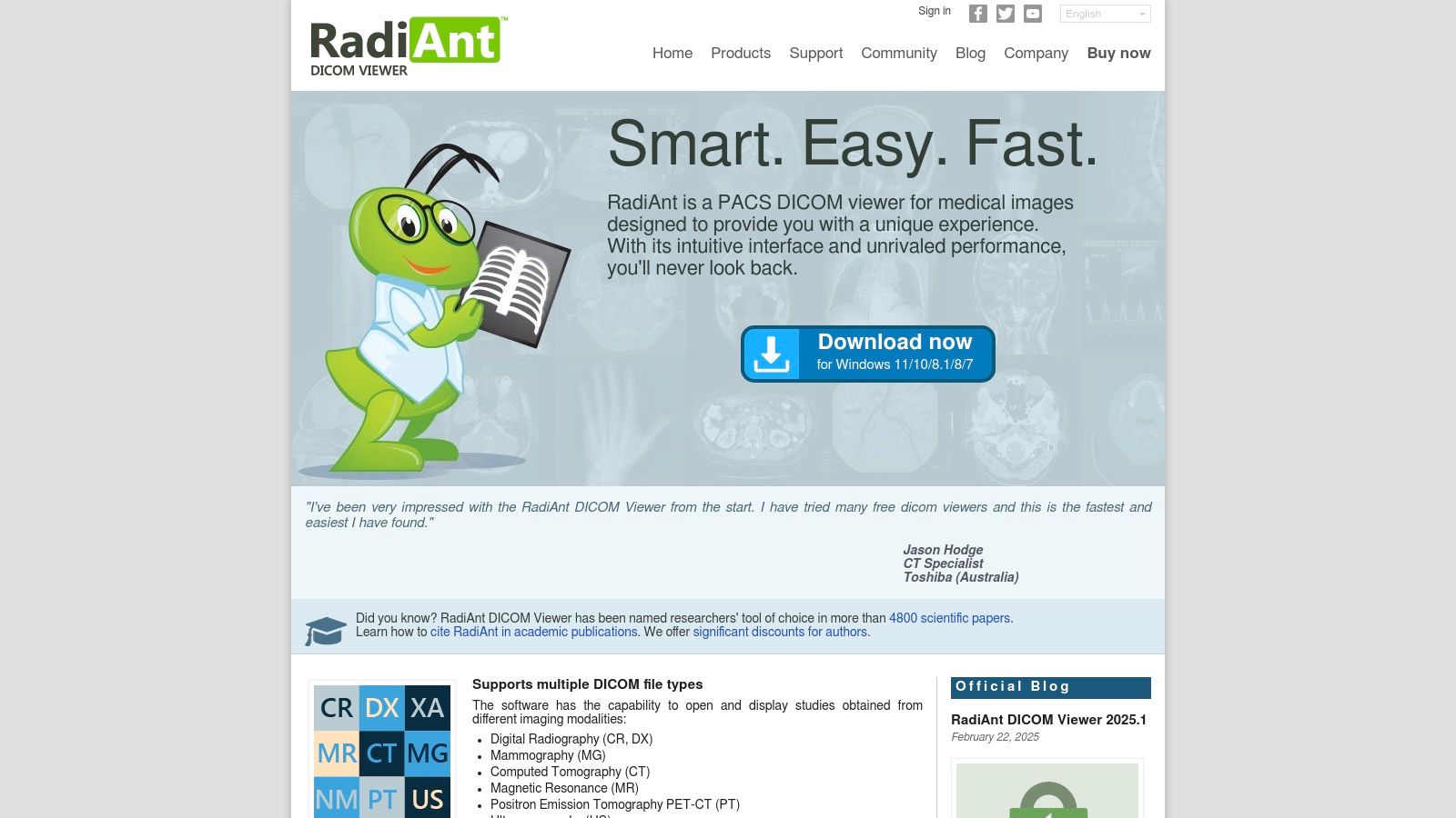 This Windows-based viewer handles large datasets with impressive efficiency thanks to its optimized architecture. Consider a scenario where you need to review a series of high-resolution CT scans during a team meeting – RadiAnt allows you to navigate and manipulate images smoothly without the frustrating lag common in other viewers. This responsiveness is essential in fast-paced medical environments where every second counts.
Beyond its speed, RadiAnt offers an impressive range of clinical tools:
This Windows-based viewer handles large datasets with impressive efficiency thanks to its optimized architecture. Consider a scenario where you need to review a series of high-resolution CT scans during a team meeting – RadiAnt allows you to navigate and manipulate images smoothly without the frustrating lag common in other viewers. This responsiveness is essential in fast-paced medical environments where every second counts.
Beyond its speed, RadiAnt offers an impressive range of clinical tools:
- MPR (Multiplanar Reconstruction) with Oblique Planes: Allows visualization of anatomical structures from multiple angles, providing better understanding of complex pathologies. This feature is especially useful for surgical planning and studying anatomical relationships.
- Advanced Measurement Tools: Includes ROI, angle, and Cobb angle measurements, giving researchers and clinicians quantifiable data for analysis and tracking. These tools are particularly helpful for growth assessment, orthopedic measurements, and tumor volume calculations.
- DICOM Networking (C-STORE SCU/SCP, Q/R): Enables smooth integration with PACS and other DICOM modalities in hospital networks, improving workflow and data accessibility. This functionality is essential for efficient image sharing and archiving.
- DICOM CD/DVD Import and Anonymous Export: Makes data exchange simple while protecting patient privacy. This feature proves invaluable for researchers collaborating on projects or clinicians seeking second opinions.
- Fast performance with efficient memory usage: Handles complex imaging data smoothly even on basic hardware.
- Clean, intuitive user interface: Reduces learning time so users can focus on images rather than software.
- Budget-friendly licensing options: Provides excellent value, making it accessible to more users.
- Consistent updates with new features: Shows ongoing development commitment and responsiveness to user needs.
- Windows only (though usable on Mac via virtualization): Limited native compatibility with other operating systems.
- Less advanced 3D capabilities than premium alternatives: May not meet needs for specialized 3D rendering applications.
- Not FDA-cleared for primary diagnosis in the US: Cannot be used as the main diagnostic tool in US clinical settings.
4. MicroDicom
MicroDicom is free for Windows, which strikes an excellent balance between functionality and user-friendliness. Its comprehensive toolset makes it valuable for various users, from doctors reviewing patient scans to researchers analyzing medical imaging data. While it may not have all the advanced features of premium commercial viewers, its zero-cost approach and accessibility make it highly valuable for many practical applications. Practical Applications and Use Cases:
- Quick Image Review: MicroDicom shines at providing fast, easy access to DICOM images. Its clean interface gives users immediate access to essential viewing tools like windowing, zooming, and panning. This is perfect for clinicians who need to quickly review images without the complexity of a full PACS system.
- Educational Settings: The free availability makes MicroDicom an excellent choice for medical students and trainees learning about medical imaging. Students can practice with DICOM files and learn basic image manipulation without expensive software licenses.
- Research and Analysis: The software offers basic measurement tools (distance, angle, area) and limited 3D reconstruction via MPR, making it useful for many research applications. While not as robust as specialized research platforms, it’s a good fit for smaller projects or initial analyses.
- Image Sharing and Collaboration: The lightweight nature of the software makes sharing DICOM images with colleagues simple, especially for those who might not have access to advanced viewers.
- DICOM Image Viewing: Supports most common DICOM modalities with standard viewing tools like windowing, zooming, and panning.
- Basic Measurements: Includes tools for measuring distance, angle, and area within images.
- Limited 3D Capabilities: Offers MPR reconstruction for basic 3D visualization.
- DICOM Header Information: Shows complete DICOM header data for thorough image information.
- Image Processing and Annotations: Provides basic image processing filters and annotation tools.
- Free: No licensing costs whatsoever.
- Lightweight: Minimal system requirements allow it to run on older hardware.
- Simple Interface: Easy to learn and navigate, even for users with limited technical skills.
- Wide Modality Support: Handles most common DICOM modalities effectively.
- Limited Advanced Visualization: Lacks sophisticated features found in commercial viewers.
- No PACS Integration: Doesn’t function as a PACS server or client.
- Windows Only: Not available for Mac or Linux systems.
- Infrequent Updates: Updates are less regular compared to paid alternatives.
5. 3D Slicer
 3D Slicer stands out as the leading open-source platform for advanced DICOM visualization and analysis. While it functions as a basic viewer, its true value lies in its comprehensive toolkit for image processing, segmentation, and 3D rendering—making it essential for medical imaging research and development.
Going beyond simple viewing, 3D Slicer offers a solid foundation for manipulating and analyzing medical image data. Researchers, medical device manufacturers, and medtech startups can use its capabilities for:
3D Slicer stands out as the leading open-source platform for advanced DICOM visualization and analysis. While it functions as a basic viewer, its true value lies in its comprehensive toolkit for image processing, segmentation, and 3D rendering—making it essential for medical imaging research and development.
Going beyond simple viewing, 3D Slicer offers a solid foundation for manipulating and analyzing medical image data. Researchers, medical device manufacturers, and medtech startups can use its capabilities for:
- Developing and testing new image processing algorithms: The Python interface and extensive plugin ecosystem enable fast prototyping and integration with existing research workflows. This makes 3D Slicer perfect for experimenting with new segmentation techniques, registration methods, and image analysis processes.
- Creating customized visualizations for research publications and presentations: With advanced rendering capabilities, researchers can generate high-quality 3D models and visualizations of anatomical structures, surgical plans, and other medical data to improve communication and understanding.
- Pre-operative surgical planning and simulation: Though not FDA-cleared for clinical use, 3D Slicer can be used in research settings to explore potential surgical approaches and simulate procedures using patient-specific imaging data.
- Educational tool for medical imaging and informatics: Academic institutions can use 3D Slicer to teach students about medical image data, image processing techniques, and custom analysis script development.
- Advanced segmentation tools with semi-automated and AI-assisted options.
- Comprehensive 3D visualization capabilities.
- Cross-platform compatibility (Windows, macOS, Linux).
- Extensive plugin ecosystem for specialized tasks.
- Integration with programming environments for custom analysis workflows.
- Extremely powerful and versatile platform.
- Free and open-source with strong community support.
- Cross-platform availability.
- Integrates with other research tools and frameworks.
- Steep learning curve for new users. The interface can overwhelm those used to simpler.
- Interface is less clinical-oriented, focusing more on research functions.
- Not FDA-cleared for diagnostic use. Its application is mainly in research and development settings.
- Requires more computing resources than simpler viewers due to its advanced processing capabilities.
- Download the appropriate version for your operating system from the official website.
- Explore the extensive documentation and tutorials available online. The Slicer community forum provides valuable troubleshooting support.
- Start with the built-in tutorials to learn the interface and basic functions.
- Use the plugin ecosystem to extend the software’s capabilities for your specific research needs.
6. Weasis
 Weasis stands out as a versatile, open-source DICOM viewer that excels in integration and cross-platform functionality. Built on Java, it runs smoothly across Windows, macOS, and Linux, making it an ideal solution for teams working in mixed computing environments. This accessibility is particularly valuable for medtech startups, academic institutions, and research facilities operating with budget constraints but needing reliable imaging tools.
What sets Weasis apart is its robust integration with PACS and HIS/RIS systems. This capability is essential for hospital IT departments looking to incorporate the viewer into their existing infrastructure without major disruptions. Medical device manufacturers and healthcare technology companies can use Weasis as a flexible platform to test and demonstrate their products’ compatibility within simulated hospital environments. Companies specializing in DICOM communication can also customize Weasis thanks to its open-source nature.
The software supports numerous image formats beyond DICOM, expanding its usefulness for researchers working with various medical imaging data types. Users appreciate the customizable interface that can be tailored to specific workflows, improving efficiency. The inclusion of basic 3D visualization and measurement tools makes Weasis a comprehensive yet lightweight viewing solution.
Features:
Weasis stands out as a versatile, open-source DICOM viewer that excels in integration and cross-platform functionality. Built on Java, it runs smoothly across Windows, macOS, and Linux, making it an ideal solution for teams working in mixed computing environments. This accessibility is particularly valuable for medtech startups, academic institutions, and research facilities operating with budget constraints but needing reliable imaging tools.
What sets Weasis apart is its robust integration with PACS and HIS/RIS systems. This capability is essential for hospital IT departments looking to incorporate the viewer into their existing infrastructure without major disruptions. Medical device manufacturers and healthcare technology companies can use Weasis as a flexible platform to test and demonstrate their products’ compatibility within simulated hospital environments. Companies specializing in DICOM communication can also customize Weasis thanks to its open-source nature.
The software supports numerous image formats beyond DICOM, expanding its usefulness for researchers working with various medical imaging data types. Users appreciate the customizable interface that can be tailored to specific workflows, improving efficiency. The inclusion of basic 3D visualization and measurement tools makes Weasis a comprehensive yet lightweight viewing solution.
Features:
- Cross-platform compatibility (Windows, macOS, Linux) through Java
- Integration capabilities with PACS and HIS/RIS systems
- Support for various image formats (including but not limited to DICOM)
- Customizable user interface with different layouts
- Basic 3D capabilities and measurement tools
- Standalone and web-based deployment options
- Multi-language support
- Free and open-source, eliminating cost barriers for startups and research institutions
- Works on all major operating systems, enabling collaborative workflows
- Can be deployed as a web-based viewer, supporting remote access and telehealth applications
- Strong integration options with hospital systems, simplifying data management
- Java dependency sometimes creates compatibility issues, requiring specific Java versions
- Performance may lag behind native applications when handling very large datasets
- Interface can be less intuitive than commercial alternatives, requiring more time to learn
- Limited advanced visualization features for complex 3D rendering or sophisticated image analysis
- Download the appropriate Weasis package for your operating system from the website.
- Ensure you have a compatible Java Runtime Environment (JRE) installed. Check the documentation for specific version requirements.
- For web-based deployment, follow the configuration guidelines in the Weasis server documentation.
- Explore the customization options to optimize the interface and tools for your specific needs.
7. Orthanc: A Lightweight DICOM Server and Web Viewer
Orthanc stands out among DICOM viewers by functioning as a complete, open-source, lightweight DICOM server with an integrated web viewer. While you can use it simply as a viewer, its real power lies in how it facilitates DICOM workflows within hospitals and research environments, making connections between different systems much easier. This makes it particularly valuable for organizations building research databases, secondary archives, or custom PACS solutions. Medical device manufacturers can use Orthanc as a testing platform for DICOM conformance and integration. Healthcare technology companies can incorporate it as the backend for their imaging applications, while researchers can use it to create and manage image repositories for studies. Hospital IT departments often find Orthanc useful for creating departmental PACS solutions or specialized image archives. For academic institutions, its open-source nature makes it excellent for educational purposes. MedTech startups looking to save costs can use Orthanc to speed up development while keeping infrastructure expenses down. Companies working with DICOM communication can develop customized solutions for their clients using this versatile platform.
Features:
Medical device manufacturers can use Orthanc as a testing platform for DICOM conformance and integration. Healthcare technology companies can incorporate it as the backend for their imaging applications, while researchers can use it to create and manage image repositories for studies. Hospital IT departments often find Orthanc useful for creating departmental PACS solutions or specialized image archives. For academic institutions, its open-source nature makes it excellent for educational purposes. MedTech startups looking to save costs can use Orthanc to speed up development while keeping infrastructure expenses down. Companies working with DICOM communication can develop customized solutions for their clients using this versatile platform.
Features:
- Full DICOM server functionality (Storage, Query/Retrieve, etc.) with web-based access
- REST API for effortless integration with third-party systems and advanced viewers
- Anonymization and modification of DICOM files for research and data privacy
- Plugin architecture for extending functionality and customizing workflows
- Built-in simple web viewer with basic manipulation tools like pan, zoom, and window/level
- Cross-platform Compatibility: Runs on Windows, macOS, and Linux.
- Free and Open-Source: No licensing fees, full access to the source code.
- Ideal for Research and Archiving: Excellent for building custom research databases and secondary archive solutions.
- Lightweight: Minimal system requirements make it suitable for deployment on a variety of hardware.
- Limited Viewer Functionality: The integrated viewer is basic and lacks advanced features found in dedicated diagnostic viewers. It’s not intended for primary diagnostic interpretation.
- PACS-Centric Focus: While versatile, it’s primarily designed for PACS functionality, which may require some technical expertise to configure for other uses.
- Steeper Learning Curve: Setting up and optimizing Orthanc requires more technical knowledge compared to plug-and-play viewers.
- Start with the provided documentation and community forums for installation and configuration guidance.
- Explore the available plugins to extend functionality and tailor Orthanc to your specific needs.
- Consider using the REST API to integrate Orthanc with a more advanced DICOM viewer for complex visualization tasks.
- If used in a production environment, ensure appropriate security measures are in place.
Top 7 DICOM Viewer Solutions: Quick Feature Comparison
| Product | Core Features ★ | UX / Performance 🏆 | Value Proposition 💰 | Target Audience 👥 | Unique Points ✨ |
|---|---|---|---|---|---|
| OsiriX MD | Advanced 3D/4D visualization, DICOM PACS, AI-assisted tools | Intuitive Mac UI; excellent with large datasets | Premium, high-end diagnostic tool | Medical professionals on macOS | FDA cleared; high-fidelity rendering |
| Horos | 3D/4D rendering, DICOM DB, plugin support | User-friendly; active community support | Free & open-source alternative | Researchers, educators, and clinicians | Cost-effective solution mirroring premium features |
| RadiAnt DICOM Viewer | Fast loading, MPR, extensive measurement tools | Extremely fast; clean and intuitive interface | Affordable licensing | Windows-based healthcare professionals | Noted for speed and efficiency |
| MicroDicom | Basic DICOM viewing, essential measurements, limited MPR | Lightweight; straightforward interface | Completely free | Clinicians and researchers on Windows | No licensing fees; minimal system requirements |
| 3D Slicer | Advanced segmentation, comprehensive 3D visualization | Powerful but steep learning curve | Free & robust for research | Academics and research professionals | Highly extensible; programmable for custom workflows |
| Weasis | Cross-platform (Java), integration with PACS/HIS, customizable UI | Clean interface; versatile deployment (desktop/web) | Free & open-source | Hospitals & IT teams needing integration | Flexible deployment and multi-format support |
| Orthanc | Full DICOM server, REST API, built-in web viewer | Lightweight but requires technical know-how | Free & ideal for PACS solutions | IT specialists and hospitals optimizing workflows | Complete PACS solution with API-driven integration |
Choosing the Right DICOM Viewer for Your Needs
Finding the perfect DICOM viewer among many available options depends heavily on your specific requirements and available resources. For medical device manufacturers, researchers, or hospital IT staff, selecting the appropriate tool can dramatically improve your workflow and diagnostic capabilities. When comparing tools like OsiriX MD, Horos, RadiAnt, MicroDicom, 3D Slicer, Weasis, and Orthanc, consider these key factors:- Implementation and Getting Started: Each viewer has different complexity levels. Some require dedicated IT support for setup, while others offer straightforward installation. Consider your team’s technical skills and available implementation time. Look for viewers with intuitive interfaces and comprehensive documentation to simplify setup and training.
- Budget and Resources: Free, open-source options like Horos and Weasis provide solid functionality for basic viewing needs. If you need advanced features, 3D rendering, or specialized analysis capabilities, paid solutions like OsiriX MD or RadiAnt might be worth the investment. Evaluate your budget constraints against the specific features that will deliver the most value to your work.
- Integration and Compatibility: Smooth integration with your existing PACS and EMR systems is essential for efficient workflows. Verify that your chosen viewer works with your operating system and supports the specific DICOM standards relevant to your applications. Also consider how easily the viewer allows for data import/export to facilitate collaboration with colleagues.
- Advanced Visualization and Analysis Needs: Research teams and device manufacturers often require sophisticated tools for 3D rendering, image segmentation, and quantitative analysis. Evaluate each viewer based on these specific capabilities, including support for specialized imaging modalities and availability of plugins or extensions.
Planning a DICOM viewer build?
Use PYCAD's DICOM viewer development checklist to scope viewer features, DICOM infrastructure, performance, and compliance requirements before development starts.