
Over the last years I have spoken with many dentists across general dentistry, orthodontics, endodontics, implant work, and other specialties. I have also worked closely with vendors and dental software teams in Europe, the US, Latin America, and Asia. The same phrase keeps coming up: digital dentistry. Almost nobody means the same thing.
I have spent years helping dental companies ship imaging tools and AI into real clinics and labs. This series is written from that work, not from a vendor brochure.
In long meetings with dental software vendors I hear how they define the stack, what they bundle, and what they defend as “workflow.” With dentists I hear what they want chairside, what they refuse to buy again, and where the software slows a full day of patients. With engineers I spend a lot of hours every week on the same systems from the inside: data formats, performance, integrations, and what breaks when two products that should talk never do.
I am writing this series to show what that field looks like from those rooms. Digital dentistry is messy. It is also where real progress in dentistry is happening.
One thing before you read on. Clinics, labs, vendors, and software teams all show up in this guide. The patient still sits at the center. Understanding the stack, and trying to improve it, only matters if care gets better.
If this series works, you leave with a clearer picture of the path, a few things you had not named before, and a little more patience when the next handoff fails mid-conversation. If it also pulls more people into fixing those breaks, even better.
What digital dentistry means
First, let’s go over the boring wikipedia style definition of digital dentistry.
In plain terms, digital dentistry is the shift from film, plaster, and paper-only charts toward digital capture, digital design, digital manufacture, and software that stores and moves the case.
That includes:
- Capture: intraoral cameras, sensors, panoramic units, CBCT, intraoral scanners
- View and share: software that opens 2D radiographs, CBCT volumes, and scan meshes
- Design: dental design software on meshes for restorations, guides, and appliances.
- Make: mills, printers, and lab production systems that take those designs
- Records: practice management (PMS), imaging databases, and lab case systems
- Automation: tools that assist charting, detection, or design on those data types
A scanner alone is not digital dentistry. The scanner is one capture device inside a longer path. The same is true for a mill, a CBCT unit, or a single AI feature. The practice or lab feels the whole path, including handoffs that fail between products that never talk. I hear that often: the image exists, the mesh exists, and somebody still emails a ZIP or asks whether automatic DICOM–STL alignment is even possible.
Who this guide is for
This series is for people who buy, build, or run dental software and hardware in real clinics and labs:
- Clinicians and practice owners choosing or living with imaging, scanners, and PMS links
- Labs that receive scans, designs, and change requests from many clinics
- Vendors selling hardware, software, or both
- Engineers and product teams building dental software
We assume you already know the basics — a bitewing, a CBCT volume, an intraoral scan mesh. The focus here is how those pieces connect in a real clinic or lab, and where that connection usually breaks.
How different stakeholders see digital dentistry
Clinicians care about chair time, image quality, and whether the chart opens with the patient. They feel PMS friction, license limits across operatories, and support that disappears after install. On implant-planning calls I keep hearing the same clinical need: not one pretty 3D picture, but continuous views around the bone, soft tissue you can trust when CBCT is merged with an intraoral scan, and clear nerve / safety context. A “digital” purchase that slows a full day of patients is a failed purchase, even if the brochure looked modern.
Vendors (hardware and software sellers) care about attach rates, bundles, and renewals. Sensors, CBCT units, and scanners often ship with a preferred imaging suite. Sales language stresses seamless workflow. Reviews later stress lock-in and surprise licenses. Dental software companies we ship with, care about something else again: can their users open the study, can AI run without every clinic buying a GPU workstation, and can custom anatomy (individual teeth, vessels, lesions) land inside the product they already sell.
Engineers building dental software care about data formats, APIs, performance on large CBCT, multi-user sessions, and integrations that survive PMS version churn. They hear “integrates with Dentrix” and ask which Dentrix. They think in pipelines: ingest → store → view → annotate → export → bill. When a scribe or AI product wants to write back into the chart, the serious ones insist on a human review step before SOAP notes or perio charting hit the PMS — narrative notes alone are not the product.
Labs care about clean inputs and clear instructions. They live on IOS meshes, bite records, shade notes, remakes, and portals that differ by clinic and brand. Digital dentistry for a lab is less about one CBCT viewer and more about whether the case arrives complete and whether design changes are tracked without phone tag.
Four groups. Four clocks. One case file crossing all of them.
Where these views meet — and where they pull apart
They meet on outcomes everyone wants: fewer remakes, clearer images, faster case turnaround, records that match the appointment.
They pull apart on who pays the tax when software is awkward.
- Clinicians pay in chair time and staff workarounds.
- Labs pay in remakes and incomplete scans.
- Vendors pay when churn rises after year two.
- Engineers pay when “simple integration” becomes a permanent connector project.
The same words hide different pain. “Open” for a clinician may mean “I can use another sensor.” For an engineer it may mean “documented APIs and export that does not strip metadata.” For a lab it may mean “I can open this mesh in the tools we already run.” For a vendor it may mean a controlled partner program. Until those definitions are spoken, buying meetings talk past each other. I have sat in both seats on those calls — building the software and listening to the dentist who has to live with it.
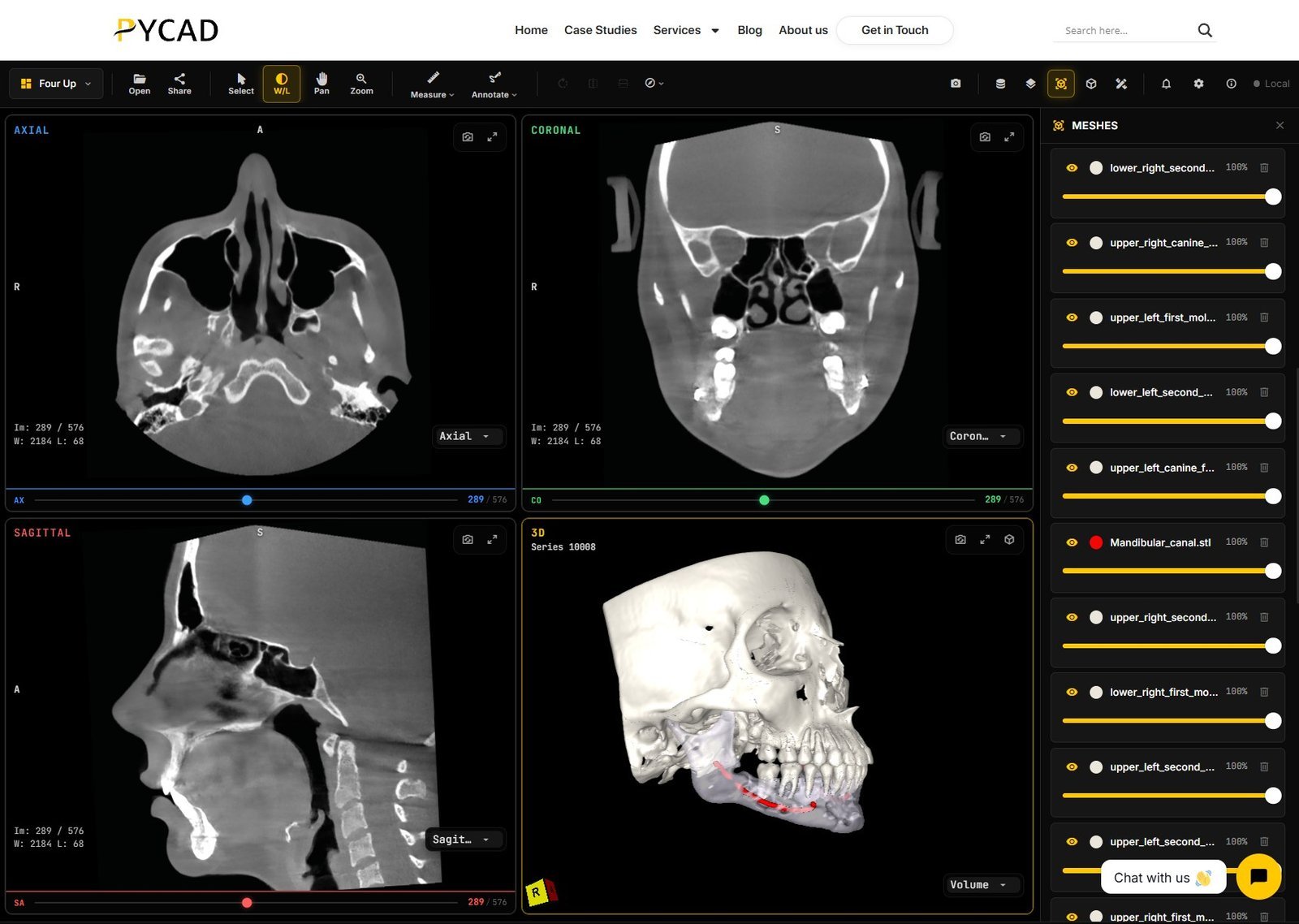
The stack in one picture
Hold one mental model for the rest of the series:
- Data — what the case is made of (2D radiographs, CBCT, IOS meshes, dental EHR fields)
- Capture / view — devices and software that create and show those data
- Design — dental design on meshes (crowns, guides, appliances)
- Make — mill, print, finish
- Communicate / review — how clinics, labs, and specialists hand the case back and forth
- PMS / systems — scheduling, charting, billing, and where images attach
- AI — helpers that sit on EHR text, 2D images, CBCT, or IOS, each with different inputs and failure modes
Most public “complete guides” stop at scan → design → mill/print. That middle is real. It is not the whole path clinics and labs fight with every week — and it is not the whole path dental software teams ask us to build.
What reviewer dentists keep complaining about
Nick Fotache at RevUp summarized hundreds of dentist reviews on Dentaltown into buying criteria for imaging software (The Most Common Problems Dentists Have With Dental Imaging Software, May 2026). Treat that piece as a checklist, not a brand war. The themes match what I hear on calls.
Themes that show up again and again:
- PMS integration that works with your PMS version, not only the brand name on the brochure
- Hardware that quietly picks the software (buy the sensor or CBCT, inherit the suite)
- Multi-user and licensing friction in busy multi-chair days
- Full-day performance that demos never show (slow loads, crashes under load)
- Cloud vs local trade-offs when the internet drops — and the opposite problem: AI that only runs well if every chair has a serious GPU
- Support quality after the sale
- Hidden costs (updates, seats, screens, add-ons)
- Updates that break workflows that already worked
If you are choosing imaging or a wider digital path, write those questions down before the demo. Ask them with your actual PMS build, chair count, and worst-case image volume in mind.
Open vs closed ecosystems
In dentistry, the machine purchase is often the software decision. Sensors, panoramic units, and CBCT systems commonly ship inside a vendor suite. Switching software later can mean orphaned hardware, new sensors, or painful migration of years of images.
Open architecture platforms exist so practices can change software without throwing out every sensor. Closed bundles exist because one vendor owns support and the story is simpler on day one. Neither side is automatically virtuous. The buying question is exit path: if this relationship ends in five years, what still works, and what must you replace.
Clinics feel that as flexibility. Labs feel it when each clinic sends cases from a different locked portal. Engineers feel it when exports are incomplete or undocumented — or when the only way to get patient ID and tooth number out of an old imaging brand is a brittle database scrape instead of a clean DICOM path. Vendors feel it as attach rate. Same stack layer, different scorecard.
What this series goes deep on that others don’t
Many “digital dentistry 101” articles stop at the familiar triangle: scan → dental design → mill or print. Useful. Incomplete.
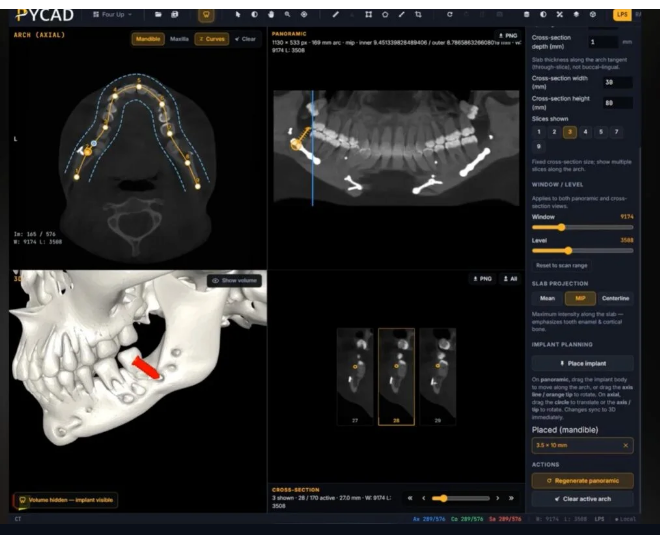
From our calls and builds, we will go deeper on layers those guides skim:
- Dental data as first-class — 2D radiographs, CBCT, IOS, and dental EHR fields as different objects with different jobs (OPG/RVG workflows are not the same product as CBCT implant depth)
- CBCT across specialties — how implant, ortho, endo, and other clinical jobs use the same modality differently
- Capture and clinical surfaces — including how volumes and scans are viewed, shared, and handed off between people (browser share links, who uploads the CBCT, phone vs desktop)
- Design → manufacture → communicate / review — the loop after the pretty design screen, including lab and specialist review and mesh–volume alignment
- PMS integration reality — middlemen, cost, read vs write, missing fields, version skew, human review before write-back
- AI that differs by data — helpers on charts vs bitewings vs CBCT vs IOS are not one product category; quality also depends on the scan and on whether labels were checked by a clinician
This article is Part 1 of that series. Upcoming parts (titles only, coming soon):
- Digital dental data (2D, CBCT, IOS, dental EHRs)
- Capture & clinical surfaces
- CBCT across dental specialties
- Design → manufacture → communicate/review
- PMS integration
- AI for dentistry
- Digital dentistry: a look ahead
What’s coming next
Next up: Digital dental data (2D, CBCT, IOS, dental EHRs) — what each data type is for, where it lives, and why treating them as one blob breaks integrations. Coming soon.
Then capture and clinical surfaces, CBCT by specialty, design-to-make-to-review, PMS, AI, and a look ahead, in that order.
Soft next step
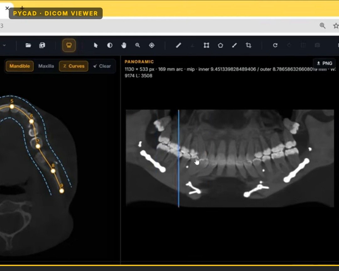
When Part 2 is live, start there. If you want a feel for CBCT and dental viewing in the browser while you wait, our dental viewer demo is at pycad.co/demos/dental-dicom-viewer. This series stays on the map first; the later parts carry the depth — with the same field notes behind them.