
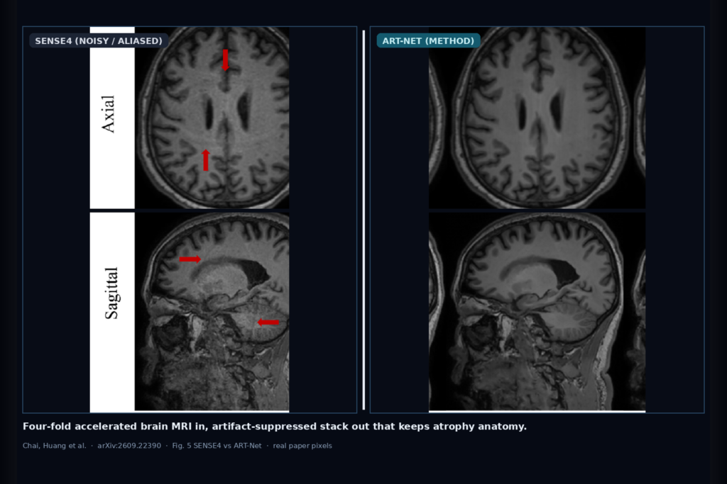
Four-fold SENSE brain MRI shortens the acquisition, but the console stack often still carries noise and residual aliasing that look like tissue edges. Chai, Huang, Xu, Zhou, Pan, Yeom, Gong, and Gong take that hang problem on in arXiv:2609.22390. Their ART-Net sits after conventional SENSE4 reconstruction and refines the magnitude volume toward a paired fully sampled T1w (AF0) while tracking segmentation and reader scores that matter for atrophy reading.
The study is a retrospective paired design: 80 participants scanned with both fully sampled T1-weighted imaging and four-fold SENSE on the same exam. Train / validation / independent test splits are 45 / 5 / 30 at the participant level. Input is the registered SENSE4 volume. Output is an image-domain refinement, not a raw multicoil k-space rebuild.
What hangs on the viewer
Hang the SENSE4 T1w stack in. Hang the ART-Net cleaned T1w stack out beside it, in the same AF0-aligned frame of reference the authors used for scoring. For atrophy or reader workflows that care about medial temporal and gray-white boundaries, the useful overlays are the cleaned series itself plus any FastSurfer-style labels you already run on brain volumes.
AF0 in the paper means the fully sampled reference acquisition (acceleration factor 0). ART-Net is the proposed method. SENSE4 is the four-fold accelerated conventional reconstruction that still shows noise and residual aliasing.
How it works in plain words
ART-Net is an Anatomy-aware Residual Attention Network: a residual U-Net backbone with generalized self-attention blocks, trained against AF0 with adversarial, perceptual, Smooth L1, SSIM, total-variation, and COBRAI (correlation-based residual artifact) terms. SENSE4 volumes are registered to AF0 with ANTs before training and evaluation. The checkpoint is picked by highest mean PSNR on the five-participant validation set. The thirty-participant test set stays held out.
Comparators in the same SENSE4-to-AF0 post-processing setting include DAGAN, a DAGAN-style generator variant, SwinGAN, and ablations that drop COBRAI or swap the attention block for CBAM. The network never needs raw multicoil k-space or hand-drawn artifact masks; it works on conventionally reconstructed magnitude images.
What the numbers say
On the independent test cohort (n = 30), ART-Net reaches PSNR 31.03 ± 2.88 dB and SSIM 0.963 ± 0.022, against SENSE4 at 29.89 ± 2.53 dB and 0.953 ± 0.022. LPIPS falls to 0.0161 ± 0.0082 from SENSE4 0.0245 ± 0.0070. FID falls to 2.38 ± 1.10 from 7.90 ± 2.91. Against DAGAN, DAGAN_generator, and SwinGAN under matched training, ART-Net leads on PSNR, SSIM, and LPIPS.
FastSurfer labels on reconstructed volumes versus AF0 give ART-Net medial temporal Dice 0.8824 ± 0.0827 and whole-brain Dice 0.8857 ± 0.0885, the highest among the reported deep-learning methods for those groups. Central nuclei Dice is 0.9455 ± 0.0601. Basal ganglia Dice is close to CBAM-ART (0.8758 ± 0.0889 vs 0.8775 ± 0.0869).
Two radiologists scored gray-white boundary, basal ganglia, and cerebellum-brainstem on a 1-5 scale for all thirty test participants. ART-Net overall mean is 3.58 ± 0.65, second only to AF0 at 4.53 ± 0.77 and ahead of SENSE4 at 2.03 ± 0.61. Domain scores for ART-Net are 3.98 ± 0.50 (gray-white), 3.35 ± 0.63 (basal ganglia), and 3.42 ± 0.62 (cerebellum-brainstem), each higher than the other refinement methods (all P < 0.001 in the paper’s pairwise tests). Interreader κ on overall score is 0.917.
Where it fails and what not to trust
Single center, one scanner, one field strength, one sequence, and one acceleration factor. Validation used only five participants. The model postprocesses SENSE4 magnitude images, so registration bias between separate AF0 and SENSE4 acquisitions can still leak in. The cohort is not disease-specific; the authors caution that better Dice and reader scores show segmentation agreement and conspicuity, not proof that disease-related atrophy, subtle lesions, or abnormal signal are preserved.
This is not a cleared medical device. Rebuild on your own SENSE protocols, vendors, and atrophy cohorts before you hang ART-Net output next to a clinical read. Treat the cleaned stack as a research series with a human gate.
For a viewer or clinic AI shop
Wire registered SENSE4 T1w in; hang ART-Net magnitude T1w out in the same geometry you use for AF0-aligned reading. Keep provenance on the series: model name, SENSE factor, arXiv:2609.22390. If you already run FastSurfer or similar labels for medial temporal atrophy rails, score the cleaned volume against your AF0 or clinical reference the same way the paper does.
Prefer this path when you already own conventional SENSE4 DICOM and cannot reach raw multicoil data. Audit medial temporal Dice, reader scores, and failure cases on your scanner before you advertise a shorter exam with unchanged anatomy reads.
Rebuild from arXiv:2609.22390. PDF: https://arxiv.org/pdf/2609.22390.
Sources
- Chai, C., Huang, B., Xu, L., Zhou, J., Pan, B., Yeom, K. W., Gong, Q., Gong, N.-J. Anatomically Faithful Artifact Suppression in SENSE Accelerated Brain MRI. arXiv:2609.22390, 2026. https://arxiv.org/abs/2609.22390. PDF: https://arxiv.org/pdf/2609.22390.
- Task framing: registered SENSE4 T1w magnitude → ART-Net refined T1w toward paired AF0 (fully sampled) reference. Metrics in body: PSNR/SSIM/LPIPS/FID, FastSurfer Dice (medial temporal, whole-brain), blinded reader scores.