
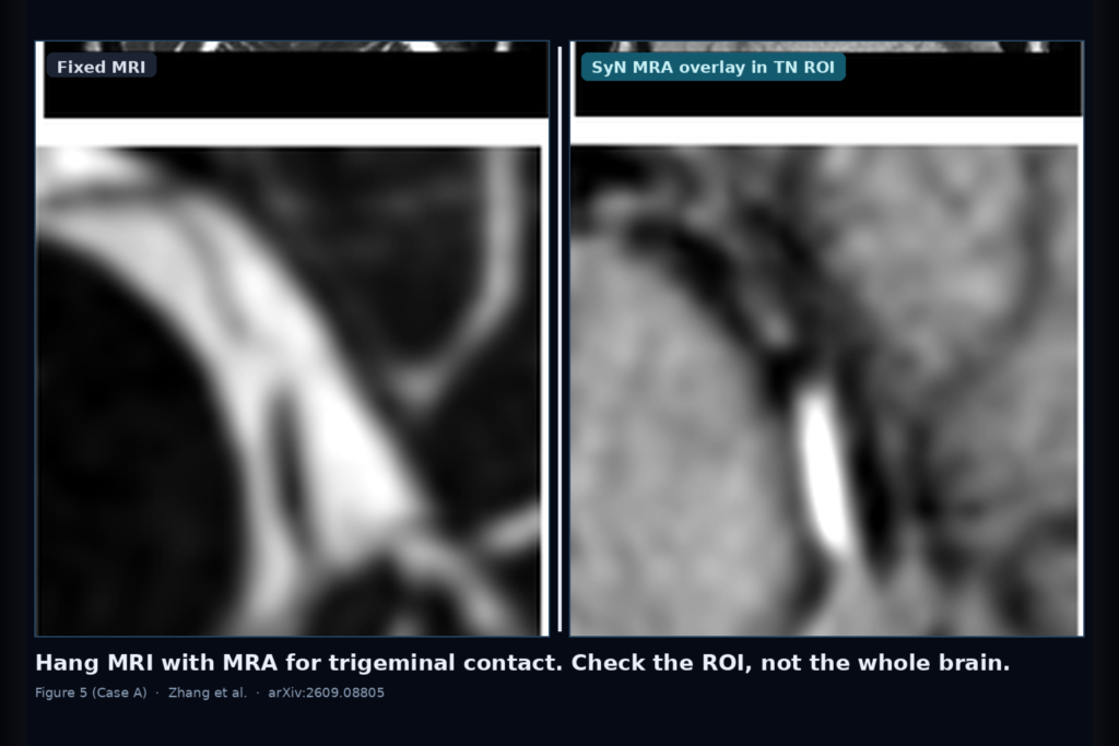
Preoperative trigeminal neuralgia work often hangs a CISS-type structural MRI next to a TOF-MRA and asks the reader to mentally fuse them. The nerve lives on MRI. The candidate offending vessel lights up on MRA. Microvascular decompression planning wants both in the same space, inside a small cisternal ROI, not as a pretty whole-brain warp. Many fusion viewers still score registration as if the whole head were the endpoint. That is the wrong test.
Xupeng Zhang, Xihang Wang, Michael Xie, Haoyuan Liang, Hau Ern Lien, Oishika Das, James Feghali, Risheng Xu, and Peirong Liu (Johns Hopkins ECE / Data Science and AI Institute, Neurosurgery, and BME) posted arXiv:2609.08805 on 8 September 2026. The paper is Evaluation Principles for MRI-MRA Registration in Trigeminal Neuralgia: An ROI-Centered Neurovascular Benchmark. Code: https://github.com/jhuldr/TN-Reg-Benchmark.
They treat TN MRI-MRA fusion as a local, vessel-aware, contrast- and FOV-aware visualization problem. The cohort is 149 MVD patients with clinician-annotated bilateral trigeminal ROIs (298 ROIs). Six pipelines go through the same ROI-centered readout: ANTs Affine, ANTs SyN, ConvexAdam, FireANTs, EasyReg, and SynthMorph. Whole-brain friendliness is not the score that decides whether the overlay is safe to hang.
What the benchmark actually measures
MRI is fixed. MRA is moving. After each method warps MRA into MRI space, the team crops a matched 48 cubed trigeminal ROI on a 0.47 mm isotropic grid and scores that crop, not the full volume. Image-based checks include vessel-background separability on the warped MRA (Vessel AUC) and local multimodal intensity correspondence (ROI NMI). Downstream vessel checks run VesselFM once on each warped MRA, then measure how the predicted vessel tree sits relative to the clinician-labeled vessel segment inside the ROI.
Those labels are partial on purpose. Annotators marked the vessel segment that matters for the nerve, not an exhaustive vascular tree. That property breaks naive one-sided distances. Annotation-to-prediction distance rewards methods that grow large predicted trees until they cover the labeled segment. Prediction-to-annotation distance punishes the same sprawl. In the paper the two directions reverse method rankings, and predicted vessel volume swings by more than an order of magnitude across pipelines. Symmetric distance sits in between, but it is still a segmentation-derived localization readout, not a pure registration error.
Local image similarity and vessel separability do not co-rank methods either. EasyReg and SynthMorph lead on median Vessel AUC. FireANTs leads on ROI NMI. If your viewer dashboard only shows one of those numbers, you can crown the wrong winner for the hang you actually care about.
What hangs on the scan
For a planning viewer the useful hang is structural MRI with TOF-MRA resampled into the same frame, then a tight zoom on the cisternal trigeminal ROI. Cyan clinician vessel annotation and red post-registration vessel prediction (or a simple MRA intensity overlay) sit on that crop so a reader can see whether contact looks anatomically plausible. Figure 5 in the paper is the qualitative map of that idea: whole-brain slice with a yellow ROI box, the matched 48 cubed intensity crop, and the overlay row where cyan and red either agree or drift apart.
Three operating regimes show up clearly. High local MRA contrast with good FOV coverage can put image-level vessel visibility and downstream localization in the same place. Mid-contrast cases can keep measurable vessel-background separability while the predicted vessel sits millimeters away from the annotated segment. Low-contrast ROIs push Vessel AUC toward chance and often fail localization. In this cohort, 63 percent of ROIs fell in the Low-contrast tier, so a single unstratified AUC summary will bury method differences that only appear when local vessels are bright enough to judge.
Where whole-brain scores and local distances lie
Deformable refinement is not free lunch. In Good-FOV ROIs, ANTs SyN shaved a small amount off annotation-to-prediction distance relative to affine (median paired change about -0.18 mm). In Bad-FOV ROIs that benefit disappeared. A reliable affine initialization stays the prerequisite when MRI and MRA coverage do not match. Methods that assume a strong pre-alignment need that affine stage named in the pipeline, not hidden.
Reader review on 100 stratified ROIs adds the clinical check. Locally favorable one-sided distances can sit next to globally implausible warps. SynthMorph often looked broken at whole-brain scale in the review set, yet still posted the lowest median annotation-to-prediction distance in the aggregate table, because a wrong global warp can still park some predicted vessel voxels near the tiny annotated segment. If your QC only watches that one-sided ROI distance, you can green-light an overlay a neurosurgeon would reject on the full hang.
ANTs SyN was the most balanced profile across the reported metrics: competitive Vessel AUC (highest median in the High-contrast tier, around 0.76), moderate prediction volume, and anatomically plausible overlays more often than the volume-heavy alternatives. It is slower (about 20 minutes per pair in their setup) but batchable. ConvexAdam needed external affine pre-alignment. EasyReg and SynthMorph were detection-sensitive but grew large predicted trees that inflated reverse distances.
Caveats for anyone shipping a fusion hang
Vessel annotations are clinical snippets, not full trees, so distance metrics without volume reporting will mislead. TOF-MRA prefers arterial flow; low local contrast may mark venous offenders as much as bad acquisition, and the blinded vessel-typing in the paper is hypothesis-generating, not intraoperative ground truth. Segmentation-derived distances mix registration geometry with VesselFM sensitivity. FOV mismatch attenuates deformable gains. Single-institution MVD cohort, mostly Siemens 3T CISS plus multi-slab TOF. Fail closed when the warped MRA leaves the TN sampling grid empty, when local contrast is near chance, when whole-brain review looks shifted even if the ROI distance looks fine, and when your only dashboard number is a whole-brain similarity score.
For a DICOM viewer or clinic platform
Treat this paper as an evaluation checklist for MRI-MRA fusion in trigeminal planning, not as a single winning network. Hang MRI and registered MRA together. Zoom the cisternal ROI. Report local vessel separability with contrast tier, ROI intensity correspondence, symmetric vessel distance with predicted volume, and a whole-brain plausibility check a human can fail. Prefer a reliable affine before deformable polish, and do not let a locally cute vessel distance override a globally broken warp. If your product already fuses skull-base MRI with TOF for MVD consults, ROI-centered QC is the difference between a trusted overlay and a misleading one.
Rebuild from arXiv:2609.08805 and https://github.com/jhuldr/TN-Reg-Benchmark. As of 14 September 2026 the abstract, PDF, and GitHub repo respond.
Sources
- Zhang, X., Wang, X., Xie, M., Liang, H., Lien, H. E., Das, O., Feghali, J., Xu, R., Liu, P. Evaluation Principles for MRI-MRA Registration in Trigeminal Neuralgia: An ROI-Centered Neurovascular Benchmark. arXiv:2609.08805, posted 8 September 2026. https://arxiv.org/abs/2609.08805. PDF: https://arxiv.org/pdf/2609.08805. Code: https://github.com/jhuldr/TN-Reg-Benchmark.
- ANTs Affine / SyN; ConvexAdam; FireANTs; EasyReg; SynthMorph; VesselFM (methods compared or used for downstream vessel readout as cited in the paper).