
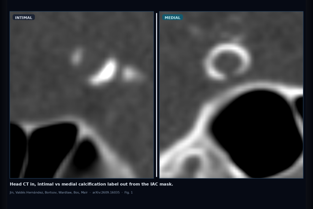
Hang an intimal vs medial subtype label on intracranial arterial calcification masks from routine head CT, without asking a radiologist to re-score every scan by eye. That is the clinic ask Jin, Valdés Hernández, Bortsov, Wardlaw, Bos, and Mair take on in arXiv:2609.16035, posted around mid September 2026. The paper is Automated Distinction of Intimal and Medial Intracranial Arterial Calcification from CT Head. Code is at github.com/bjin96/iac-subtyping.
IAC shows up on everyday non-contrast head CT and tracks with stroke and other neurovascular disease. Presence and volume alone miss the shape story: intimal calcium sits with atherosclerosis, while medial calcium is linked to wall stiffening and haemodynamics. Radiologists already separate the two with a visual score. The authors ask whether that subtype hang can come from the mask itself, and whether auto-segmented masks are good enough to keep the label usable.
What hangs on the viewer
Upstream is an IAC segmentation mask on head CT, either drawn by hand or produced by automated segmentation (here, ViTiac-S ensembles fine-tuned on Rotterdam Study CT). Downstream you get an intimal vs medial subtype call per internal carotid artery side, plus a joint patient-level call that can also land in a mixed class when the two sides disagree. For a DICOM viewer or neurovascular AI rail, that means you can hang a subtype badge beside the calcification overlay once the mask is non-empty, without opening a separate visual-score worksheet.
Scans with no IAC cannot be subtyped. Tiny calcifications under 4 mm3 were excluded because shape features, especially embeddings, become unstable. Manual masks with IAC came from 1766 participants; automated masks with IAC from 1704, after dropping the 100 series used to fine-tune the segmenter.
How it works in plain words
Three feature paths feed the same lightweight logistic regression with 5-fold cross-validation (scikit-learn). The Automated Visual path turns Kockelkoren-style cues into numbers: 3D connected components, 2D skeletonisation on axial, coronal, and sagittal slices, then circularity, thickness, and morphology averaged across calcification objects. The Sphericity path scores how sphere-like each 3D object is, builds a per-scan sphericity distribution, and turns that into a Wasserstein difference against intimal and medial reference distributions from the training fold. The Deep Learning path samples point clouds from the mask and pulls 128-d shape embeddings from MedShapeNet’s point-cloud encoder (original embedding cut from 4096).
Left and right internal carotid arteries are scored separately. For the joint “total” label, features or embeddings from both sides are concatenated. Labels follow the Rotterdam visual assessment: binary intimal/medial per side, and a predominant subtype plus mixed at the patient level.
What the numbers say
On manual masks, the embedding method leads overall. Weighted F1 (mean ± SD) reaches 71.5 ± 3.7 on the right artery and 69.4 ± 0.7 on the left. Joint artery classification drops to 59.8 ± 1.7 weighted F1, largely because the mixed class is hard. Automated Visual and Sphericity land in a similar band (right artery weighted F1 about 68.9 and 68.6). Column F1A in Table 1 shows weighted F1 when the same classifiers run on auto ViTiac masks; for the embedding method that is 71.0 ± 3.7 (right), 69.5 ± 1.8 (left), and 57.9 ± 1.7 (total). Bootstrapped mean differences between manual and auto weighted F1 were not significant.
Pairwise agreement is highest between Automated Visual and Sphericity (Cohen κ about 0.56–0.58 per side). Both agree less with the embedding method, which may be picking up complementary shape cues. Confusion matrices show Automated Visual leaning toward calling medial cases intimal; Sphericity and Deep Learning balance the two subtypes better. All three struggle on mixed.
Where it fails and what not to trust
Mixed predominant subtype (different labels on left vs right ICA) is the weak class across methods. Calcifications under 4 mm3 are out of scope. No IAC means no subtype. This is not a full diagnostic claim or a treatment recommendation: it is automated subtype quantification against a visual-score reference on Rotterdam Study CT. MedShapeNet embeddings trade hand-crafted explainability for flexibility. Rebuild and measure on your own scanners and labeling protocol before quoting clinic numbers.
For a viewer or clinic AI shop
Wire IAC mask in (manual or auto), intimal/medial subtype out, with an optional ViTiac-style segmenter upstream for a full CT-to-subtype rail. Prefer hanging the label on clear ICA calcifications first. Surface mixed as uncertain rather than forcing a binary badge. Audit auto-mask vs manual-mask agreement on your hold-outs, and keep the visual score available when governance still wants a human subtype call. Large-scale association work and prospective outcome studies are still needed before subtype badges drive care pathways.
Rebuild from arXiv:2609.16035. Code: https://github.com/bjin96/iac-subtyping.
Sources
- Jin, B., Valdés Hernández, M. del C., Bortsov, R., Wardlaw, J.M., Bos, D., Mair, G. Automated Distinction of Intimal and Medial Intracranial Arterial Calcification from CT Head. arXiv:2609.16035, 2026. https://arxiv.org/abs/2609.16035. PDF: https://arxiv.org/pdf/2609.16035.
- Code: github.com/bjin96/iac-subtyping.
- Dataset: Rotterdam Study non-contrast head CT (institutional; data use agreement). Prior visual score: Kockelkoren et al. PLOS ONE 2017. Related IAC segmentation: Bortsova et al.; Jin et al. ViTiac.