
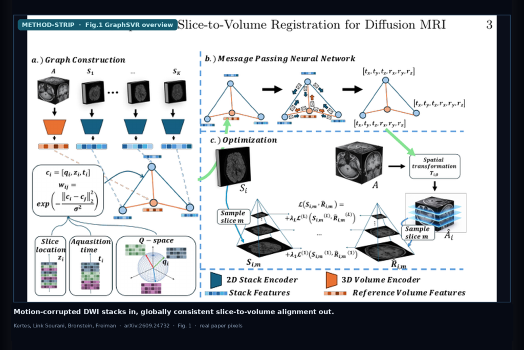
Hang a motion-corrupted diffusion stack into a DICOM viewer or tractography pipeline and the shop question is not whether eddy ran. It is whether the stacks sit in one anatomical frame when the patient jerked between slice groups, especially on sparse 12/24/32-direction protocols where eddy’s smooth-motion assumption breaks. Noga Kertes, Daphna Link Sourani, Alex M. Bronstein, and Moti Freiman (Technion; Bronstein also ISTA) take that 4D hang seriously in arXiv:2609.24732, posted 21 September 2026. They call the method GraphSVR: a q-space-aware graph over stacks that predicts globally consistent stack-wise rigid transforms, optimized zero-shot against a T1/T2 anatomical reference with no paired ground-truth motion. Under severe synthetic motion, grid and rotation error drop by about 73% versus FSL eddy, and grid error stays under ~3 mm. Code: https://github.com/nogakertes/GraphSVR.git.
What hangs on the viewer
Upstream is a DWI acquisition of K diffusion-weighted measurements with encodings qi, plus a few b=0 volumes, often with multiband factor 4 so each readout is a slice group (stack) of interleaved slices. Inter-stack misalignment is the pain: EPI is fast per slice, but the subject can still move between stacks, so the 4D volume jerks when you scrub directions or feed tractography. Downstream hangables for a DICOM or diffusion shop include: stack-wise rigid transforms Ti in SE(3) that map an anatomical reference A into each stack frame; a re-sampled volume where every stack sits in one anatomical coordinate system; and QC plots of predicted trajectories against whatever motion model you already trust.
GraphSVR’s shop claim is not a new eddy binary. It hangs acquisition-aware SVR on top of the stacks you already have, using only the anatomical reference you already scan, and it is aimed at the sparse, jerky cases where eddy under-corrects.
How it works in plain words
Each stack Si is a graph node with features from a lightweight 2D ResNet-style encoder (slice embeddings summed). A 3D CNN encodes the anatomical reference A into a reference node. Edges come from a k-nearest-neighbor graph in a joint acquisition space: z-normalized diffusion direction qi, physical slice locations zi, and acquisition time ti. Edge weights use an RBF kernel. A reference node connects to all stacks with unit weight. An attention-based message-passing network (two layers, four heads, hidden 64 in their setup) aggregates across the graph and an MLP head regresses 6-DoF rigid parameters per stack.
Optimization is per case and self-supervised. For each stack, GraphSVR warps A by Ti, samples slices at the known zi locations, and maximizes a multi-scale mix of local mutual information (LMI) and normalized gradient fields (NGF), with exponentially decaying pyramid weights. Adam runs up to 400 iterations with ReduceLROnPlateau and early stopping. No paired ground-truth motion is required. The first time point is held motion-free and aligned to the reference frame.
What the numbers say
Data: seven healthy volunteers on a Siemens Prisma 3T at Technion’s May-Blum-Dahl MRI Research Center. Evaluation uses two simulation strategies because real scans lack intra-acquisition ground-truth motion. Real-image recombination draws slice groups across five consecutive scans of one subject who changed pose between scans (internally still during each scan), preserving realistic DWI appearance with known stack-wise transforms (T1-registered approximate GT). Fully synthetic runs fit a DTI model on a motion-free scan, impose controllable rigid trajectories, reorient tensors, add Rician noise (SNR=30), and subsample to 12/24/32 directions. All sims use multiband factor 4 and two b=0 volumes: 21 recombination cases and 42 synthetic cases.
Baseline is FSL eddy with the linear eddy-current model, susceptibility correction off, and motion estimates re-referenced to the first time point so frames match. Metrics are grid error (GE, mm) and rotation error (RE, rad) versus ground-truth transforms, summarized as per-case median across stacks.
Table 1 (fully synthetic, by motion severity) is the shop number. Under severe motion, eddy GE is 11.018±8.119 mm and RE 0.119±0.087 rad; GraphSVR reaches 2.952±3.395 mm and 0.031±0.037 rad (paired Wilcoxon, p<0.05). That is the ~73% cut the abstract reports. Grid error stays below 3 mm even in the severe setting. Under minor/moderate motion GraphSVR also wins or stays competitive (severe is where eddy collapses). Table 2 (by direction count) shows GraphSVR GE stays near 2.2–2.4 mm across 12/24/32 directions while eddy swings higher, including 4.341±2.915 mm at 32 dirs in their summary. Fig. 2 (recombination, 12/24/32 dirs) shows significantly lower GE than eddy at every sparsity level under abrupt pose changes that violate smooth-motion assumptions. Fig. 3 (moderate synthetic trajectory) shows GraphSVR tracking ground truth through rapid pose changes where eddy lags.
Ablation Table 3 (4 synthetic cases, 24 dirs, moderate motion): full q-space + time + location edges give GE 1.649±0.260 mm; zeroing all relational features jumps to 3.202±2.417 mm. The graph is doing real work.
Where it fails and what not to trust
This is research software on simulated motion from seven healthy volunteers. It is not a cleared medical device, and it has not been stress-tested on pediatric or clinical motion-prone cohorts yet. Ground-truth transforms come from simulation (recombination and synthetic DTI); real clinical scans still lack intra-acquisition GT motion, so the 73% figure is simulation-backed, not a bedside guarantee. The eddy comparison disables susceptibility correction for fairness and uses the linear field model; shops that rely on eddy’s full distortion pipeline should re-benchmark with their own flags. Hyperparameters (k=4, pyramid levels, α=β=1) were tuned on a held-out subset. Validate stack-wise Ti and the re-sampled 4D volume on your scanner, multiband factor, and direction schedule before you trust overlays or tractography from GraphSVR-corrected stacks.
For a viewer or clinic AI shop
Wire motion-corrupted DWI stacks plus a T1/T2 anatomical reference in; hang stack-wise rigid transforms and a globally consistent 4D volume out. Prefer GraphSVR when you already run eddy but still see jerky inter-stack misalignment on sparse-direction or non-smooth motion cases, and when you can afford a per-case zero-shot optimize on a GPU (they used one NVIDIA L40, 46 GB). Keep a QC path that overlays predicted vs eddy trajectories, reports GE/RE when you have a simulation harness, and lets a reader scrub directions on the corrected volume. Start from the preprint and the public repo. Rebuild from arXiv:2609.24732. PDF: https://arxiv.org/pdf/2609.24732. Code: https://github.com/nogakertes/GraphSVR.git.
Sources
- Kertes, N., Link Sourani, D., Bronstein, A.M., Freiman, M. GraphSVR: q-Space-Aware Graph-Based Slice-to-Volume Registration for Diffusion MRI. arXiv:2609.24732, 2026. https://arxiv.org/abs/2609.24732. PDF: https://arxiv.org/pdf/2609.24732.
- Severe motion (Table 1): eddy GE 11.018±8.119 mm / RE 0.119±0.087; GraphSVR GE 2.952±3.395 mm / RE 0.031±0.037 (~73% reduction). Grid error stays under ~3 mm in severe setting.
- Fig. 2: real-image recombination boxplots (init / eddy / GraphSVR) at 12/24/32 directions. Fig. 3: moderate synthetic trajectory GT vs GraphSVR vs eddy.
- Code: https://github.com/nogakertes/GraphSVR.git. Data: 7 healthy volunteers, Siemens Prisma 3T; multiband factor 4; sparse 12/24/32 directions.