
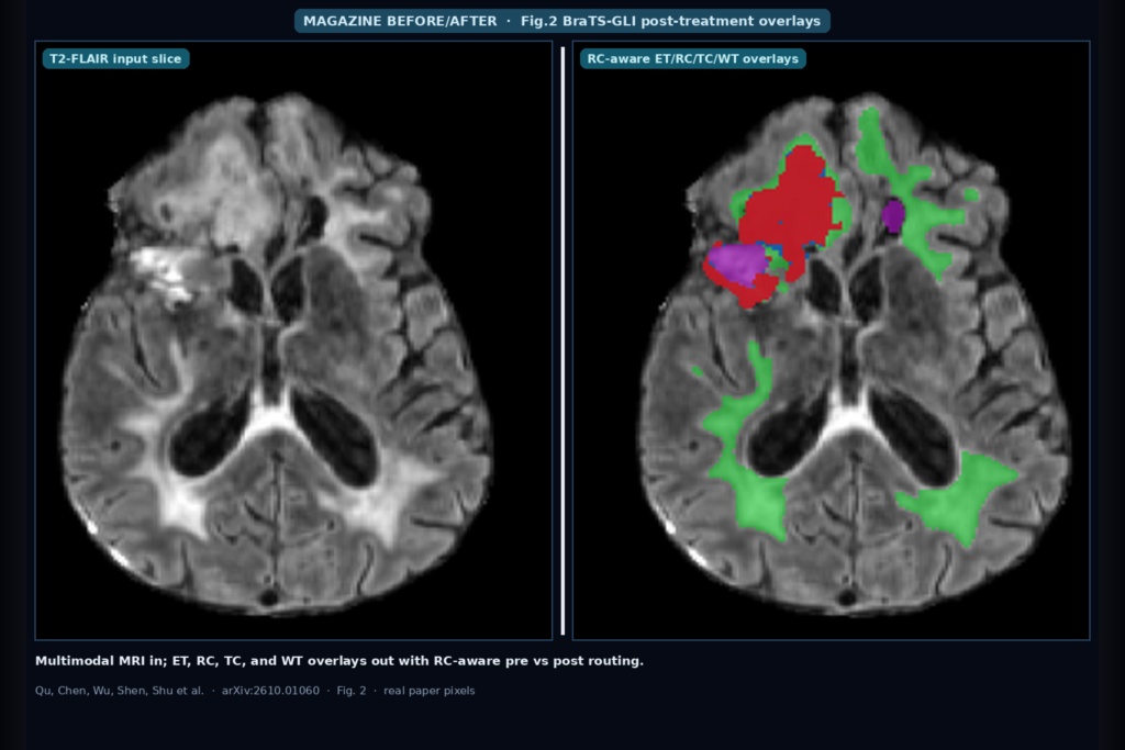
Hang a pre-treatment or post-treatment multimodal glioma MRI study into a neuro DICOM viewer and the clinic ask is the same: can you get reproducible enhancing tumor (ET), tumor core (TC), whole tumor (WT), and, after surgery, resection cavity (RC) overlays without a radiologist redrawing every timepoint.
BraTS-GLI 2025 puts both cohorts in one task, but RC only exists after resection, so a naive pooled model can fire false RC on pre-op cases or miss the cavity after surgery.
Lin Qu, Ziqi Chen, Anqi Wu, Jinyao Shen, and Hai Shu (East China Normal University / NYU) take that hang seriously in arXiv:2610.01060, submitted 1 October 2026 to eess.IV. Their RC-aware nnU-Netv2 routes each case by treatment status, trains with empty-target handling for missing RC, and ranked second on the official BraTS-GLI 2025 blind test under the team name ECNU NYU.
What hangs on the viewer
Upstream input is four co-registered, 1 mm isotropic, skull-stripped MRI volumes per case: T1, T1Gd, T2, and T2-FLAIR, stacked as a 4-channel tensor at 240×240×155.
Downstream hangables for a viewer plugin or clinic AI shop are the four overlapping region overlays that BraTS-GLI trains on: ET, TC, WT, and RC. Official categorical labels underneath are background, NETC, SNFH, ET, and RC.
At inference each sigmoid channel is thresholded at 0.5, then decoded back into those categorical labels with RC winning on overlaps. Tiny 26-connected WT and RC components under 50 voxels are dropped before export.
The clinic twist is treatment status. Pre-treatment cases have no anatomical RC, so that channel is an all-zero target during training. Post-treatment cases can carry a real cavity next to residual ET/TC/WT.
The submission therefore does not run one undifferentiated network on every case. Known treatment status in the challenge case ID routes pre-treatment studies to a pre-only custom-loss nnU-Netv2, and post-treatment studies to an RC-aware joint model. Exactly one network runs per case. No probability averaging, voting, multi-fold fusion, or multi-architecture ensemble.
How it works in plain words
All compared nnU-Netv2 trainers share the same self-configuring preprocessing, planning, PlainConvUNet backbone, deep supervision, and default augmentation. Differences live in the training objective and in how empty region channels are handled when pre- and post-treatment cases share a mini-batch.
Single-cohort training (pre-only or post-only) uses a lesion- and boundary-aware custom loss: half binary cross-entropy, half a lesion-aware Dice surrogate that mixes voxel Dice with a Gaussian-smoothed target-weighted Dice, plus a boundary-weighted BCE term. The authors treat these coefficients as empirical weights for this study, not as a universal recipe.
Joint pooled training adds the RC-aware objective. Standard and boundary-weighted BCE still run on all four region channels. Soft Dice runs only on channels that have a non-empty target in the current mini-batch.
Channels that are empty across the mini-batch, including RC on every pre-treatment sample, get a controlled false-positive penalty instead of soft Dice. That is the knob aimed at stopping spurious RC activation before surgery while still supervising real cavities after surgery.
Routing at inference is not an ensemble. Pre-treatment cases go to the pre-only custom-loss branch. Post-treatment cases go to the RC-aware joint branch. Sliding-window inference and test-time mirroring follow nnU-Netv2 defaults.
The paper explicitly avoids synthetic tumor generation, on-the-fly GliGAN augmentation, and multi-model fusion for the submitted pipeline. First place in BraTS-GLI 2025 used a three-model nnU-Net ensemble with GliGAN; this second-place route bets on task structure instead.
What the numbers say
Labeled BraTS-GLI 2025 training data covers 1251 pre-treatment cases from 1133 patients and 1621 post-treatment cases from 731 patients, with 34 patients shared across cohorts.
For post-challenge ablations the authors re-split at the patient level (pre held-out 274 cases; post held-out 315 cases) so the same patient never leaks across train and test in either cohort.
On the official blind test, ECNU NYU ranked second. Mean lesion-wise Dice was 0.7878 (ET), 0.8709 (RC), 0.7923 (TC), and 0.8715 (WT). Mean NSD@1.0 was 0.8309, 0.8712, 0.7980, and 0.8336 on the same regions.
Arithmetic means across those four regions sat about 0.012 below the first-place GliGAN-augmented nnU-Net ensemble, while RC Dice (0.8709) stayed ahead of the two third-place entries (MIST 0.8400; EGASegNet 0.8262).
On the internal post-treatment held-out set, the full RC-aware joint configuration beat the joint baseline by +0.042 mean lesion-wise Dice and +0.043 NSD@1.0 (paired bootstrap 95% CIs excluding zero; n=315). Naive joint custom-loss training was the weakest post-treatment setup, so pooling alone plus a single-cohort custom loss is not enough.
Against Swin UNETR and SegMamba under the same held-out protocol, the routed branches led most region-metric cells while using about 30.8 M parameters versus roughly 62 to 67 M for those backbones.
Where it fails and what not to trust
This is a challenge pipeline on BraTS-GLI labels, not a cleared neuro-oncology device. Treatment status must be known at inference for routing; if your PACS hang does not carry a reliable pre vs post flag, you need another gate before you pick a branch.
The authors note that the pre-only custom-loss branch looked strong on the official validation leaderboard but did not show a uniform gain over the pre-only baseline on their later internal split, so do not treat that branch as universally better for every pre-treatment draw.
Empty-target Dice handling is tied to the full joint loss package; the ablations do not isolate one term as the sole causal driver of the RC gains. Qualitative Fig. 2 is one post-treatment held-out case (BraTS-GLI-02846-100). Joint baselines can miss the cavity or place spurious RC inside tumor, while the RC-aware joint recovers the cavity and keeps nested WT/TC/ET structure. Predictions still vary across the test set.
Component cleanup under 50 voxels will drop small true islands if your clinic cares about tiny residuals. No public code URL is listed in the preprint text we used; start from the arXiv page until the authors publish a repo.
For a viewer or clinic AI shop
Wire T1, T1Gd, T2, and T2-FLAIR in; hang ET, RC, TC, and WT overlays out, with categorical BraTS-GLI labels if your viewer expects NETC/SNFH/ET/RC.
Gate inference on treatment status so pre-op cases never ask an RC-aware post model for a cavity that should not exist. Prefer a single routed nnU-Netv2 branch per case when you want competitive BraTS-GLI-style overlays without an ensemble or on-the-fly synthetic tumor generator in the serving path.
Keep a human confirm on post-treatment RC and residual ET before volumes enter planning or response assessment.
Rebuild from arXiv:2610.01060. PDF: https://arxiv.org/pdf/2610.01060. Team: ECNU NYU (Qu/Chen/Wu/Shen/Shu).
Sources
- Qu, L., Chen, Z., Wu, A., Shen, J., Shu, H. RC-aware nnU-Netv2 for Pre-treatment and Post-treatment Glioma Segmentation Using Multimodal MRI. arXiv:2610.01060, 2026. https://arxiv.org/abs/2610.01060. PDF: https://arxiv.org/pdf/2610.01060.
- Data: BraTS-GLI 2025 labeled training; pre 1133 patients / 1251 cases; post 731 patients / 1621 cases; 34 shared patients. Internal held-out: pre 274 cases, post 315 cases. Inputs: T1, T1Gd, T2, T2-FLAIR co-registered 1 mm skull-stripped.
- Official blind test (rank 2, ECNU NYU): LW Dice ET/RC/TC/WT 0.7878 / 0.8709 / 0.7923 / 0.8715; NSD@1.0 0.8309 / 0.8712 / 0.7980 / 0.8336. Paired post held-out vs joint baseline: +0.042 LW Dice, +0.043 NSD@1.0. Routed branches ~30.8 M params; one model per case; no GliGAN / ensemble in submission. Fig. 2 overlays: WT green, TC blue, ET red, RC purple.