
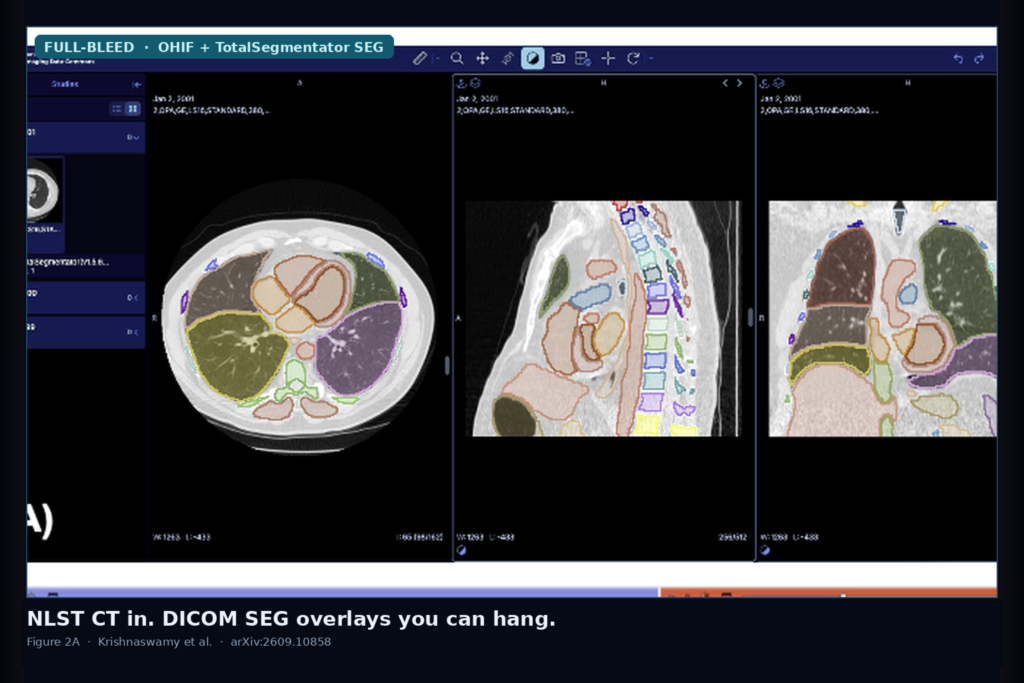
You have NLST screening CTs in DICOM. The useful overlays lived elsewhere: Sybil boxes in a custom JSON, NLSTseg lesions in NIfTI, TotalSegmentator organs in research volumes that do not travel with SeriesInstanceUID provenance. Krishnaswamy, Thiriveedhi, Pai, Clunie, Octaviano, Bridge, Pieper, Kikinis, and Fedorov (Brigham and Women’s / Harvard Medical School and collaborators) posted arXiv:2609.10858 on 9 September 2026. The paper is Annotating anatomy and pathology in the National Lung Screening Trial computed tomography images. It ships three IDC analysis-result collections that put those overlays back into DICOM SEG and Structured Report objects you can hang in OHIF, 3D Slicer, or any SEG-capable stack.
For a viewer shop, that is the whole point. NLST already gave you the images. What was missing was interoperable image-derived content: lesion boxes, pixel lesions, organ masks, and radiomics, all with composite context and coded semantics instead of filename folklore.
What you get to hang
Three collections, all as analysis results in the NCI Imaging Data Commons:
- NLST-Sybil: 970 DICOM SR TID1500 instances for 581 patients / 970 CT series, carrying 9,280 planar lesion bounding boxes converted from Sybil’s JSON via highdicom. Finding coded as Lesion (SCT 52988006), finding site Lung (SCT 39607008).
- NLSTSeg: 601 DICOM SEG series for 601 patients covering 705 lesions (tumors and nodules), converted losslessly from NIfTI with dcmqi itkimage2segimage. Lung-lobe location rides in AnatomicRegionSequence; tumor vs nodule in SegmentedPropertyTypeCodeSequence. Matching TID1500 SRs hold first-order and shape radiomics from pyradiomics (one case dropped when SEG and CT dimensions disagreed).
- TotalSegmentator-CT-Segmentations: TotalSegmentator v1.5.6 organ SEGs plus shape and first-order radiomics SRs for 26,194 NLST patients (about 126,051 CT series after filtering unsuitable series). Labels mapped to SNOMED concepts for segment metadata.
Table 1 in the paper summarizes the counts. Everything references the original NLST DICOM CTs, so SeriesInstanceUID / SOPInstanceUID linking is native, not a sidecar spreadsheet.
How the conversion stays honest
Sybil boxes become four SCOORD points in TID1500 Measurement Report instances, one SR file per annotated series. NLSTseg NIfTIs become DICOM SEG with patient/study composite context copied from the referenced CT. TotalSegmentator runs on IDC-filtered NLST series after DICOM-to-NIfTI conversion, then dcmqi / highdicom write SEG + TID1500 pairs. Radiomics use IBSI feature names and UCUM units. The authors validate objects with dciodvfy, spot-check in IDC-hosted OHIF and in 3D Slicer with QuantitativeReporting / dcmqi, and publish Looker Studio dashboards for cohort filtering.
They also cross-check collections against each other. On series that have both Sybil boxes and NLSTseg masks, in-plane tumor-fraction overlap and out-of-plane recall flag axial mismatches (Figure 6). Comparing NLSTseg lobe labels to TotalSegmentator lobe SEGs on 575 shared series flags about 26% location disagreements, plus clear TotalSegmentator failure modes such as aorta masks swallowing tumor (Figures 7-8). Those QC dashboards are part of the release, not an afterthought.
Where it still fails
This is a data and interoperability paper, not a new segmenter. TotalSegmentator errors remain TotalSegmentator errors; the DICOM wrap does not fix wrong aorta or lobe labels. Sybil and NLSTseg cover cancer-screen-positive subsets (hundreds of patients), not the full 26k. Four NLSTseg patients were dropped for ID or spreadsheet inconsistencies. Radiomics failed on one dimension-mismatched case. Browser OHIF and Slicer paths depend on SEG/SR support you already ship or extend. If your product only loads NIfTI masks and ignores DICOM SEG, this release does not help until that rail exists.
For a viewer shop
Treat these collections as ready secondary series: NLST CT in, SEG/SR overlays out, loadable beside the primary images without a dataset-specific JSON or NIfTI adapter. Use them to demo organ overlays at screening-CT scale, to regression-test SEG and TID1500 loaders, and to build lobe-aware lesion cohorts with the published dashboards. Fail closed when the series was filtered out of TotalSegmentator, when composite context is missing, when your viewer cannot render SR planar graphics, and when you treat AI organ masks as ground truth without the QC filters the authors already show how to run.
Rebuild from arXiv:2609.10858, code at https://github.com/ImagingDataCommons/idc-nlst-plus (MIT), and the IDC / Zenodo collection links in the paper’s Data Availability. As of 13 September 2026 the abstract, PDF, GitHub repo, and IDC filters respond.
Sources
- Krishnaswamy, D., Thiriveedhi, V., Pai, S., Clunie, D., Octaviano, I., Bridge, C. P., Pieper, S., Kikinis, R., Fedorov, A. Annotating anatomy and pathology in the National Lung Screening Trial computed tomography images. arXiv:2609.10858, posted 9 September 2026. https://arxiv.org/abs/2609.10858. PDF: https://arxiv.org/pdf/2609.10858. Code: https://github.com/ImagingDataCommons/idc-nlst-plus.
- Mikhael et al. Sybil lung cancer risk model and lesion box annotations (cited source dataset).
- Chen / Lin et al. NLSTseg pixel-level lung cancer lesion segmentations on NLST LDCT (cited source dataset).
- Wasserthal et al. TotalSegmentator (v1.5.6 used for organ SEGs).
- NCI Imaging Data Commons (IDC) and NLST CT collection via TCIA / IDC.