
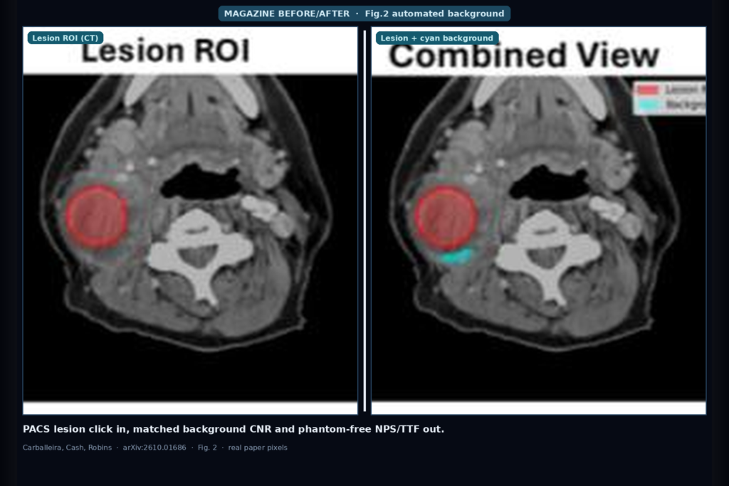
Hang a head-and-neck CT into a clinic physics or DICOM-viewer quality workflow and the shop question is not another phantom spreadsheet. It is whether a lesion click on a PACS screenshot can land on the matching DICOM slice, whether background ROIs for CNR stay tissue-matched without an operator redrawing them, and whether noise texture and spatial resolution can be read from the same patient anatomy that the radiologist actually reads. Rafael Carballeira, Hayley A. Cash, and Marthony L. Robins (Dartmouth Thayer / Dartmouth Hitchcock / MUSC) take that hang seriously in arXiv:2610.01686, submitted 1 October 2026 to physics.med-ph. They call the system AnatomIQ: an open-source Python toolkit with automated CT background detection, phantom-free NPS and TTF on patient anatomy, a Gradio web UI, and a PACS-screenshot click-to-select path that maps lesion coordinates into the DICOM stack.
What hangs on the viewer
Upstream inputs are a PACS display capture plus the corresponding CT DICOM stack. The operator defines the DICOM crop region once, clicks the lesion on the screenshot, loads the stack, and confirms registration with a visual overlay. Downstream hangables for a physics shop or viewer plugin include lesion and cyan background ROI overlays, CNR / SNR / Rose detectability, a dose-normalized figure of merit (CNR squared over SSDE) with protocol recommendations, plus on-demand NPS (noise texture) and TTF (resolution from trachea or vessels used as natural circular edges).
Analysis runs about 2 to 3 minutes per case. CNR lands in under 10 seconds. NPS is about 5 to 15 seconds and TTF about 10 to 25 seconds when requested. That is roughly 50% faster than manual ROI placement in their description, and it removes the operator redraw that breaks reproducibility across readers.
How it works in plain words
Five modules sit behind the Gradio UI: a DICOM handler with HU conversion, a background detector, a quality-metrics block (CNR, SNR, dose metrics, NPS, TTF), publication-style figures, and the web front end. Background detection is the piece that replaces freehand ROIs. Instead of assuming subcutaneous fat, the algorithm picks a tissue-specific HU window from the lesion itself: fat lesions use about −190 to −30 HU; soft-tissue lesions use lesion mean ± 30 HU; denser enhanced tissue uses lesion mean ± 40 HU. Dual spatial constraints keep the background local: an inner exclusion buffer (default 5 mm, slider-adjustable, larger for big masses) avoids partial-volume spill from the lesion edge, and an outer limit keeps background noise comparable to the lesion neighborhood. Morphological cleanup drops tiny components, fills small holes, and opens thin fascia or vessel walls so noise statistics come from a homogeneous patch. Each candidate is scored on area (≥100 mm²), homogeneity (CV under 0.3 preferred), and a GOOD / WARNING / FAILED flag.
NPS builds on that same uniform-tissue mask. The toolkit searches low-variance patches, starting large (256×256) and downscaling toward 32×32 only when anatomy will not give enough clean windows, then ensemble-averages (default M = 16) with detrending and Hann windowing in an AAPM TG-233 style periodogram. TTF skips a dedicated edge phantom: Hough circle detection finds high-contrast circular anatomy (trachea, vessels, airways, contrast >200 HU), averages radial edge profiles into an ESF, differentiates to an LSF, and Fourier-transforms to a TTF curve with TTF50 / TTF10 and a structure quality score.
Core formulas stay shop-familiar. CNR is absolute lesion-minus-background HU over background sigma. Rose detectability is CNR times the square root of lesion area. FOM is CNR²/SSDE. Dose recommendations use the noise ∝ 1/√dose relationship to project CNR at alternate SSDE levels. CNR bands follow Rose-style thresholds: ≥5 adequate, 3–4.9 marginal, <3 inadequate.
What the numbers say
Validation treats head-and-neck as a near-worst-case site: small FOV, mixed tissue, scarce large uniform regions. Automated backgrounds still met size and homogeneity criteria across sites they tested. On a uniform CT phantom the NPS pipeline kept full 256×256 patches (M = 16) and produced a symmetric peak at 0.098 mm⁻¹ with average NPS 58.5 HU²·mm². On an in-vivo head-and-neck case it downscaled to 32×32, peaked at 0.066 mm⁻¹ with average NPS 646.9 HU²·mm², and the 2D NPS looked anisotropic along tissue planes rather than pipeline-artifactual. TTF on a resolution phantom insert gave TTF50 = 0.156 mm⁻¹ and TTF10 = 0.277 mm⁻¹. On the patient trachea (quality 0.89, contrast 988 HU) it gave TTF50 = 0.172 mm⁻¹ and TTF10 = 0.298 mm⁻¹, below a 0.50 mm⁻¹ Nyquist and consistent with a soft-tissue-oriented smooth kernel. Phantom numbers are internal controls for the pipeline, not a claim that phantom and in-vivo peaks must match numerically.
Where it fails and what not to trust
This is a research toolkit with a provisional patent filed and optional commercial licensing mentioned, not a cleared medical device for every protocol. CNR acceptability bands are applied uniformly today; published detectability thresholds vary by body region and task, and the authors flag anatomy-aware thresholds as future work. NPS peak frequencies at different patch sizes are not apples-to-apples (frequency resolution scales with patch width). Head-and-neck forced the 32×32 ensemble; liver-scale organs should tolerate larger patches, but that anatomy-aware sizing is planned rather than shipped as automatic. Re-validate HU windows, SSDE tagging, and overlay registration on your own PACS crop and DICOM path before you trust dose recommendations. No public GitHub URL appears at the end of the preprint; start from the arXiv abstract and PDF.
For a viewer or clinic AI shop
Wire PACS screenshot plus CT stack in; hang matched cyan background ROIs, CNR/SNR/Rose/FOM, dose tips, and optional in-vivo NPS/TTF curves out. Prefer this pattern when your shop still burns minutes redrawing background ROIs for CNR audits, when IR/DLIR noise texture matters more than a single sigma, and when you want resolution from trachea or vessels without booking a phantom slot. The Gradio path (screenshot crop, lesion click, DICOM transfer, visual verify, metrics tabs) is the same hang a DICOM-viewer team already knows how to embed as a side panel or micro-app.
If you integrate, keep the human confirm step on coordinate transfer and on any dose change suggestion. Do not treat a single CNR number as a substitute for task-based detectability on your scanner fleet. Rebuild from arXiv:2610.01686. PDF: https://arxiv.org/pdf/2610.01686.
Sources
- Carballeira, R., Cash, H.A., Robins, M.L. AnatomIQ: An Open-Source Toolkit for Automated Background Detection in Medical Imaging. arXiv:2610.01686, 2026. https://arxiv.org/abs/2610.01686. PDF: https://arxiv.org/pdf/2610.01686.
- Timing: ~2–3 min/case; CNR <10 s; NPS 5–15 s; TTF 10–25 s; ~50% faster than manual ROI. CNR bands: ≥5 adequate, 3–4.9 marginal, <3 inadequate (Rose).
- NPS: phantom peak 0.098 mm⁻¹ (avg 58.5 HU²·mm²) vs in vivo 0.066 mm⁻¹ (avg 646.9). TTF: phantom TTF50/TTF10 = 0.156/0.277 vs trachea 0.172/0.298 (quality 0.89, contrast 988 HU). No public code URL in the preprint; start from arXiv. Provisional patent filed; optional commercial licensing mentioned.