
Ultrasound is where a lot of clinics actually make quantitative calls: ejection fraction, fetal biometry, organ size, lesion borders. Those numbers sit on a contour. If the contour breaks when the probe, vendor, or operator changes, the measurement is noise. Chao Qin, Fahad Shahbaz Khan, Salman Khan, Sarim Ather, Siddiq Anwar, Rao Muhammad Anwer, and Shadab Khan (MBZUAI with STMC Al Ain, King’s College Hospital London Dubai, and ADIA Lab) posted arXiv:2609.19230 on 16 September 2026. The paper is Open ultrasound foundation model for robust segmentation and clinical measurement across heterogeneous settings.
They ship two pieces. SonoCorpus pools 456,963 images and 1,626,085 expert masks from 53 public datasets across 24 clinical applications and 17 countries. SonoBase is an interactive, memory-conditioned segmentation model pretrained on that corpus. Across fifteen evaluation sets that introduce new organs, devices, operators, and geographies, it beats SAM2, MedSAM2, and concept-promptable MedSAM3 on every dataset in their tables, and matches per-dataset specialists trained on the same data. On fully external data it stays above what those promptable baselines score on their own in-distribution tests.
What hangs on the viewer
Input is a B-mode frame or a short ultrasound clip plus a point or box prompt. Output is a structure overlay you can edit, then optionally propagate across adjacent frames with the memory bank. The backbone is SAM2-style promptable segmentation with an image-pyramid hybrid encoder tuned for ultrasound texture instead of natural-image priors. That is the practical hang for a DICOM or PACS-adjacent viewer: click once on the chamber, head, gland, or lesion, get a mask, correct a few slices, let temporal memory finish the clip.
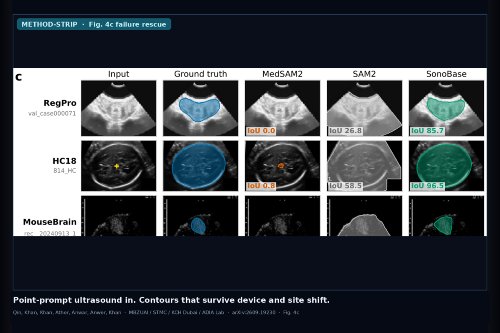
They evaluate more than pixel overlap. Cardiac ejection fraction from SonoBase contours on CAMUS lands within published inter-observer variability (6.63% absolute error with box prompts). At the defibrillator-candidacy EF threshold they report fewer misclassifications than the promptable baselines (13% versus 18 to 42%). Fetal head circumference error is 1.81 mm and gestational-age error is about 1.2 days, both below the inter-observer bands they cite. Where a baseline produces no usable mask (IoU under 10), which happens on about one in four of their 5,223 test cases, SonoBase recovers a clinically useful overlay (IoU above 50) on 81% of those failures, including handheld probes run by minimally trained users in Sierra Leone and Tanzania (ACOUSLIC).
Why ultrasound-specific pretraining matters here
General promptable medical models still treat ultrasound as another modality in a mixed bag. Speckle, dropouts, and probe motion look nothing like CT or MRI, so a contour that looks fine on a clean teaching set can vanish on a point-of-care handheld or a blind sweep. SonoCorpus is the authors’ answer to that fragmentation: videos dominate the mix, scanners include GE, Philips, Siemens and others, and the evaluation split keeps a true external tier never seen in training. Five labeled examples can adapt the model to a new setting in their few-shot tables, and the same training recipe transfers to newer SAM3-style backbones, which puts the gain on the ultrasound pretraining data more than on a one-off architecture trick.
For a viewer shop that already hangs SEG overlays on US series, this is one foundation checkpoint, interactive prompts, optional temporal fill, then derived measurements that a human still owns. Fail closed when the overlay collapses on a low-contrast border, when the study is not ultrasound, or when the downstream number sits near a therapy threshold and the operator never reviewed the contour.
Where it still fails
Catastrophic rescue is strong but not total. About 19% of baseline failures stay unresolved under their IoU rules, and some datasets (including the hardest handheld ACOUSLIC slice of the story) show lower rescue rates than prostate or fetal-head sets. Box prompts usually help more than a single point when the structure is large or the contrast is weak. Clinical measurement still depends on a correct anatomic target: a beautiful mask on the wrong chamber is still a wrong EF. Fairness and site audits in the paper use the axes available in public data; they are not a full multi-center reader study. Treat the released checkpoints as a starting platform, not a cleared device.
For a viewer or clinic AI shop
Hang this as an interactive ultrasound rail: series in, point or box prompt, structure overlay out, human edits in, memory propagation across the clip, then EF or biometry from the approved contour. Prefer it when your install mixes cart and handheld probes, when operators vary, or when you are tired of shipping a new specialist network for every organ. Keep a QC gate before any number that changes therapy. If the overlay IoU against a quick second contour is poor, if the clip is outside the trained anatomy set, or if someone wants silent auto-EF without review, do not ship the number.
Rebuild from arXiv:2609.19230. As of 18 September 2026 the abstract and PDF respond. The authors release checkpoints, optimizer states, data-split indices, deduplication hashes, and starter code for full and LoRA fine-tuning.
Sources
- Qin, C., Khan, F. S., Khan, S., Ather, S., Anwar, S., Anwer, R. M., Khan, S. Open ultrasound foundation model for robust segmentation and clinical measurement across heterogeneous settings. arXiv:2609.19230, posted 16 September 2026. https://arxiv.org/abs/2609.19230. PDF: https://arxiv.org/pdf/2609.19230.
- SAM2 / MedSAM2 / MedSAM3 (cited promptable baselines).
- CAMUS, HC18, ACOUSLIC and the other public ultrasound sets listed in SonoCorpus.