
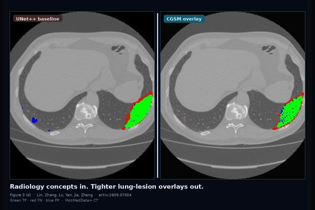
Hang a COVID-era chest X-ray or a MosMed-style CT slice with a short structured report. Coarse location text alone often leaves fuzzy margins and tiny opacities under-segmented. CGSM turns that report into radiology concepts (ground-glass, consolidation, crazy-paving, fuzzy margins, distribution) that a clinician panel already scored, then hangs a lesion overlay guided by those concepts. Changheng Lin, Wenjie Zhang, Yushan Lu, Xinyue Yan, Xiao Jia, and Wei Zhang (Shandong University) posted arXiv:2609.07004 on 7 September 2026 (v3 on 10 September). The paper is CGSM: Concept-Guided Segmentation Model for Precise Pulmonary Lesion Delineation.
Image-only U-Nets miss radiology priors that describe margin sharpness, texture, and where disease sits in the lung. Prior vision-language segmenters help, but the paired text in public COVID datasets is usually location-heavy and weak on boundary attributes. That gap shows up as red false negatives and blue false positives around small lesions in qualitative grids.
What goes in, what comes out
Input is a 224×224 CXR or CT slice plus a paired structured report from the dataset. Output is a pixel lesion mask you can hang as an overlay. Concepts are not free-typed at inference in the paper’s main setup. An LLM (Gemini-3 in their write-up) proposes disease-specific phrases for the modality, five clinicians score each phrase 1–10, and phrases averaging below 6 are dropped and regenerated. Those curated concepts feed a concept encoder at train and test time alongside the report text.
Two public cohorts carry the numbers. QaTa-COV19 has 9,258 COVID CXRs with expert pixel masks (5,716 / 1,429 / 2,113 train/val/test). MosMedData+ has 2,729 CT slices (2,183 / 273 / 273) with lesion masks. Both ship paired structured reports in the LViT-style packaging the authors follow.
How concepts steer the overlay
Three encoders run in parallel. ConvNeXt-Tiny reads the image into a four-level feature pyramid. A frozen CXR-BERT encodes the report tokens. The same frozen CXR-BERT plus a small trainable projection head turns the concept phrases into concept tokens.
CVAM (Concept-Visual Alignment Module) pulls each concept token toward supporting visual evidence with contrastive learning, so “fuzzy margins” has to latch onto edges that look soft rather than floating as a bag-of-words tag. The CM-Decoder then uses those aligned concepts as modulation (scale and shift) while fusing image and text features through hierarchical multi-head cross-attention before the segmentation head. Training mixes cross-entropy, Dice, and the CVAM loss on an A100 with AdamW and ImageNet-init for the vision backbone.
Numbers that support the viewer hook
On QaTa-COV19, CGSM reaches 91.59 Dice and 84.49 mIoU, ahead of MMI-UNet at 90.88 / 83.28. On MosMedData+, it reaches 79.08 Dice and 65.40 mIoU versus 78.42 / 64.50 for MMI-UNet. Ablations show both CVAM and the CM-Decoder help; the full stack beats concept tokens alone. Figure 3 paints the practical story: green true positives, red false negatives, blue false positives. CGSM columns sit closer to ground truth on both CXR rows and CT rows, especially around small scattered lesions where UNet++ and several VL baselines bleed red and blue.
Where it still fails
Everything is 2D at 224×224, so through-plane consistency and native DICOM resolution are out of scope. The concept bank is disease- and modality-scoped; swap in a new pathology and you rebuild and re-review concepts. The report is still required as text input in this pipeline, unlike text-free modules such as IGRA. COVID-era QaTa and MosMed distributions do not stand in for arbitrary interstitial or oncologic lung disease. No public code link appears in the preprint skim. Calibration, uncertainty, and DICOM-SEG export are not the paper’s job.
For a viewer shop
Treat CGSM as a recipe for report-conditioned pulmonary lesion overlays when you already have structured text and can afford a curated concept list reviewed by clinicians. Fail closed when you only have the pixels, when the concept bank was built for a different disease, on full 3D volumes without a slice policy, and when you need millimeter surgical margins rather than overlap-oriented COVID lesion maps.
Rebuild from arXiv:2609.07004. As of 13 September 2026 the abstract and PDF respond. The preprint is the source of record until a camera-ready version exists.
Sources
- Lin, C., Zhang, W., Lu, Y., Yan, X., Jia, X., Zhang, W. CGSM: Concept-Guided Segmentation Model for Precise Pulmonary Lesion Delineation. arXiv:2609.07004, posted 7 September 2026 (v3 10 September 2026). https://arxiv.org/abs/2609.07004. PDF: https://arxiv.org/pdf/2609.07004.
- Li, Z., et al. LViT: Language meets Vision Transformer in Medical Image Segmentation. IEEE TMI 2023.
- Boecking, B., et al. CXR-BERT / Making the Most of Text Semantics to Improve Biomedical Vision-Language Processing (cited text encoder).
- Liu, Z., et al. ConvNeXt (visual backbone).
- QaTa-COV19 and MosMedData+ public COVID lesion segmentation datasets (as cited).