
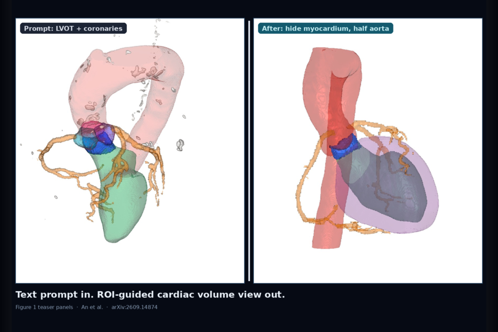
You ask a volume viewer to show the left ventricular outflow tract from the left ventricle to the aorta, keep the coronary arteries visible, and leave the myocardium in the scene. Then you ask it to segment the three aortic valve cusps. Then you say hide the myocardium and cut aorta opacity in half. In most clinical DVR and iso-surface setups, that sequence means transfer-function edits, viewpoint nudges, and a lot of trial and error. Haill An, Suhyeon Kim, Minjun Kang, Eunwoo Lee, Bin Sheng, Lei Bi, and Younhyun Jung (Gachon University and collaborators) posted arXiv:2609.14874 on 14 September 2026. The paper is MedVA: An End-to-End Neuro-Symbolic Agentic System for Medical Volume Visualization.
MedVA turns the natural-language request into an ROI intent graph, pulls masks from pretrained medical segmenters sitting on the original volume, then adjusts visibility with a volume-based objective instead of guessing from a screenshot. For a DICOM viewer or clinical platform team, the hook is not another chat overlay. It is a concrete rail from clinical wording to hung ROIs and opacity that you can audit.
Three agents between the prompt and the render
The first agent is neuro-symbolic intent formulation. An MLLM drafts a ROI set from the user text. Symbolic reasoning over FMA 5.1.0 and NCI 26.07d then expands or corrects that set so broad clinical phrases do not leave related structures out. Table 2 in the paper shows the difference on prompts such as pancreas with surrounding vessels: primary ROIs stay bold, and the symbolic pass adds context vessels that MLLM-only wording missed.
The second agent is multi-model ROI identification. MedVA calls TotalSegmentator 2.13.0 and VoxTell 0.1.1 on the original volume, not on a 2D rendering gallery. Across thirteen representative ROIs, mean Dice sat around 87% for both models, with neither winning every structure. The agent picks per label. On a chest volume it preferred VoxTell for airway when TotalSegmentator spilled into peripheral bronchi. On a liver volume it mixed four TotalSegmentator masks with five VoxTell masks, including hepatic veins that only one model returned.
The third agent is objective-driven visualization optimization. Instead of asking an MLLM whether an ROI looks occluded in a screenshot, MedVA scores ROI visibility along rays in the volume, compares full-scene visibility to isolated-ROI visibility, and drives a visualization-parameter-adjustment loop. Across three volumes and both DVR and ISR modes, that volume-based loop averaged about 11.9 seconds, 4.0k tokens, and 1.8 iterations, cutting optimization time by roughly half versus a rendering-based loop in their comparison.
What hangs on the cardiac and body cases
Figure 1 walks a cardiac volume through the exact prompt sequence above. LVOT, coronaries, myocardium, aorta, and left ventricle appear first. Aortic cusp labels arrive on the second request. After hide-myocardium and half-aorta-opacity, the coronaries and cusps stay readable without a manual 1D transfer-function session. Case studies on pelvic and abdominal volumes show the same pattern for skeletal substructures, tumor versus hepatic vessels, and requests that should drop the rib cage when the clinical focus moves inside the liver.
A formative user study with nine participants scored 51 of 54 multiple-choice questions correctly (94.4%). Laterality, containment, and lobar or segmental location questions were all correct. The three misses involved finer relationships with surrounding anatomy. Treat that as usability evidence for the interaction pattern, not as a multi-center reader trial.
Where it still fails
MedVA only knows the clinical concepts and segmentation labels its knowledge bases and models cover. Anatomy or pathology outside that coverage will not magically appear in the ROI graph. Runtime still concentrates in ROI identification and visualization optimization (about 89% of end-to-end time in their breakdown), so a live viewer needs budgets, caching, and a cancel path. The system is evaluated on selected volumes and scripted interactions. It is not a drop-in replacement for every modality, every vendor transfer-function preset, or every intraoperative latency target. When TotalSegmentator and VoxTell disagree on a vessel or tumor margin, someone still has to decide which mask hangs as truth.
For a viewer or clinic AI shop
Treat MedVA as a volume-visualization control plane: clinical text in, ROI intent graph out, masks from named segmenters on the source volume, then opacity and occlusion updates driven by a measurable visibility score. Hang the intent graph beside the render so a reader can see which structures were primary versus context. Hang the chosen segmenter name per ROI. Fail closed when the symbolic expansion invents an ROI your models cannot segment, when both segmenters disagree beyond a QC threshold, when the volume is not the modality the segmenters were trained for, or when a user needs sub-second interactive TF scrubbing that this agent loop will not beat. If your platform already stores SEG-style labels and can drive DVR or ISR parameters from an API, this paper is a practical blueprint for putting natural-language volume requests on the same case without letting the MLLM own the anatomy.
Rebuild from arXiv:2609.14874. As of 15 September 2026 the abstract and PDF respond. Public code and weights are not linked as a release URL in the preprint text we used.
Sources
- An, H., Kim, S., Kang, M., Lee, E., Sheng, B., Bi, L., Jung, Y. MedVA: An End-to-End Neuro-Symbolic Agentic System for Medical Volume Visualization. arXiv:2609.14874, posted 14 September 2026. https://arxiv.org/abs/2609.14874. PDF: https://arxiv.org/pdf/2609.14874.
- TotalSegmentator 2.13.0 and VoxTell 0.1.1 (cited ROI identification models).
- FMA 5.1.0 and NCI Thesaurus 26.07d (cited symbolic grounding resources).
- Prior agentic visualization systems compared in the paper (AVA, ChatVis, NLI4VolVis, and related work).