
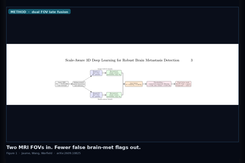
Hang a multimodal brain MRI with T1c and T2-FLAIR in the viewer and the hard problem is not drawing a pretty contour. It is catching small metastases without flooding the worklist with false flags. Sylvain Jaume, Hongming Wang, and Simon K. Warfield (MIT CSAIL, Harvard, and Boston Children’s Hospital / Harvard Medical School) posted arXiv:2609.10825 on 9 September 2026. The paper is Scale-Aware 3D Deep Learning for Robust Brain Metastasis Detection in Multimodal MRI. Their move is simple enough to hang behind a DICOM rail: run two compact 3D U-Nets on different spatial fields of view, fuse the whole-volume probability maps late, then threshold and filter connected components.
Large patches give context but shrink the relative size of tiny lesions. Small patches raise lesion occupancy and lose neighborhood cues. Prior multi-scale work often mixes resolution, modality, and architecture changes at once. This study holds resolution and modalities fixed (1 mm isotropic T1c + T2-FLAIR from BraTS-METS) and asks what changes when you only vary FOV and how you fuse the probabilities.
What goes in, what comes out
Input is a preprocessed BraTS-METS multiparametric volume using T1c and T2-FLAIR only. Output is a binary lesion mask after sliding-window inference, weighted late fusion, probability thresholding, and a minimum-component volume filter (primary setting 0.010 mL). The mask is shaped for BraTS Task 1 export (foreground mapped to the challenge label).
The internal development cohort (DEV97) has 97 patients and 667 connected-component ground-truth lesions. Lesions were stratified by physical volume from under 0.010 mL up to at least 0.500 mL before evaluation. Patch optimization used a seed-42 90/10 split inside positive and negative patch sets; that split is patch-level, not patient-level, so whole-volume lesion metrics on DEV97 are development measures, not a clean patient-held-out test.
How dual FOV fusion cuts spurious flags
Both branches share the same compact 3D U-Net: encoder channels 32/64/128, 256-channel bottleneck, instance norm, ReLU, and a one-channel tumor logit head. Branch A sees 96 cubed patches. Branch B sees 64 cubed patches made by center-cropping the lesion-centered 96 cubed patches, so FOV is the controlled variable rather than a different sampling grid. Each network trains separately with Dice-focal loss, Adam at 1e-4, batch size 2, and axis flips. At inference, overlapping sliding windows rebuild aligned whole-volume maps P96 and P64 (50% overlap; strides 48 and 32 voxels).
Fusion is a weighted average, P = α P96 + (1 − α) P64. On DEV97 the selected setting is α = 0.60 (60% large-context, 40% small-context) with probability threshold 0.60 and min volume 0.010 mL. That operating point reached lesion-level sensitivity 0.582, precision 0.602, F1 0.592, and 2.639 false positives per patient. Versus the 96 cubed endpoint alone, that is a relative F1 lift of about 17% and a cut of roughly 57% in FP/patient (from about 6.2 down to 2.6). A same-FOV 64+64 ensemble control improved over a single 64 cubed model but still trailed cross-FOV fusion on F1 and false-positive burden in paired patient bootstrap tests. So averaging two independently trained nets helps, and complementary spatial context helps further.
Size still dominates recall. Under the selected fusion, sensitivity climbs from 0.057 below 0.010 mL to 0.891 at or above 0.500 mL. Fusion did not magically rescue the tiniest stratum relative to the large-FOV baseline; its main gift is precision and fewer false flags. An exploratory cross-FOV agreement filter trimmed FP/patient a bit more (about 2.64 to 2.27) without a meaningful F1 gain. Code for the detector path is linked at github.com/ailabrepo/BraTSDetector.
Where it still fails
Very small metastases remain the weak spot. Only 4 of 70 lesions under 0.010 mL were detected at the primary operating point, and the 0.010 mL component filter itself trades away some of that stratum. False-positive audits of high-confidence unmatched components show compact lesion-like mimics plus peripheral or boundary anatomy and a few large irregular non-lesional blobs. Official BraTS 2026 validation was much weaker than the internal numbers (ET all-lesion instance F1 0.385), and the challenge container later applied the same retained weights under both FOV checkpoint names, so that score is not a clean external replica of the separately trained internal ensemble. The same-FOV control used a shorter five-epoch schedule, so FOV effects are supportive rather than fully isolated. Inputs stay on T1c and FLAIR; there is no modality ablation. If you need near-perfect milliliter-scale mets, radiologist-adjudicated FP labels, or a multi-class ET/RC/WT product, this preprint is not that shipping stack.
For a viewer shop
Treat the dual-FOV rail as a detection overlay path: T1c + FLAIR volume in, two sliding-window U-Nets at 96 cubed and 64 cubed, late probability fusion out, then hang connected-component candidates with volume and centroid metadata next to the series. Prefer the fused map when the clinical pain is false flags on a multi-met worklist, not when you only care about maximizing recall of sub-0.01 mL seeds. Fail closed outside BraTS-METS-style 1 mm isotropic pre/post treatment MRI, when only one contrast is available, when you need subregion labels the binary detector never predicted, and when external instance-F1 must match the internal development curve this paper already shows it does not.
Rebuild from arXiv:2609.10825. As of 12 September 2026 the abstract and PDF respond. Check the GitHub link above for code and checkpoint status; the preprint is the source of record until a camera-ready version exists.
Sources
- Jaume, S., Wang, H., Warfield, S. K. Scale-Aware 3D Deep Learning for Robust Brain Metastasis Detection in Multimodal MRI. arXiv:2609.10825, posted 9 September 2026. https://arxiv.org/abs/2609.10825. PDF: https://arxiv.org/pdf/2609.10825. Code: https://github.com/ailabrepo/BraTSDetector.
- Maleki, N., et al. BraTS-METS / BraTS 2025–2026 brain metastases challenge materials (cited dataset).
- Moawad, A. W., et al. BraTS-METS Challenge 2023 preprint (cited).
- Ronneberger, O., et al. U-Net. MICCAI 2015; Çiçek, Ö., et al. 3D U-Net. MICCAI 2016.
- Isensee, F., et al. nnU-Net. Nat. Methods 2021.