
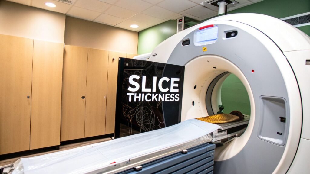
CT slice thickness is the z-axis width of each reconstructed (or acquired) slice — the thickness of one cut through the loaf. It is a protocol choice: thinner sees small things and builds cleaner MPRs; thicker is quieter and cheaper on dose. This page is that trade-off. It is not “what resolution is.”
If you meant spatial vs contrast vs temporal as pillars → resolution of a CT scan. If you meant partial volume as an artifact → artifacts in computed tomography. If you meant a calculator → CT slice thickness calculator (the tool stays a tool).
The trade-off
| Thinner (e.g. <1 mm) | Thicker (e.g. >3 mm) | |
|---|---|---|
| Spatial detail | Small structures stay separate | Fine detail can vanish |
| Noise | Grainier (fewer photons per slice) | Smoother |
| Partial volume | Less averaging of a pea into the bread | More — a 4 mm lesion on a 5 mm slice can disappear |
| MPR / 3D | Isotropic, usable coronals | Stair-step, blocky |
| Dose / time | Often more mAs, more data | Lower dose, faster review |
ALARA still applies: the thinnest slice that answers the question, not the thinnest the tube can do. A follow-up of a known 4 cm mass does not need 0.6 mm.
How a slice is made
Pre-patient collimators are lead shutters next to the tube. They shape the fan into a ribbon. That ribbon width is the nominal thickness. Tighter ribbon = thinner cut along z.
The beam then hits a detector array — thousands of elements in rows. Final thickness is collimation plus how those rows are grouped. Single-slice scanners in the 1990s had almost no grouping choice. CTA then sat on slices thicker than 3 mm and smeared vessels along z.
MDCT put multiple detector rows on the gantry. One rotation can grab many slices. That unlocked 0.5–0.625 mm acquisitions, whole-organ scans in a few seconds, and the later trick: scan thin, stack thick for review.
Isotropic voxels
A voxel is a 3D pixel. It is isotropic when slice thickness equals in-plane pixel size — a cube. Cubes reformat without a squash. Coronal and sagittal MPRs then look like the original axials. Rectangular bricks (thick z, fine xy) make stair-steps. That is why 0.625 mm axials exist even when the radiologist reads 3 mm.
Acquired vs reconstructed
Acquired thickness is hardware: collimators + detector grouping at scan time. Modern default is as thin as the scanner will do.
Reconstructed thickness is software after the fact. Stack those thin axials into 3 mm or 5 mm for a quieter primary series. Keep the thin set for MPR, 3D, and a second look at a tiny finding. Same scan, two thicknesses. You do not re-irradiate the patient to get the thick series.
Protocol millimetres
| Exam | Typical thickness | Why |
|---|---|---|
| Cardiac CTA | 0.5–0.75 mm | Coronaries are millimetres wide; stenosis hides on thick z |
| HRCT chest (ILD) | 1.0–1.25 mm | Interstitium is a web; 5 mm averages it away |
| Complex fracture (wrist, ankle) | 0.625–1.5 mm | Needs MPR the surgeon can trust |
| CT urography / stones | 2–3 mm | Small calcifications without a screening-level dose |
| Non-contrast head (stroke) | 2.5–5 mm | Bleed / large infarct, fast; not a temporal-bone study |
| Routine abdomen / pelvis | 3–5 mm | Organs and larger masses; less noise, less dose, fewer images |
Patient size changes the reconstructed choice more than the acquired one. A larger abdomen starves photons; stacking to a thicker review series raises SNR without another trip through the gantry.
Dose, noise, partial volume
Thinner slice = smaller volume = fewer photons = more grain. The usual compensation is more mAs. That is the dose cost. Iterative reconstruction and deep-learning reconstruction take some of that cost back — they are why low-dose thin-slice lung screening is usable. They do not delete the trade-off; they shrink it.
Partial volume is the pea mashed into a thick slice of bread: two tissues in one voxel become one HU. A 4 mm nodule on a 5 mm slice can read as “a bit of lung.” The catalog of how that looks, and the other artifacts, is artifacts in computed tomography. The mm choice that causes it is this page.
Need a number for a protocol sheet? Use the slice-thickness calculator. This article does not replace that tool.
Building a viewer that has to show acquired and reconstructed thickness as two facts, not one slider, is product work. Case studies.
Keep Reading
Related Articles
Explore the full Quantitative Imaging HubAll services, tools, and guides on this topic — in one place.
Visit Hub →