
One End-Diastole Cardiac MRI Frame In, a Full 30-Frame Beating-Heart Cine Out Without ECG Gating by Warping the Real Pixels With a Phase-Conditioned Flow Model
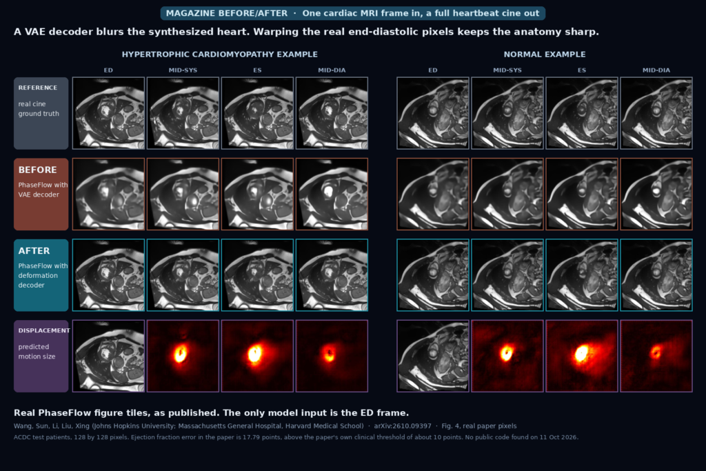
Give PhaseFlow one end-diastolic cardiac MRI frame and it generates a 30-frame cine of the heartbeat with no ECG gating. It predicts a motion field and warps the real pixels, which keeps the image sharp, and it takes its timing from a mean phase curve for the patient’s pathology group. Tested on ACDC with 30 test patients and one training run, the ejection fraction error is 17.79 points, above the paper’s own clinical threshold of about 10, and I found no public code. Wang et al., arXiv:2610.09397.
Handheld Endoscope or Microscope Video In, One Wide-Field Tissue Mosaic Out at About Ten Frames per Second From a Modality-Tuned Optical Flow Model
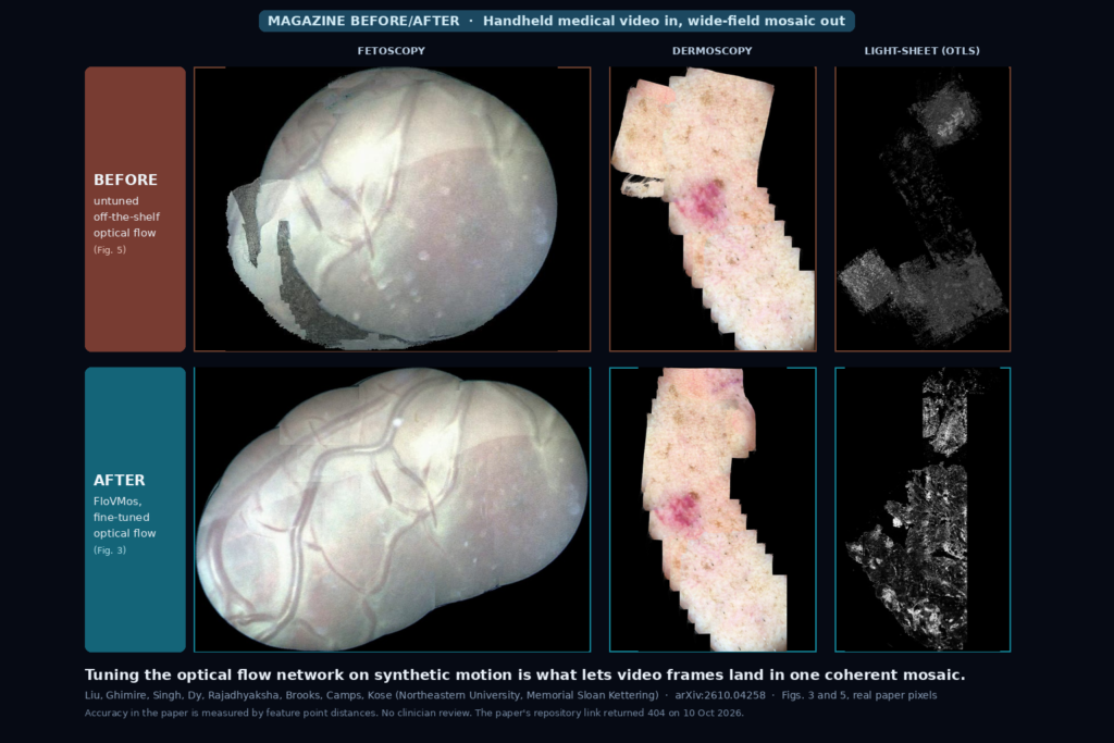
Feed FloVMos video from a handheld endoscope, dermoscope or confocal microscope and it stitches the frames into one growing wide-field mosaic, at about ten frames per second on 1 megapixel frames in the authors’ research code. The trick is fine-tuning an optical flow network per device on synthetic motion, so no hand-labelled flow is needed. Several real test sets are one or two videos, accuracy is a feature distance check, a GPU is required, and the repository linked in the paper returned a 404 when I checked. Liu et al., arXiv:2610.04258.
Undersampled Cardiac MRI In, Sharper Reconstruction Out From a Frozen Pretrained Vision Encoder With About Half the Trainable Weights
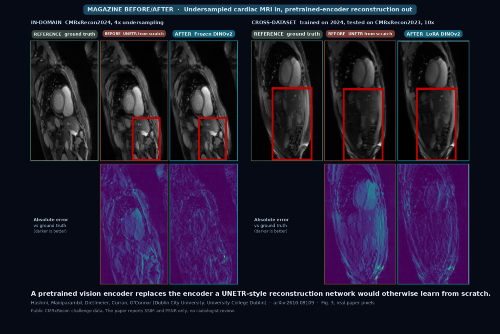
Hand an undersampled cardiac MRI slice to a UNETR-style network whose encoder is a frozen pretrained CLIP, BiomedCLIP or DINOv2 model, and the reconstruction beats the same network trained from scratch, with about 49 percent fewer trainable parameters. The gap holds with 5 percent of the training data and across two challenge datasets. It is a 2D study on public CMRxRecon data scored with SSIM and PSNR only, with no reader study, and the linked GitHub repository held only a README when I checked. Hashmi et al., arXiv:2610.08109.
Eight X-ray Projections In, a Generated 3D Lung CT Volume Out With No Training but About 22 Minutes of GPU Time
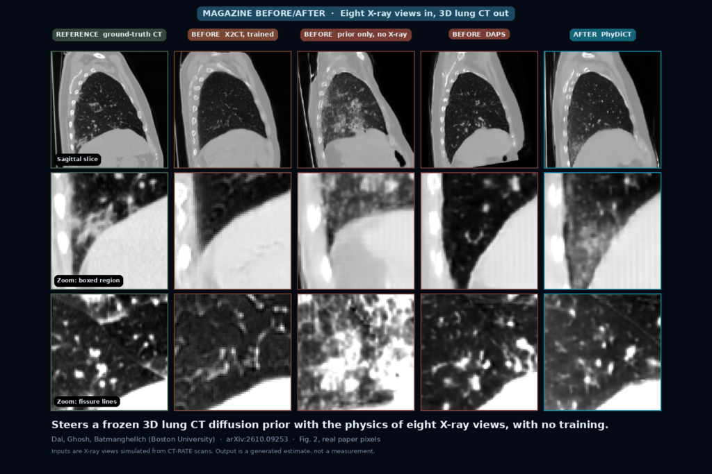
Give it eight simulated X-ray projections around the chest and it steers a frozen, text-conditioned lung CT diffusion model until its own rendered X-rays match, returning a 3D volume with no paired training. It takes about 22 minutes per volume on an RTX 6000, is built for lung CT only, and was tested on X-rays simulated from CT-RATE scans, so the output is a plausible estimate, not a measurement. Apache 2.0 code. Dai et al., arXiv:2610.09253.
Two Coronary Angiogram Masks In, One Connected 3D Artery Tree With Radii Out in About a Tenth of a Second
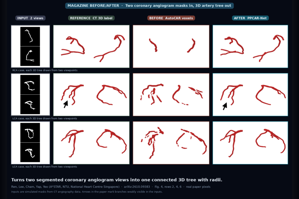
Give it one to seven segmented coronary angiogram views and their gantry angles, and it returns one connected 3D artery tree with a radius along every branch in about 0.12 s on an RTX 4090, with no point matching between views. Trained and scored on simulated masks from CT. MIT code and checkpoints public. Ren et al., arXiv:2610.09383.
CT Slice In, No-Reference Quality Score Out That Catches Dose Noise but Should Not Pick Your Denoiser
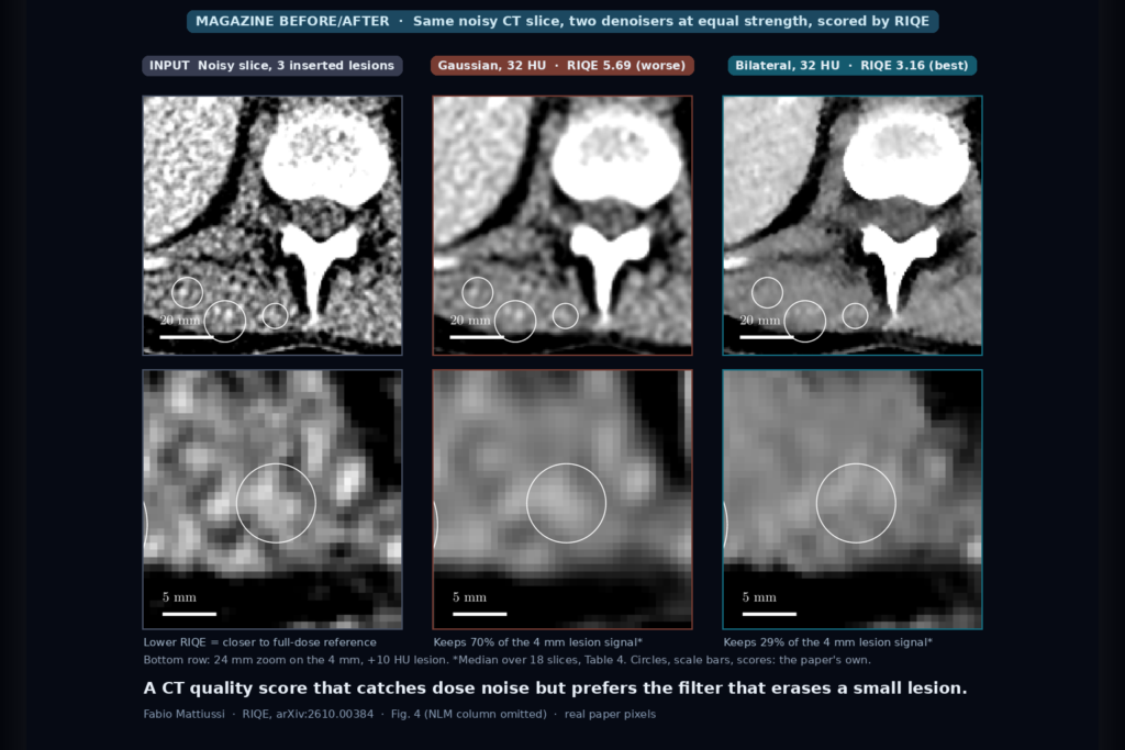
Give it one CT slice and it returns a no-reference quality score fitted on public full-dose scans. It flags simulated dose reduction, added noise and blur, but it prefers an edge-preserving filter that wipes out most of a small lesion. MIT code, CC BY model. Mattiussi, arXiv:2610.00384.
Brain T1 Plus Tumor Mask In, Slice-Consistent Healthy Tissue Fill Out for Registration and Parcellation
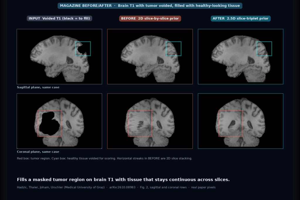
Give it a brain T1 and a mask over the tumor, and it paints in healthy-looking tissue that stays continuous across slices, so atlas registration and parcellation can run on a filled copy. Code and checkpoint public. Hadzic et al., arXiv:2610.08983.
Prostate MRI Near Rectal Gas or Metal In, Undistorted High-Resolution DWI and ADC Out
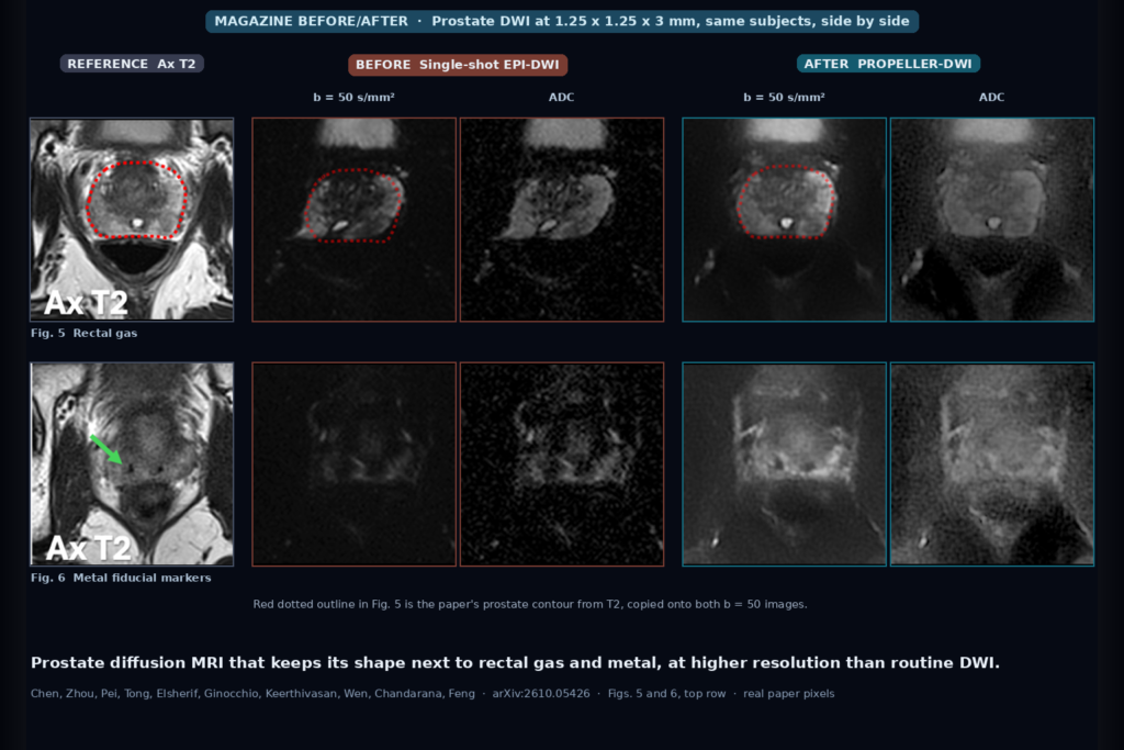
A PROPELLER diffusion sequence with self-supervised reconstruction keeps the prostate’s shape on DWI and ADC next to rectal gas and fiducial markers, at 1.25 mm in-plane. Siemens prototype, no public code. Chen et al., arXiv:2610.05426.
3D T1 Brain MRI In, Closest Real Scans Out, Searchable by Whole Brain or Any of 103 Regions
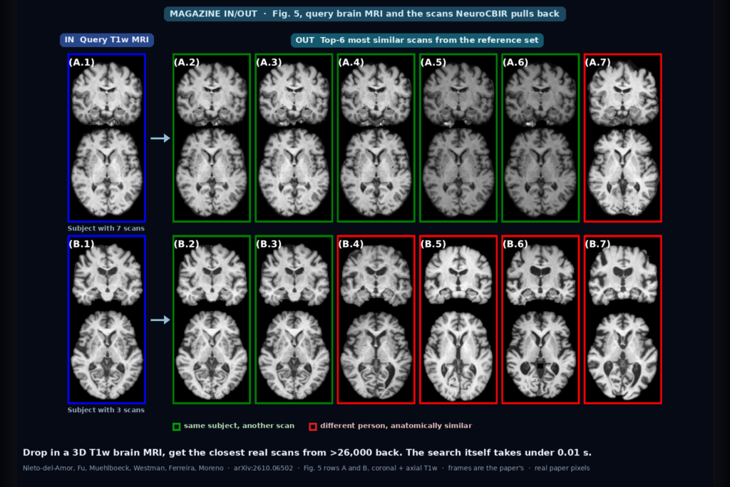
Drop in a 3D T1w brain MRI and get back the most anatomically similar real scans from a reference set of more than 26,000, by whole brain or by region. Code, weights, and embeddings public. Nieto-del-Amor et al., arXiv:2610.06502.
On-Board Radiotherapy CBCT In, Cleaner CT-Like Slices Out, Trained Without Paired Scans
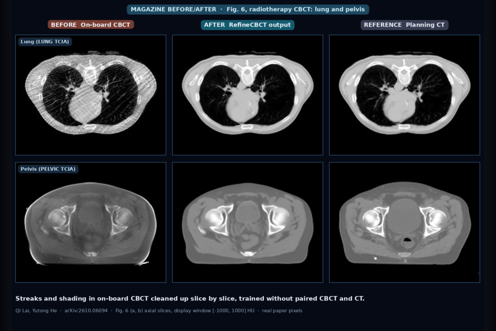
Feed it an on-board radiotherapy CBCT slice and get back one with fewer streaks and CT numbers closer to the planning CT. RefineCBCT learns without paired CBCT and CT. Code public. Lai and He, arXiv:2610.06094.