Imagine a diagnostic tool so powerful it can see what the human eye might miss, catching diseases at their earliest stages with an astonishing level of precision. This isn't science fiction. This is the new reality being created by machine learning in medical imaging—a practical, life-saving technology that is already changing patient care for the better.
A New Era of Sight in Healthcare
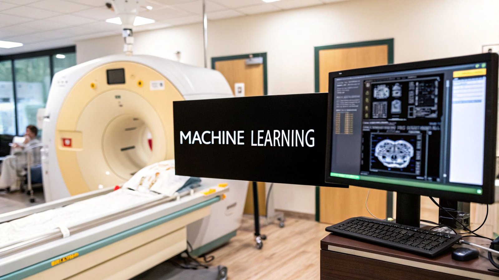
Medical diagnostics is in the middle of a massive transformation, moving away from subjective observation and toward data-driven certainty. The engine driving this change is machine learning, a type of artificial intelligence that teaches computers to find intricate patterns in enormous amounts of data. When we apply this to medical imaging, we’re essentially giving clinicians a powerful new form of sight.
Think of it like training a world-class radiologist, but on a scale that’s impossible for any single person. An algorithm can sift through millions of scans—MRIs, CTs, and X-rays—learning to pinpoint the most subtle signs that point to the start of a disease. This ability is completely reshaping how we diagnose and treat patients.
Empowering Clinicians with Deeper Insights
The goal of machine learning isn't to replace the invaluable expertise of doctors and technicians. Far from it. Instead, it acts as a trusted partner, amplifying their skills and making their work more efficient. By automating repetitive tasks and highlighting potential areas of concern, these tools free up clinicians to focus on what they do best: handling complex cases and caring for their patients.
This human-AI collaboration brings a few major advantages to the table:
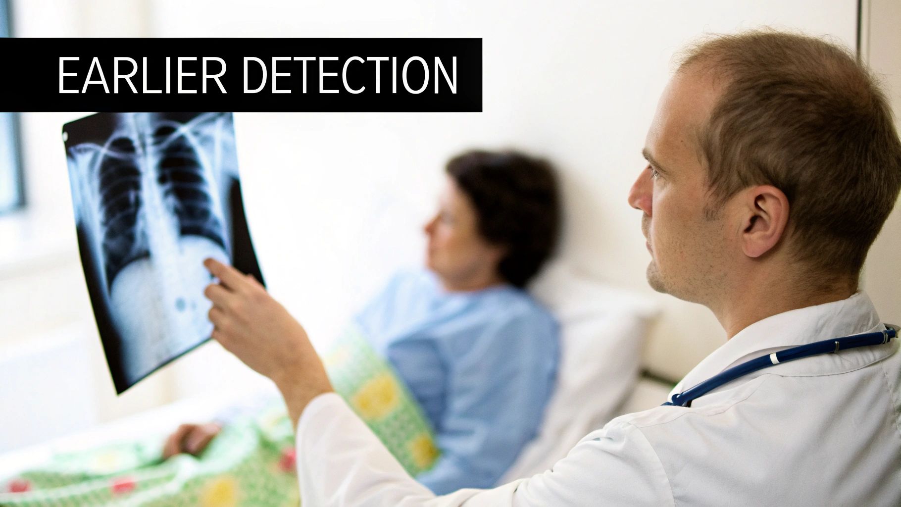
- Earlier Detection: These algorithms can spot the faint signals of diseases like cancer or neurological disorders long before they would typically be visible to the human eye.
- Greater Accuracy: By analyzing data with computational precision, machine learning minimizes the chance of human error and inconsistencies in diagnosis.
- Personalized Treatment: The insights pulled from imaging data can help craft treatment plans uniquely suited to a patient's specific condition, leading to better outcomes.
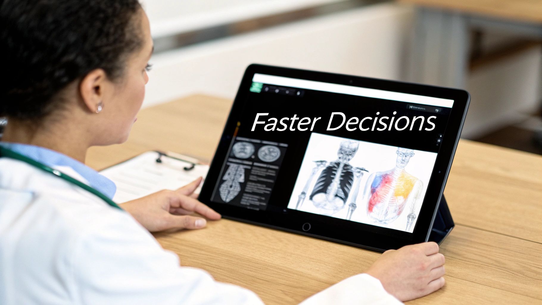
At its heart, machine learning in medical imaging is about turning visual information into life-saving intelligence. It’s about giving doctors a clearer, more detailed map of a patient's health so they can make faster, more confident decisions.
We're right at the forefront of this movement. At PYCAD, we build the custom web DICOM viewers that make these AI insights accessible and actionable. By weaving these intelligent tools directly into medical imaging web platforms, we're not just supporting healthcare professionals—we're actively shaping what comes next.
You can learn more by exploring our insights on the future of medical imaging. Feel free to also check out our work on our portfolio page.
How a Machine Learns to See Inside the Body
How does a collection of circuits and code learn to spot a tiny, potentially cancerous nodule on a lung scan? It’s not magic. The process is a lot like training a new medical student, but it happens on a massive, superhuman scale. We’re essentially teaching an algorithm to recognize patterns, one image at a time.
This journey into artificial sight kicks off with a concept called supervised learning.
Imagine you have a huge library of tens of thousands of chest X-rays. A team of expert radiologists pores over every single one, meticulously labeling them: "this image shows a fracture," "this one is healthy," or "this one contains a suspicious lesion."
The machine learning model is then fed this entire labeled dataset. It studies each image and its corresponding label, slowly learning the subtle visual cues—the specific pixel patterns, textures, and shapes—that define a fracture versus healthy bone. Over and over, it tweaks its internal logic to get better at making the right call.
The Power of Deep Learning and Neural Networks
As effective as that is, the real leap forward in medical imaging comes from a more advanced approach: deep learning. Deep learning models, known as neural networks, are inspired by the layered structure of the human brain, which allows them to learn from data in a much more complex and intuitive way.
The star player in this field is the Convolutional Neural Network (CNN).
Think of a CNN as a digital detective armed with a specialized magnifying glass, examining an image piece by piece. It doesn’t just see the whole picture at once; it scans it through multiple layers, each trained to spot specific features.
- Initial Layers: These might identify the most basic elements, like simple edges, corners, or gradients of light and shadow.
- Intermediate Layers: They start combining these basic features into more complex shapes, like circles or textures that resemble human tissue.
- Deeper Layers: These final layers assemble all the learned features to identify whole objects, like a specific organ or, crucially, an anomaly like a tumor.
This layered analysis allows a CNN to uncover incredibly subtle patterns that might be invisible to the human eye. It's an approach that's fueling incredible growth in the field.
The infographic below shows how AI is becoming a cornerstone of modern diagnostics—boosting speed, accuracy, and the very way we see inside the body.
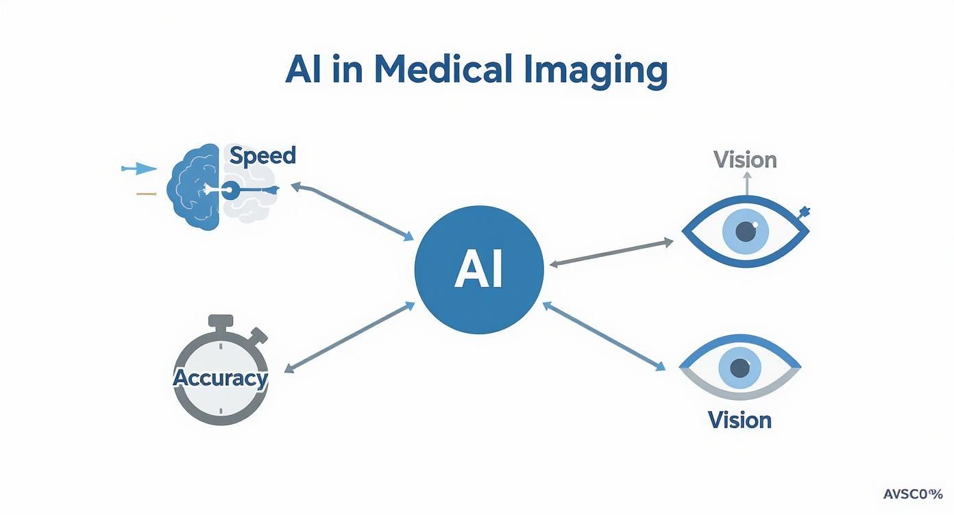
This visual really drives home how AI acts as a multiplier for clinical skills, making the diagnostic process faster and far more precise.
From Raw Data to Actionable Insight
Of course, an AI is only as good as the education it receives. The quality of the training data is everything. That’s why the careful preparation of imaging data is such a critical first step. You can dive deeper into this essential stage in our guide on data preprocessing for machine learning.
This isn't just theory; it’s a rapidly expanding market with a profound real-world impact. The global AI in medical imaging market was valued at around USD 1.36 billion and is projected to explode to approximately USD 19.8 billion by 2033. That’s a compound annual growth rate of 34.67%—a testament to its value.
Deep learning, powered mainly by CNNs, accounts for over 57% of this market's revenue, showing just how dominant it is for image recognition tasks.
By training on vast, expertly labeled datasets, a machine learns to "see" with a level of detail and consistency that complements human expertise. It becomes a tireless assistant, capable of analyzing thousands of scans without fatigue.
Ultimately, this whole process comes together to create a tool that fits right into a clinical setting. At PYCAD, we specialize in this final, crucial step. We build custom web DICOM viewers and integrate them into medical imaging web platforms, making sure these powerful AI insights are delivered to clinicians in a way that’s intuitive and immediately useful. You can see examples of our work on our portfolio page.
Real-World Victories in Patient Diagnosis
The true measure of machine learning in medical imaging isn’t found in complex algorithms or raw processing power—it’s seen in the lives it changes for the better. This technology has broken out of the research lab and is now making a tangible difference in clinics everywhere, giving doctors the tools to diagnose diseases faster and more accurately than ever before. These aren't just far-off promises; these are stories of real impact, happening right now.
Picture an oncologist carefully reviewing a CT scan. Working silently alongside them is an AI assistant, flagging a minuscule cluster of cells—the earliest, faintest whisper of cancer. This seamless teamwork makes it possible to intervene at the most critical moment, radically improving a patient's chances. This is the new reality unfolding in oncology, where machine learning serves as a second set of tireless, ever-vigilant eyes.
A Collaborative Partner in Specialized Care
Let's be clear: this technology isn't about replacing the irreplaceable expertise of a clinician. Think of it instead as a powerful and dedicated partner, one that enhances their skills and allows them to perform at their absolute best. By handling the tedious, data-heavy tasks, it frees up doctors to focus on what matters most: complex decision-making and genuine patient care.
The applications are spreading across a whole host of medical fields:
- Neurology: Smart algorithms are now able to comb through brain scans to spot subtle patterns linked to neurodegenerative diseases. This can help forecast the progression of conditions like Alzheimer's or multiple sclerosis, giving families and doctors precious time to plan therapies.
- Cardiology: In the past, measuring heart function from echocardiograms or cardiac MRIs was a time-consuming manual process. Machine learning now automates these complex calculations, delivering consistent, objective data to cardiologists in a fraction of the time.
- Radiology: AI tools can intelligently sort a radiologist's massive worklist, pushing scans that show signs of critical issues—like a stroke or a pulmonary embolism—straight to the top. This simple act ensures the most urgent cases get the immediate attention they need.
This powerful partnership is elevating the entire standard of care. It transforms overwhelming streams of imaging data into clear, actionable insights that guide life-saving decisions every single day.
The clinical applications of this technology are growing at a breathtaking pace, paving the way for earlier detection and highly personalized treatment plans for countless diseases. The table below highlights just a few examples of how these tools are making a difference across various medical specialties.
Clinical Applications of Machine Learning in Imaging
| Specialty | Application | Patient Benefit |
|---|---|---|
| Oncology | Automated tumor detection & segmentation in CT/MRI scans. | Earlier cancer diagnosis and more precise treatment planning. |
| Cardiology | Quantifying blood flow & heart chamber volume from scans. | Faster, more accurate assessment of cardiovascular health. |
| Neurology | Identifying early signs of Alzheimer's or MS in brain MRIs. | Proactive disease management and better therapeutic planning. |
| Radiology | Prioritizing urgent cases (e.g., stroke) in the worklist. | Reduced wait times for critical diagnoses, leading to faster care. |
| Ophthalmology | Screening for diabetic retinopathy from retinal images. | Prevents blindness through early, accessible screening. |
As you can see, the impact is both deep and wide, demonstrating how these intelligent systems are becoming indispensable tools in modern medicine. AI-driven models have shown incredible results in spotting liver, skin, and prostate cancers early on, boosting diagnostic accuracy and accelerating intervention.
In one striking example, the UK's National Health Service rolled out AI-powered 3D heart scanning technology across dozens of its hospitals, leading to fewer invasive procedures and significant cost savings. You can learn more about these impactful AI imaging findings and what they mean for the future of patient care.
From Pixels to Patient Outcomes
Turning a digital image into a better health outcome takes more than just a clever algorithm. It requires a seamless, almost invisible integration into the day-to-day clinical world. Doctors need tools that present AI-driven findings intuitively, right where they already work. This is where the visualization platform becomes absolutely crucial.
The ultimate goal is to bridge the gap between complex computational analysis and the decisive moment of clinical judgment. An effective interface turns AI insights into a natural extension of the clinician’s own diagnostic process.
At PYCAD, our entire mission is to build that bridge. We specialize in creating custom web DICOM viewers and integrating them into medical imaging web platforms. Our solutions are meticulously designed to present complex AI outputs—like heatmaps showing suspicious regions or precise 3D models of organs—in a format that is instantly understandable and interactive.
We make sure the profound value of machine learning in medical imaging isn’t lost in translation. Instead, it’s placed directly into the hands of the people who save lives. Take a look at our portfolio page to see how we bring these powerful innovations to life.
Transforming the Radiology Workflow
In a clinical setting, time isn't just money—it's a critical factor that can reshape a patient's entire journey. The speed and precision of a diagnosis can make all the difference, and this is where machine learning is stepping in as a genuine game-changer for radiology departments. This isn't science fiction; it's a practical evolution that’s untangling complex workflows and making them smarter and more efficient.
At its core, this shift is all about intelligent automation. Think about the repetitive, painstaking tasks that have always bogged down the diagnostic process. An AI-powered tool can now perform image segmentation—perfectly outlining an organ or lesion—in a matter of seconds. That same task could have taken a seasoned radiologist several minutes of meticulous, manual work.
Boosting Efficiency and Reducing Burnout
By taking over these routine but essential jobs, machine learning in medical imaging gives radiologists their most valuable asset back: time. Their incredible expertise is no longer spent on mundane measurements or drafting preliminary reports. Instead, they can focus their attention where it's truly needed—on the complex, ambiguous cases that demand human intuition and deep clinical judgment.
This one change sends ripples of improvement throughout the entire department:
- Intelligent Worklist Prioritization: The old "first-in, first-out" queue is becoming a thing of the past. AI algorithms can instantly scan incoming studies, flagging critical findings like a potential stroke or pulmonary embolism and bumping them to the top of the reading list.
- Faster Turnaround Times: When AI handles the groundwork, the time from scan to diagnosis shrinks dramatically. This means treatment can start sooner, which is often the most critical factor in a patient's outcome.
- Reduced Diagnostic Errors: AI acts as a tireless, data-driven second set of eyes. It can catch subtle anomalies that a radiologist might miss at the end of a long, demanding shift, adding a crucial layer of safety and reducing the risk of human error.
To make this vision a reality, the entire data pipeline has to be seamless. Getting information where it's needed instantly often relies on principles of real-time data streaming, which is fundamental for rapid, AI-powered diagnostics. For a deeper dive into this, our guide on https://pycad.co/radiology-workflow-optimization/ explains how these advancements build a more responsive and effective clinical environment.
The Critical Role of the User Interface
But here’s the thing: generating brilliant insights is only half the story. If a clinician has to jump between different screens or try to interpret a clunky data output, the technology becomes a hindrance, not a help. For machine learning to truly work, its findings must be presented intuitively, right within the radiologist's natural workflow.
This is where the user interface becomes the vital bridge between artificial intelligence and human expertise.
A great AI model with a poor interface will always be less effective than a good AI model with an exceptional one. The goal is to make AI-generated insights feel like a natural extension of the radiologist's own senses.
This seamless fusion is exactly what we live and breathe at PYCAD. We build custom web DICOM viewers and integrate them into medical imaging web platforms. Our viewers are designed from the ground up to present AI findings—like heatmaps highlighting suspicious areas or precise volumetric measurements—directly on top of the DICOM images. This simple but powerful approach ensures clinicians see everything in one unified view, helping them make faster, more confident decisions without breaking their concentration.
By obsessing over this critical point of integration, we help ensure the incredible potential of machine learning in medical imaging delivers real, tangible benefits for both clinicians and the patients who depend on them. You can see our work in action on our portfolio page.
The Road Ahead: Challenges and Breakthroughs
The future of machine learning in medical imaging is nothing short of incredible. We're on a journey toward a world where diagnostics aren't just accurate but predictive, personalized, and proactive. But like any truly groundbreaking path, this one has its share of hurdles. To bring this vision to life, we need to be clear-eyed about the obstacles and fully embrace the opportunities on the horizon.
The destination is thrilling. We're looking at next-generation ideas like predictive AI, systems that can weave together imaging data, genetic profiles, and health records to do something truly amazing: forecast someone's risk of disease years before symptoms even show up. This is the big shift—from reacting to illness to preventing it in the first place.
Facing the Real-World Hurdles
Getting there means we have to tackle some tough, real-world problems head-on. This isn't just about better code; it's about building a foundation of trust and responsibility.
Here are the big ones:
- Data Privacy and Security: Medical data is deeply personal. Building systems with ironclad security is non-negotiable. We have to protect patient confidentiality while still making data available for the large-scale analysis that these powerful models depend on.
- Algorithmic Bias: An AI model is a reflection of the data it's trained on. If that data doesn't represent the full diversity of our global population, the algorithms can inadvertently worsen existing health disparities. We have a duty to actively fight this bias.
- The 'Black Box' Problem: For a clinician to trust an AI's insight, they need to understand why it reached that conclusion. This is the entire point of explainable AI (XAI)—peeling back the layers to make the AI's decision-making process transparent and easy to follow.
These aren't just roadblocks; they're guideposts. They show us how to build a more fair, reliable, and strong foundation for the future of medicine. Every challenge we solve makes the whole system better.
Building the Future, Responsibly
The answers won't come from a single developer or a single algorithm. They'll come from collaboration—from engineers, doctors, ethicists, and patients all working together. It’s a shared promise to build a future where new technology serves people with integrity.
This is exactly what drives us at PYCAD. We're all-in on pioneering this new era responsibly, making sure the tools we build are powerful, safe, fair, and transparent. We focus on bridging the gap between raw AI insights and real clinical action. Specifically, we at PYCAD build custom web DICOM viewers and integrate them into medical imaging web platforms, turning complex data into clear, actionable intelligence for healthcare professionals.
We truly believe that by working together, we can meet these challenges and unlock a healthier future for everyone. To see how we're putting these principles into practice, take a look at our portfolio page. Let's build this future together.
Your Questions, Answered
As machine learning starts to find its footing in the real world of medical imaging, it's natural for questions to pop up. This isn't just a futuristic concept anymore; it's becoming a clinical reality. Let's tackle some of the most common things both healthcare professionals and patients are asking.
Will AI Actually Replace Radiologists?
The short answer? Not a chance. The consensus across the board is that machine learning will augment, not replace, radiologists.
Think of it this way: AI is like an incredibly sharp, tireless assistant. It can spot subtle patterns, run quantitative analyses, and sift through mountains of data with a speed and consistency that humans just can't match. It offers a powerful "second opinion" and takes over the tedious, repetitive tasks.
This actually frees up radiologists to do what they do best—focus on the most complex diagnostic puzzles, consult with other doctors, and dedicate more time to direct patient care. Their role is shifting, becoming more like a super-specialist who interprets AI-driven insights within the bigger clinical picture to make that final, critical call.
What’s Really Holding Back AI Adoption in Hospitals?
It really boils down to three big challenges: data, integration, and good old-fashioned trust.
- The Data Dilemma: For an AI model to learn, it needs a massive amount of high-quality, perfectly labeled data. Sourcing this data while upholding strict patient privacy standards is a huge operational and ethical tightrope walk.
- The Integration Puzzle: Hospitals run on a complex web of existing IT systems, like PACS and EHRs. Plugging a new AI tool into this delicate ecosystem without causing chaos is a major technical feat. It has to just work.
- The Trust Factor: This is the big one. Clinicians can't rely on a "black box." They need to understand why the AI is flagging a certain spot. This is where explainable AI (XAI) comes in—it's absolutely essential for building the confidence needed to act on an AI's recommendation.
Overcoming these hurdles isn't just about writing better code. It's a three-pronged effort that requires clever technical solutions, updated regulations, and a clear, forward-thinking strategy from hospital leaders.
How Does a Custom DICOM Viewer Make AI More Useful?
Here’s the thing—standard DICOM viewers were never designed to handle the kind of sophisticated information modern AI models produce. A custom web DICOM viewer, like the ones we build here at PYCAD, is designed from the ground up to solve this exact problem. It’s the essential link between the raw power of the AI and the trained eye of the clinician.
Instead of just showing a static image, these specialized viewers can overlay the AI's findings directly onto the scan. Imagine seeing heatmaps pinpointing suspicious areas, perfectly drawn organ segmentations, or clear probability scores—all in one intuitive interface.
Suddenly, the AI’s output isn't just a bunch of abstract numbers. It’s actionable, visual information that fits right into the diagnostic workflow. This leads to quicker, more confident decisions and truly unlocks the potential of machine learning in medical imaging.
At PYCAD, our mission is to build the practical tools that bring this advanced technology into everyday clinical use. We specialize in creating custom web DICOM viewers and integrating them into full-scale medical imaging platforms that empower healthcare professionals. To see what this looks like in action, check out the projects on our portfolio page.


