Think of a medical imaging expert trying to explain their field to a curious but non-technical audience. They wouldn't start with a dry definition. Instead, they'd use a powerful analogy to make the concept click. That's the approach I'll take here.
Let's dive in.
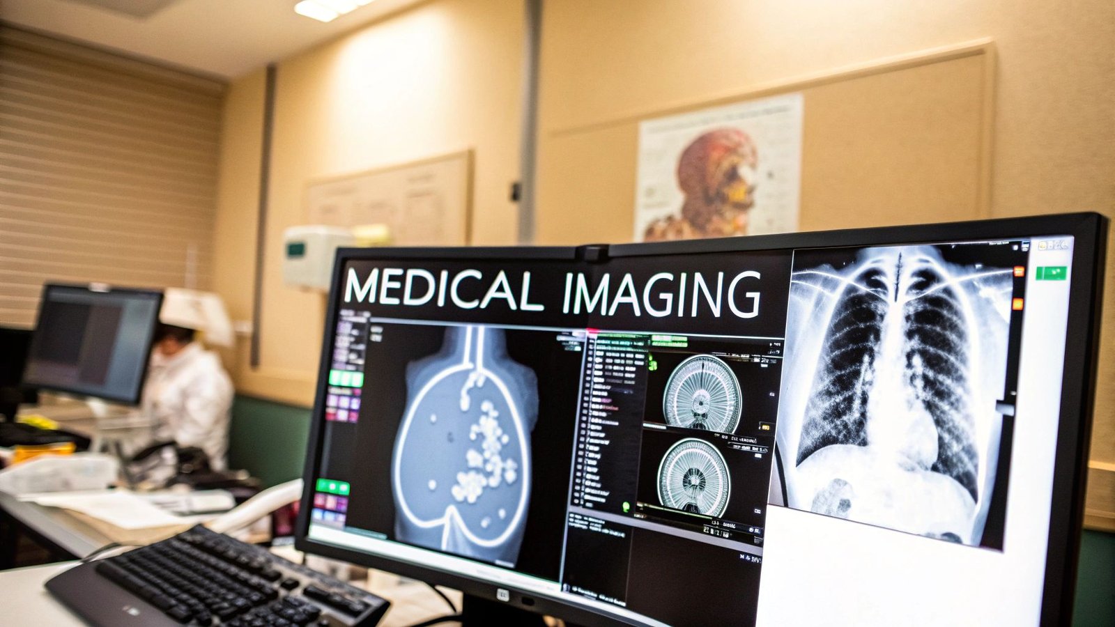
Medical image software is like a skilled interpreter, translating the complex, silent language of scans—like MRIs and CTs—into clear, visual stories that guide life-saving decisions. It’s the essential bridge between a patient's internal anatomy and a clinician's ability to understand it.
The Future of Diagnostics Through Medical Image Software
Imagine a master cartographer trying to map a new continent using only a flood of raw satellite data. Without the right tools, that data is just a jumble of numbers. This is exactly the challenge doctors face with medical scans. Medical image software is that specialized cartography kit, transforming a sea of complex data into detailed, interactive maps of the human body.
This technology gives doctors the power to navigate intricate structures, measure abnormalities with incredible precision, and plan out an entire surgery before ever making an incision. We're not just talking about looking at a static picture; this is about interacting with a dynamic, multi-dimensional model of a person's unique physiology.
From Data Points to Diagnostic Clarity
The journey from a whirring scanner to a life-changing diagnosis is nothing short of remarkable. The software meticulously processes thousands, sometimes millions, of individual data points to build a cohesive image, allowing clinicians to see what the naked eye never could.
- Image Acquisition: It starts by receiving the raw data directly from imaging machines like CT, MRI, or ultrasound.
- Data Reconstruction: Next, it assembles that raw data into coherent 2D slices or full 3D volumes that represent the body's internal structures.
- Visualization and Analysis: Finally, it provides a toolbox for clinicians to manipulate, measure, and annotate these images to pinpoint pathologies.
At its heart, this software is about empowering medical professionals with unprecedented insight. It turns abstract data into actionable intelligence that directly improves a patient's chances for a better outcome.
To put these core functions into perspective, let's break them down.
Core Capabilities of Modern Medical Imaging Platforms
This table outlines the essential functions that define today's medical image software, helping to clarify its multifaceted role in modern healthcare.
| Core Capability | Primary Function | Impact on Healthcare |
|---|---|---|
| Data Interpretation | Translates raw scan data into high-resolution 2D/3D images. | Turns complex numbers into visually clear anatomical maps for diagnosis. |
| Interactive Visualization | Allows clinicians to rotate, zoom, and slice through images. | Enables a deep, intuitive exploration of patient anatomy from any angle. |
| Quantitative Analysis | Provides tools to measure tumors, blood flow, and tissue density. | Replaces guesswork with precise, data-driven measurements for treatment planning. |
| Clinical Collaboration | Facilitates secure sharing of images and findings among specialists. | Speeds up second opinions and ensures the entire care team is on the same page. |
These capabilities work together, creating a powerful ecosystem that supports clinicians from the moment a scan is taken to the final treatment decision.
A Market Driven by Innovation
The growing reliance on this technology is clearly reflected in its market trajectory. The global medical image analysis software market was valued at a staggering USD 3.27 billion in 2023 and is projected to soar to USD 5.49 billion by 2030. This incredible expansion is fueled by the ever-increasing demand for advanced diagnostic tools, especially in critical fields like oncology and neurology.
This growth points to a much deeper shift in healthcare—a future where technology is an indispensable partner in the quest for more accurate and personalized medicine. Exploring the future of medical imaging shows just how these advancements are setting entirely new standards for patient care.
Decoding the Essential Toolkit for Clinicians
What turns a simple image viewer into a clinician's most trusted partner? It's not just one thing, but a powerful combination of features designed to turn raw pixels into life-saving insights. These aren't just bells and whistles; they're the foundational tools that empower medical professionals to see more clearly, understand more deeply, and act with unshakable confidence.
The absolute cornerstone of any serious medical imaging software is DICOM (Digital Imaging and Communications in Medicine) compatibility. Think of DICOM as the universal language of medical scans. It’s the reason an MRI from one machine can be opened, viewed, and analyzed flawlessly on software from a completely different company.
Without this shared standard, chaos would ensue. Sharing vital scans between hospitals would be a nightmare, delaying critical care and creating unnecessary risk. This universal protocol is the bedrock on which everything else is built, making it a non-negotiable for any clinical setting. To get a better handle on this foundational tech, you can find a great breakdown here: DICOM viewer is and its importance in healthcare.
Visualizing Anatomy in Three Dimensions
We all know the human body isn’t flat, so why should our view of it be? Modern software brings anatomy to life with powerful 3D visualization and rendering tools. These aren't just for show; they are absolutely essential for complex tasks like surgical planning, where understanding the spatial relationship between tissues is paramount.
Imagine a surgeon digitally rotating a patient's organ, inspecting a tumor from every angle, and mapping out the safest approach before ever making an incision. This kind of digital rehearsal drastically reduces surprises in the operating room and is directly tied to better patient outcomes.
Of course, for any of this to be effective, the information has to be presented clearly. Diving into data visualization best practices can make all the difference in turning complex data into intuitive, actionable visuals.
This infographic captures that very journey—from raw data to a clear, actionable insight.
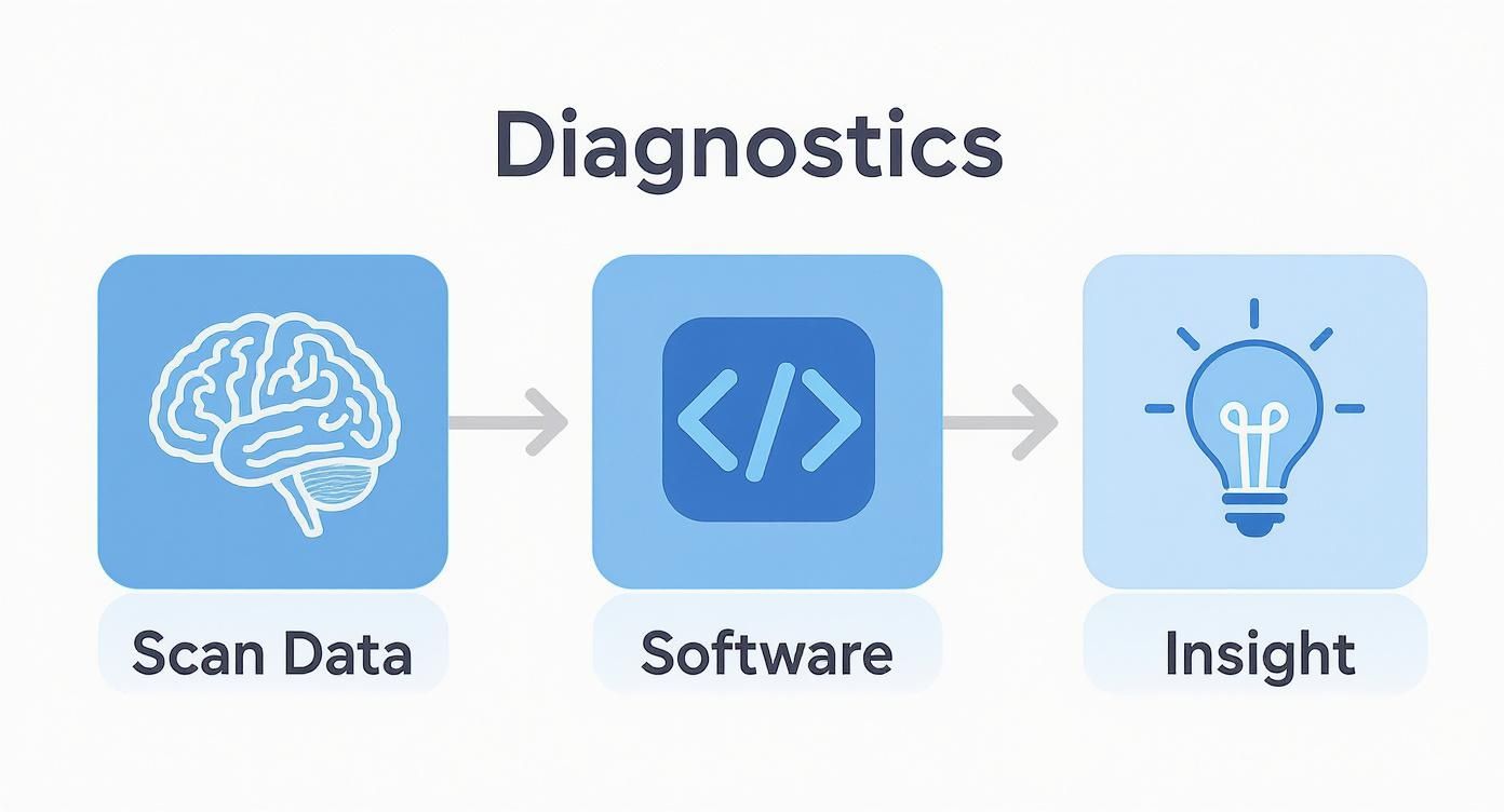
As you can see, the software is the crucial bridge, translating a flood of scan data into the clarity clinicians need to diagnose and plan treatments effectively.
Advanced Reconstruction and Collaboration Tools
Taking things a step further, Multi-Planar Reconstruction (MPR) allows clinicians to slice through a 3D model from any direction they choose. Think about looking at a patient's spine not just from the front or side, but by following its natural curve. It provides a complete anatomical picture that simple 2D views could never capture.
On top of that, medicine is a team sport. That's where precise annotation and measurement tools become indispensable. A radiologist can instantly highlight a suspicious lesion, measure its exact size, and securely share those marked-up images with an oncologist.
These features create a shared visual language among specialists, ensuring that everyone on the care team is looking at the same problem with the same understanding. This fosters seamless communication and leads to more cohesive treatment strategies.
When Off-the-Shelf Solutions Just Don't Cut It
While these core features form a great foundation, standard software doesn’t always fit the unique workflows found in specialized medicine or research. A one-size-fits-all approach often creates friction, forcing brilliant clinicians to adapt their methods to the software's limits instead of the other way around.
That’s where a custom approach makes all the difference. At PYCAD, we specialize in this area. We at PYCAD, build custom web DICOM viewers and integrate them into medical imaging web platforms. When software is built for a specific purpose, it just works better. It feels intuitive, enhances focus, and ultimately, helps clinicians do their best work. When a tool is perfectly aligned with the task at hand, it's not a luxury—it's a necessity.
The AI Revolution in Medical Imaging
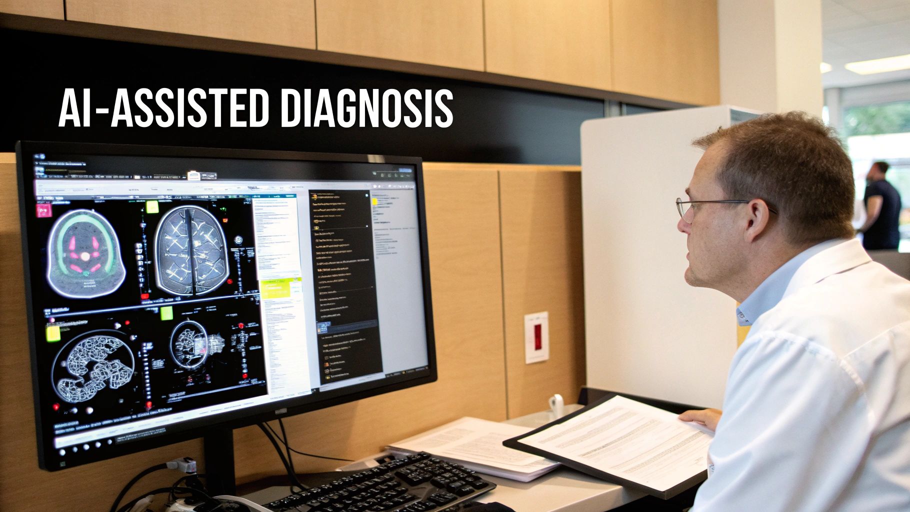
Artificial intelligence isn't science fiction anymore. It's here, right now, empowering clinicians and completely redrawing the map of what's possible in diagnostics. The real magic of AI in medical image software isn't about replacing doctors. It's about giving them a brilliant, tireless assistant that enhances their own skills, helping them work faster, more accurately, and with a whole new level of confidence.
Think of it as a second set of expert eyes, trained on millions of scans to spot patterns a human might miss. Imagine an AI algorithm poring over a chest X-ray. It can instantly flag a tiny, almost invisible nodule that might be the earliest sign of cancer, pushing it to the top of the radiologist's queue. This is human-machine partnership at its best—a powerful safety net that leads to earlier detection and better patient outcomes.
Automating the Tedious So Experts Can Focus
One of the most powerful things AI does is take over the repetitive, time-sucking tasks with incredible speed and precision. This simple shift frees up priceless time, letting clinicians focus on what they do best: complex analysis and patient care.
Here are a few ways AI is lifting the burden:
- Organ Segmentation: What used to be a painstaking manual process of tracing organs can now be done automatically in seconds. This is a game-changer for surgical planning and treatment.
- Tumor Measurement: AI algorithms provide consistent, objective measurements of tumors over time, giving oncologists clear data to see if a treatment is working.
- Workflow Prioritization: Instead of a first-in, first-out queue, AI can scan incoming studies and intelligently flag the critical ones—like a suspected stroke or pulmonary embolism—to be reviewed immediately.
By shouldering these burdens, AI-driven software allows highly trained specialists to dedicate their focus where it matters most: interpreting complex cases, collaborating with colleagues, and planning the best course of treatment for their patients.
The momentum here is impossible to ignore. The medical imaging software market is set to jump from USD 8.75 billion in 2025 to USD 12.76 billion by 2030. A huge piece of that growth comes from AI tools that automate tasks and get urgent cases in front of doctors faster, boosting diagnostic efficiency around the globe.
Building the Future with Custom AI Solutions
While off-the-shelf AI tools are helpful, the most profound breakthroughs often come from solutions built for a specific purpose. Custom development opens the door to algorithms trained for unique patient groups, rare diseases, or highly specialized research. As AI continues to reshape the field, it's worth understanding the broader trends in AI automation for business.
This is exactly where we live at PYCAD. We at PYCAD, build custom web DICOM viewers and integrate them into medical imaging web platforms, creating workflows that feel like they were made just for our clients. By embedding tailored AI capabilities directly into the software, we help organizations unlock diagnostic possibilities they never thought were possible. You can explore our work here: PYCAD portfolio. The future isn't just about using AI; it’s about building intelligent, purpose-driven tools that truly serve the people who use them.
Where Technology Meets Humanity: Real-World Impact
Theory is one thing, but the real magic happens when technology touches a human life. For medical image software, this is where the code becomes compassion, turning digital data into life-saving action. Let's move beyond the technical specs and see how these tools are empowering clinicians at the bedside every single day.
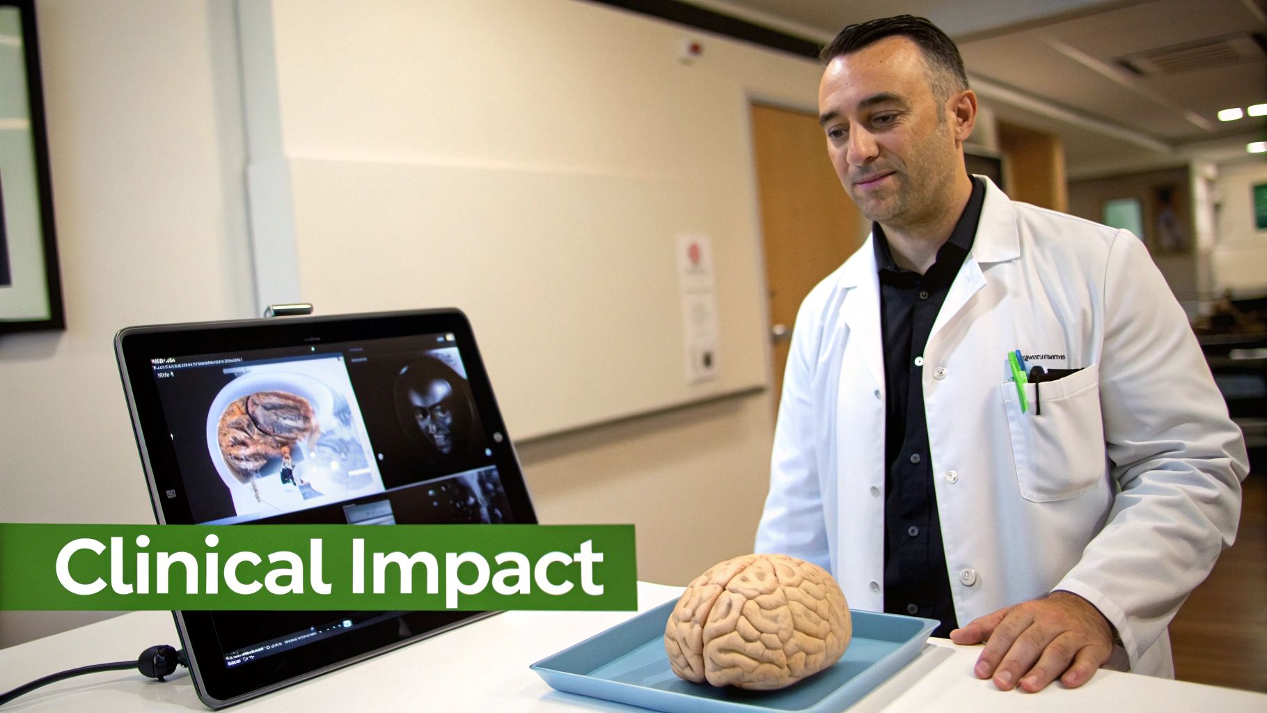
Picture a neurosurgeon facing a high-stakes operation: removing a brain tumor woven into a delicate network of blood vessels. Not long ago, they would have relied on flat, 2D film scans, piecing together a mental 3D map under immense pressure. It was an art form born of necessity.
Today, that same surgeon can build a stunningly detailed, interactive 3D model of the patient’s brain right on their screen. They can virtually peel back layers, rotate the anatomy, and map out the safest route to the tumor—all before the patient even enters the operating room. This isn't just a rehearsal; it's a mission plan that minimizes risk, shortens surgery time, and brings an entire team into perfect alignment.
Precision in Oncology and Beyond
The battle against cancer demands unwavering certainty. An oncologist needs to know, not just hope, that a treatment is working. This is where volumetric analysis in medical image software changes the game completely. Instead of just measuring a tumor's width, the software calculates its entire volume with incredible accuracy.
By comparing scans taken weeks apart, a doctor can see a tumor shrinking in three dimensions. This is the hard data, the undeniable proof that a therapy is succeeding. It allows for truly personalized care, where treatments are adjusted based on the body's actual response, not just on how a patient feels. We're moving from reacting to symptoms to proactively shaping outcomes, all thanks to smarter software.
These stories bring a core truth into focus: medical image software is the bedrock of modern, evidence-based medicine. It gives clinicians the clarity and confidence to make the best possible decisions when it matters most.
And this impact isn't limited to a few specialties. It's a wave of innovation washing over nearly every corner of medicine.
Diverse Applications Across Medical Fields
One of the most impressive things about this software is its versatility. It’s a chameleon, adapting to the unique challenges of different medical disciplines and bringing new levels of insight everywhere it goes.
- Dentistry and Orthodontics: Imagine an oral surgeon using cone-beam CT (CBCT) software to map a patient's jaw in 3D. This ensures dental implants are placed with millimeter precision, avoiding nerves and creating a perfect, lasting fit. The result? Better outcomes and fewer complications.
- Cardiology: A cardiologist can analyze a cardiac MRI to measure blood flow and pinpoint damage to the heart muscle after a heart attack. This deep functional analysis is vital for choosing the right treatment and predicting a patient's path to recovery.
- Orthopedics: Before a complex knee replacement, a surgeon can model the joint in 3D to select the ideal implant size and even rehearse the surgery. This meticulous planning leads to a better fit and improved mobility for years to come.
What connects all these examples is a simple theme: empowerment. The technology itself is powerful, but its true potential is unlocked when it's tailored to the way these experts actually work. Generic, off-the-shelf software often falls short in these highly specialized fields.
That's why we at PYCAD, build custom web DICOM viewers and integrate them into medical imaging web platforms. We believe that by creating tools designed around specific clinical workflows, we help medical professionals achieve the incredible precision their work demands. To see how our custom solutions are making a difference, we invite you to explore the PYCAD portfolio.
Choosing Your Ideal Medical Image Software
Picking the right medical image software isn't just another IT purchase. It's a foundational decision that will ripple through every corner of your clinical practice. This choice shapes how your team collaborates, how you safeguard patient data, and how ready you are for the future of medicine. To get it right, you have to look past the shiny features and focus on the core pillars of a system built to last.
The journey starts by taking an honest look at what your organization truly needs. A small, specialized clinic and a sprawling hospital network live in different worlds and have vastly different requirements. The goal is to find a solution that not only solves today's challenges but has the DNA to grow with you.
The Bedrock of Trust and Security
In healthcare, security isn’t just a feature; it's the very foundation of patient trust. Any software you’re considering must be ironclad in compliance with regulations like HIPAA and GDPR. That means airtight encryption for data, whether it's sitting on a server or flying across a network, plus granular access controls and detailed audit logs tracking every single action.
But don't just stop at the compliance checklist. You need to get a feel for a vendor's security culture. Ask them tough questions about their security protocols, how they hunt for vulnerabilities, and what their game plan is if a breach occurs. A partner who can answer confidently and openly is one you can trust with your most critical asset: your patient data.
Planning for Growth and Scalability
Your imaging needs aren't static—they’re going to grow. As you see more patients, bring in new imaging equipment, and expand your team, your software has to keep up without skipping a beat. Scalability isn’t a nice-to-have; it's a must-have for survival and success.
A truly scalable system can handle a flood of new studies and users without slowing down. It's also nimble enough to integrate new technologies as they emerge, from brilliant AI algorithms to the next generation of imaging hardware.
Market trends show us exactly why this matters. While Ultrasound is still the most common modality, its market share is expected to dip from 31% in 2016 to 29% by 2025. At the same time, CT and MRI are on the rise, with MRI's share projected to grow from 12% to 14%. This shift tells a clear story: your software must be ready for more advanced, cross-sectional imaging. It's a critical conversation to have with any potential vendor. You can dive deeper into these medical imaging market trends.
Choosing a scalable platform is like building a house with a strong foundation and a flexible floor plan. You ensure that you have the stability you need today and the room to expand and adapt for whatever the future holds.
Before you sign on the dotted line, you also need to make one of the most fundamental choices: how your software will be deployed. This decision will define everything from your budget and IT workload to how and where your team can access patient images.
On-Premise vs. Cloud Deployment: A Critical Choice
The debate between hosting software on your own servers (on-premise) or accessing it online (cloud-based SaaS) is a major fork in the road. Each path offers a different set of trade-offs, and the right one depends entirely on your organization’s resources, priorities, and culture.
To help clarify the decision, let's break down the key differences.
On-Premise vs. Cloud-Based Software Deployment
| Consideration | On-Premise Solution | Cloud-Based (SaaS) Solution |
|---|---|---|
| Data Control | Complete physical control over servers and patient data. | Data is stored on the vendor's secure servers, accessible from anywhere. |
| Initial Cost | High upfront investment in hardware, licenses, and IT infrastructure. | Lower initial cost with a predictable monthly or annual subscription fee. |
| Maintenance | Your internal IT team is responsible for all updates, security, and upkeep. | The vendor manages all maintenance, updates, and security patches. |
| Accessibility | Access is typically limited to the facility's internal network. | Secure access from any location with an internet connection, ideal for remote work. |
| Scalability | Scaling requires purchasing and configuring additional hardware. | Easily scalable by adjusting your subscription plan as your needs change. |
Ultimately, there's no single "best" answer. The right choice is the one that aligns with your budget, your IT team's capacity, and your specific security requirements.
When Your Vision Demands a Custom Fit
Sometimes, even the most feature-packed off-the-shelf software doesn't quite fit. Forcing a unique, specialized workflow into a generic box creates friction, slows people down, and stifles the very innovation you’re trying to foster. When you're pushing the boundaries of medicine, one-size-fits-all just doesn't cut it.
That's when a custom-built solution becomes the clear path forward. At PYCAD, this is our specialty. We at PYCAD, build custom web DICOM viewers and integrate them into medical imaging web platforms, creating tools that feel like a natural extension of your team's expertise. Our goal is to make technology work for you, not the other way around.
To see how we’ve helped medical innovators bring their unique visions to life, take a look at our PYCAD portfolio.
Got Questions? We've Got Answers
Stepping into the world of medical image software can feel like learning a new language. As this technology becomes more woven into the fabric of patient care, getting clear answers is the first step toward making smart decisions for your practice. Let's tackle some of the most common questions that come up, breaking them down into simple, straightforward explanations.
We'll clear up the confusion between core systems, talk about the non-negotiable issue of patient data security, and look at how you can keep your technology ahead of the curve.
What's the Difference Between a PACS and a DICOM Viewer?
This is a great question, and it's easy to see why people mix them up. They work together, but they do very different jobs.
Think of a Picture Archiving and Communication System (PACS) as the massive, secure central library for a hospital or clinic. Its entire purpose is to safely store, organize, and share a huge volume of medical images and their corresponding reports. It’s the foundational archive.
A DICOM viewer, on the other hand, is the specialized magnifying glass you use to read the books in that library. It's the software that lets you actually open, manipulate, and analyze the images from the PACS. While most PACS come with a basic built-in viewer, dedicated standalone or web-based DICOM viewers often offer far more powerful tools for visualization and deep analysis. They are the instruments of clinical insight.
This is exactly where we shine. At PYCAD, we at PYCAD, build custom web DICOM viewers and integrate them into medical imaging web platforms, giving clinicians the powerful, intuitive tools they need right at their fingertips. You can see what this looks like in our PYCAD portfolio.
How Does Medical Image Software Keep Patient Data Safe?
In healthcare, protecting patient data isn't just a feature—it's the absolute bedrock of trust. It’s not about one single lock, but a whole fortress of security measures built into the software from the ground up.
First and foremost, everything starts with strict adherence to regulations like HIPAA in the U.S. and GDPR in Europe. These aren't just guidelines; they are legally mandated standards for handling protected health information (PHI), and compliance is non-negotiable.
From there, security is built in layers with smart technology:
- Iron-Clad Access Controls: This makes sure only the right people see the right information. Using role-based permissions, a surgeon sees what they need to, a radiologist sees their own cases, and an admin has their specific view—and no one has access to data outside their role.
- End-to-End Encryption: Think of this as sending a message in a code that only the intended recipient can decipher. Data is scrambled both when it’s stored (“at rest”) and when it’s being sent over a network (“in transit”), making it completely unreadable to unauthorized eyes.
- Detailed Audit Trails: Every single click, view, and action taken inside the software is logged. This creates a permanent record of who did what and when, ensuring total accountability.
- Smart Anonymization Tools: For research and teaching, the software can strip away all personally identifiable information from images. This allows the data to drive medical progress while keeping the patient's identity completely private.
Can We Add New AI Features to Our Current Imaging Software?
Absolutely. This is one of the most dynamic and exciting developments in medical imaging today. Weaving new AI tools into the systems you already use is not only possible but is quickly becoming the smartest way for healthcare organizations to innovate. You get to add incredible new diagnostic power without the headache and expense of ripping out and replacing everything.
Whether this is possible really depends on the architecture of your current software. Modern platforms are built with what are called Application Programming Interfaces (APIs)—think of them as secure docking ports that allow different software systems to connect and talk to each other.
This modular design is the secret to future-proofing your investment. It gives you the freedom to plug in best-in-class AI tools as they emerge, whether it’s a new algorithm for detecting early-stage cancer or a tool that automates tedious measurements.
This approach lets you amplify your diagnostic capabilities in a smart, scalable way. Instead of being locked into one company's development schedule, you gain the agility to adapt and adopt the best tools on the market. This is a core skill for development partners like us. For instance, we at PYCAD, build custom web DICOM viewers and integrate them into medical imaging web platforms, frequently connecting them with specialized third-party AI to create a solution that’s a perfect fit. It’s how you ensure your platform doesn’t just keep up, but leads the way.
At PYCAD, our passion is building the next generation of medical imaging solutions. We create secure, high-performance web platforms with advanced DICOM viewers and seamlessly integrated AI, all designed to solve the real-world challenges of modern medicine. If you're ready to build a tool that truly works the way you do, let's talk about what's possible.
Discover our custom solutions by visiting our portfolio at https://pycad.co/portfolio.


