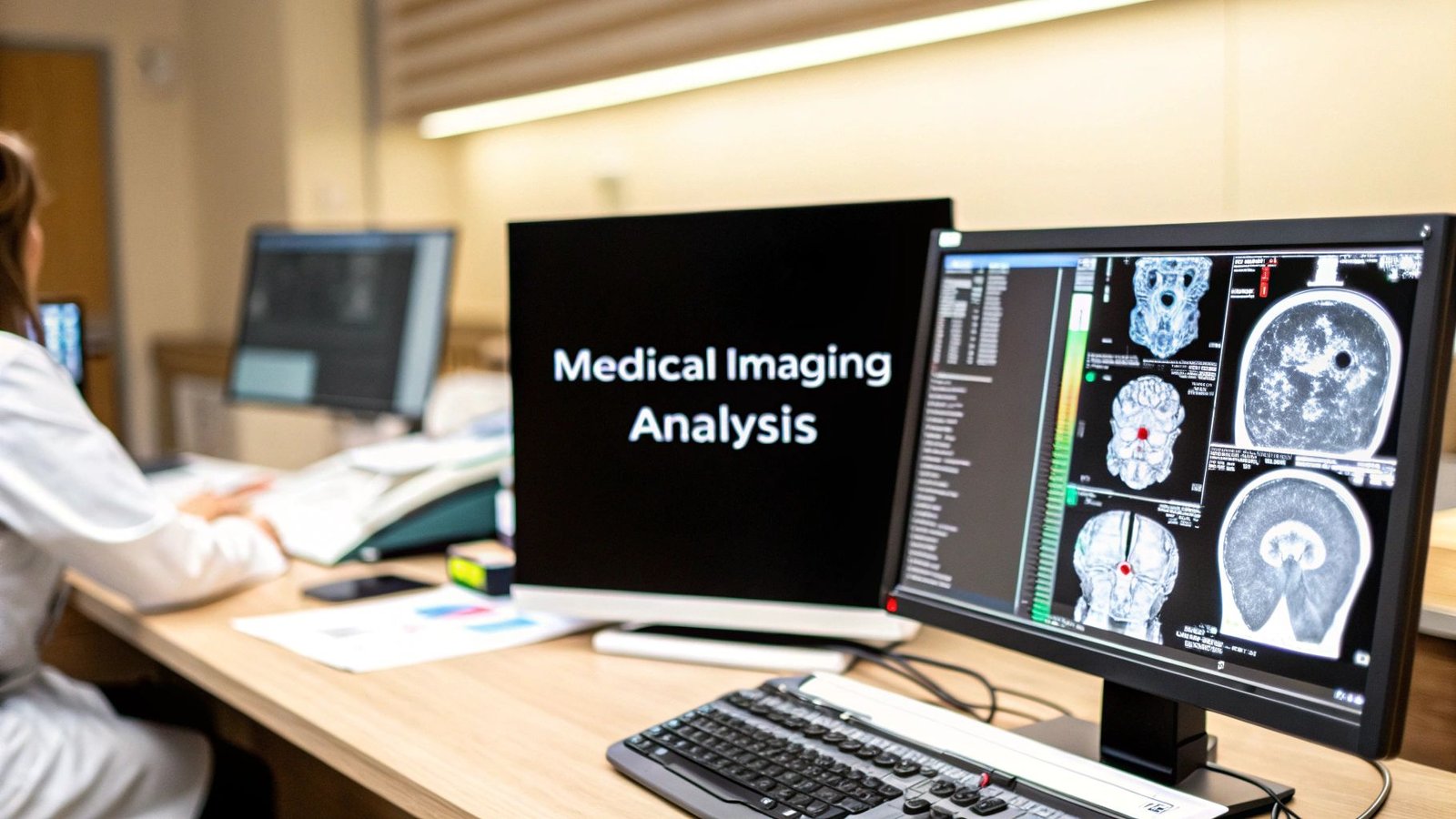
Every medical scan is a story waiting to be told—a quiet conversation between pixels and patient health. Medical imaging analysis is the art of translating these silent images into life-saving insights, giving clinicians the clarity to make earlier diagnoses and craft more precise treatments.
The Hidden World Inside Every Medical Scan
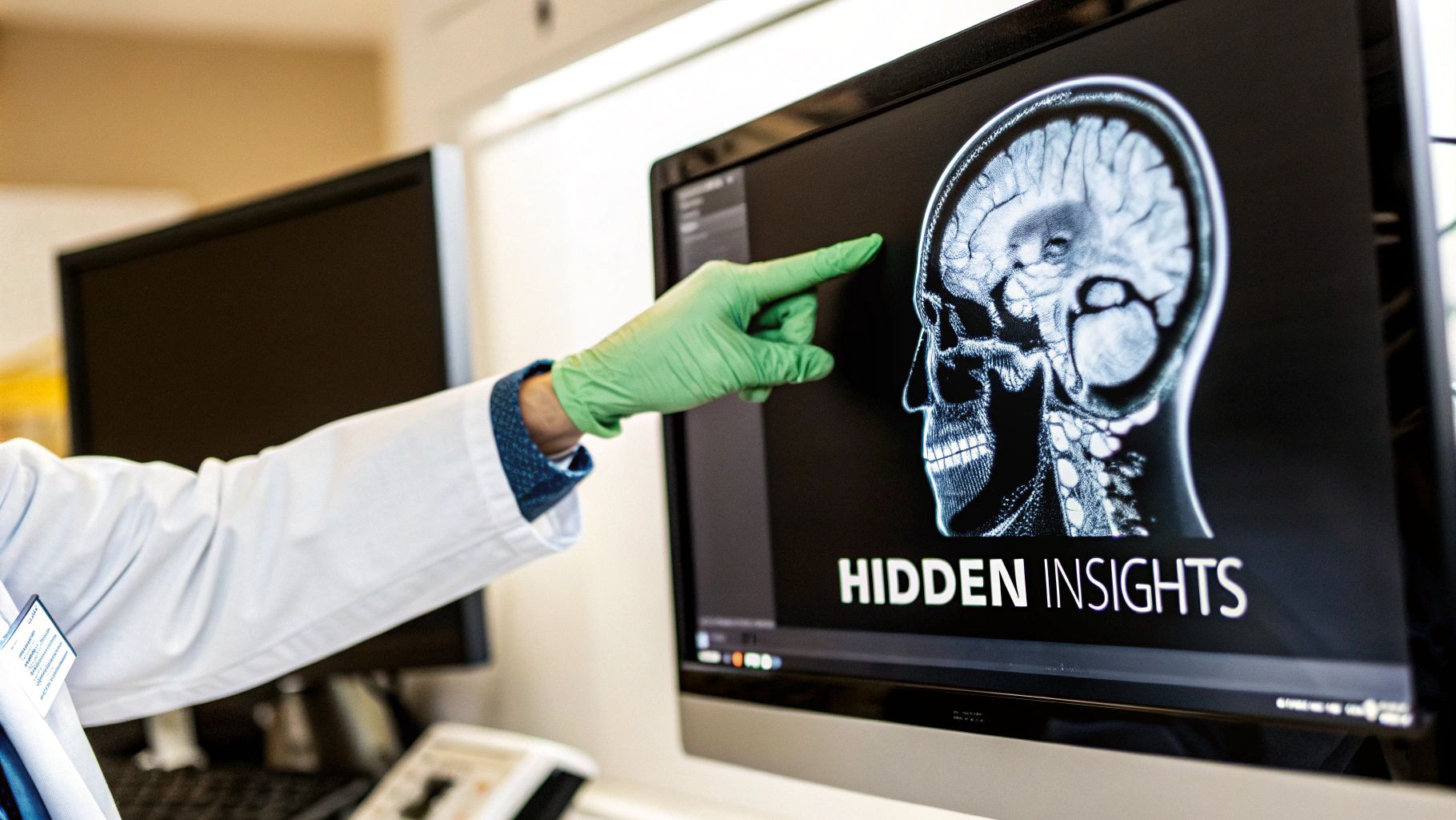
Think of a medical imaging analyst less as a technician and more as a digital cartographer. Their job is to map the incredibly complex, hidden landscapes of the human body, uncovering truths that guide a doctor’s hands and ultimately shape a patient's future. This journey, from a raw scan to a confident clinical decision, is the very heartbeat of modern medicine.
The process is far more than just looking at pictures. It's a systematic effort to pull meaningful information from every pixel. This means identifying anomalies, measuring anatomical structures with precision, tracking changes over time, and delivering objective data that backs up a diagnosis. The real goal here is to shift from subjective interpretation to data-driven certainty.
From Data Points to Diagnostic Power
At its core, medical imaging analysis turns a flood of complex visual data into focused, actionable intelligence. The entire field is built on a universal language for these images, which is essential for making sure different systems can communicate. To understand this foundational standard, you can explore our detailed guide on what DICOM is and see why it’s so critical for modern healthcare.
This transformation from image to insight is also powering a massive industry. The global medical imaging market was valued at USD 43.9 billion and is expected to climb to USD 75.8 billion by 2034. That's not just a number; it's a clear signal of the immense demand for better diagnostic tools and the central role that analysis plays in healthcare systems around the globe.
Core Pillars of Modern Medical Imaging Analysis
A truly effective analysis ecosystem isn't built on a single algorithm. It's about weaving multiple, specialized components into a seamless and intuitive workflow. To give you a clear picture, this table breaks down the fundamental pillars of any modern medical imaging platform.
| Component | Primary Role | Key Benefit |
|---|---|---|
| Data Management | Securely storing and organizing vast archives of patient scans. | Ensures data integrity, accessibility, and compliance. |
| Visualization Tools | Providing clinicians with high-performance 2D and 3D viewers. | Enables intuitive interaction with complex medical images. |
| Analysis Algorithms | Running sophisticated computations to segment, detect, and classify features. | Automates and quantifies insights, reducing human error. |
| Reporting & Collaboration | Generating clear, structured reports and enabling seamless communication. | Fosters teamwork and ensures insights lead to action. |
By bringing these elements together, we create an environment that doesn't just reveal the hidden stories within each scan but makes them easy to understand and act upon, directly improving patient care.
Translating Pixels into Clinical Knowledge
How do we teach a computer to see what a highly trained radiologist sees? It all comes down to a handful of powerful computational techniques that form the language of medical imaging analysis. Each one is designed to ask—and answer—a very specific clinical question, turning a complex grid of pixels into clear, measurable information that guides life-saving decisions.
These aren't just abstract algorithms. They're the digital tools that give clinicians the power to see with greater precision, measure with more confidence, and diagnose with enhanced certainty. Let's pull back the curtain on the four core methods that translate raw image data into profound clinical insight.
Isolate What Matters with Image Segmentation
Imagine a sculptor starting with a massive block of marble. Their first job is to carefully chip away the excess stone to reveal the intricate sculpture hidden inside. Image segmentation works on the exact same principle, but its medium is digital, and the sculpture is a specific part of the human anatomy.
It’s the process of partitioning a digital image into different regions, essentially drawing a precise outline around an object of interest. In medical imaging, this could mean tracing the exact boundaries of a tumor in a brain MRI, defining the chambers of the heart on a CT scan, or mapping the total volume of the lungs.
This digital "sculpting" is absolutely essential for quantitative analysis. By isolating a specific organ or lesion, we can accurately measure its size, volume, and shape—metrics that are critical for surgical planning, treatment monitoring, and diagnostic assessment.
For example, before a complex cardiac surgery, surgeons need a perfect map of a patient's heart valve. Segmentation provides this by creating a detailed 3D model from scan data, letting them plan their approach with millimeter-level accuracy. For a deeper look at this cornerstone technique, you can explore our comprehensive guide on medical image segmentation and its applications.
Align the Timeline with Image Registration
Now, picture two transparencies of a city map—one from today and one from five years ago. To see how the city has changed, you’d need to overlay them perfectly, aligning all the streets and landmarks. Image registration is the digital equivalent of this, aligning multiple medical scans to reveal subtle changes over time.
This technique is a game-changer for longitudinal studies, where clinicians need to track the progression of a disease or see how well a treatment is working.
- Tracking Tumor Growth: By registering a patient's current MRI with a scan taken six months ago, oncologists can precisely measure if a tumor has grown, shrunk, or stayed the same in response to chemotherapy.
- Monitoring Neurodegenerative Diseases: In diseases like multiple sclerosis, registration helps align brain scans taken over several years to spot new lesions or changes in brain volume.
- Fusing Different Modalities: It can also align images from different types of scans, like a PET scan (showing metabolic activity) with an MRI (showing anatomical structure), to give clinicians a much more complete diagnostic picture.
Find the Anomaly with Object Detection
Think of object detection as a highly trained search party, given a mission to find a specific target within a vast landscape. But instead of looking for a lost hiker, these algorithms are trained to find clinical anomalies like tumors, fractures, polyps, or microaneurysms.
Unlike segmentation, which outlines an entire structure, detection simply focuses on identifying the presence and location of a feature. It answers the question, "Is there something abnormal here, and if so, where is it?" A great example is an object detection model trained to automatically scan a chest X-ray and draw a box around any suspicious-looking nodules, flagging them for a radiologist to review more closely.
Sort and Prioritize with Image Classification
Finally, after all the details have been analyzed, we often need to put the scan into a broader category. Image classification acts like an expert librarian, sorting an entire image into a predefined class, such as 'healthy,' 'benign,' 'malignant,' or 'requires urgent review.'
This technique doesn’t pinpoint where an issue is; it gives a high-level assessment of the entire scan. It’s incredibly valuable for triage and optimizing workflows. For instance, a classification algorithm could analyze a mammogram and assign it a probability score for malignancy, helping radiologists prioritize the most concerning cases for immediate attention.
These four techniques—segmentation, registration, detection, and classification—are the backbone of modern medical imaging analysis. At PYCAD, we know these algorithms are most powerful when they’re part of a fluid clinical workflow. That is why we build custom web DICOM viewers and integrate them into medical imaging web platforms, ensuring these advanced analytical tools are accessible and intuitive for clinicians. To see examples of how these integrations transform diagnostics, you can explore our portfolio of projects.
How AI Is Supercharging Medical Diagnostics
Let's clear up a common misconception: AI isn't here to replace clinicians. It's here to become their most powerful collaborator. Think of it as a digital co-pilot for medical imaging analysis, one with the extraordinary ability to see what often remains hidden to the human eye. It can spot incredibly subtle patterns, textures, and anomalies within a scan, adding a profound new layer of perception to the diagnostic process.
This partnership between human expertise and machine intelligence is fundamentally changing what's possible in medicine. AI, especially deep learning, can analyze thousands upon thousands of images, learning the distinct visual signatures of different diseases. The process isn't unlike how a radiologist builds experience over years, but an AI model can process a volume of data that would take a single person an entire lifetime to review.
The Journey From Data To AI Co-pilot
Creating an AI you can trust with medical decisions is a meticulous journey, and it all starts with high-quality data. Developers begin by gathering huge, diverse datasets of DICOM images. These images are then painstakingly annotated by medical experts—a critical step where we essentially teach the AI to recognize what’s normal and what’s not, labeling every single pixel of interest.
From there, the real training begins. The AI learns, adapts, and refines its understanding through millions of iterations until it can reliably flag specific pathologies. This is where the magic really happens, as the algorithm moves beyond simply seeing pixels to understanding the clinical story behind them. If you're curious about the mechanics, our guide on machine learning in medical imaging is a great place to start.
This workflow is elegantly simple in its core steps.
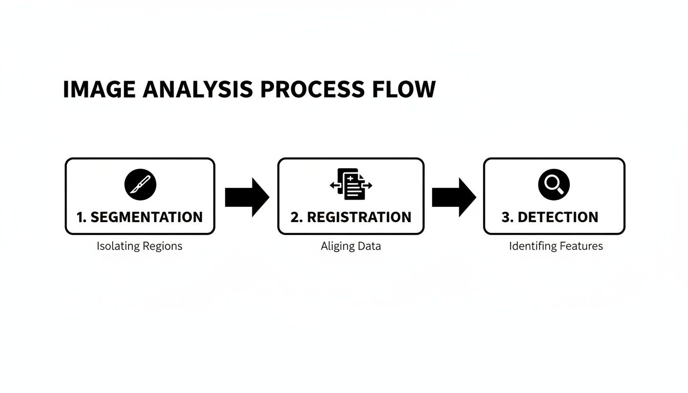
This progression from segmentation to detection shows how an AI systematically breaks down a complex image into clear, actionable insights for the clinical team.
Building Trust Through Performance Metrics
So, how do we know we can actually trust an AI’s conclusion? The answer is found in rigorous validation using key performance metrics. These aren't just abstract technical terms; they are the bedrock of an AI's reliability and its fitness for real-world clinical use.
- Sensitivity: This tells us how well the model can find patients who do have a disease (true positives). High sensitivity is absolutely crucial for screening tools, as it minimizes the risk of missing a real case.
- Specificity: This measures the model’s ability to correctly identify patients who are healthy (true negatives). High specificity is just as important to avoid false alarms that lead to unnecessary stress and follow-up procedures.
Striking the right balance between these two metrics is what separates a good research project from a great clinical tool. A model with both high sensitivity and specificity is one that clinicians can truly depend on to enhance their own diagnostic skills.
Overcoming Challenges And Looking Ahead
The road to AI-powered diagnostics isn't without its speed bumps. One of the biggest challenges we face is mitigating data bias. If an AI is trained mostly on data from one demographic, it may not perform well on others. Building diverse, representative datasets isn't just a technical goal—it's an ethical imperative to ensure these incredible tools work for everyone.
Despite the hurdles, the future is incredibly bright. The global AI in medical imaging market was valued at an astonishing USD 2.01 trillion and is projected to hit USD 22.97 trillion by 2035. Image analysis alone accounts for 51% of these applications, highlighting the immense momentum in this space.
AI is pushing us toward a future of proactive, personalized medicine. By detecting diseases earlier and with greater accuracy, we can intervene sooner, tailor treatments to the individual, and ultimately achieve better patient outcomes.
This progress isn't happening in a vacuum. To see the bigger picture of how Artificial Intelligence and automation are reshaping entire industries, including healthcare, it’s worth looking at the experts leading the charge.
At PYCAD, we're right at the forefront of this movement. We specialize in building custom web DICOM viewers and integrating them into advanced medical imaging web platforms, turning powerful AI insights into practical, everyday clinical tools. You can explore our work and see these solutions in action by visiting our portfolio page.
Building Your Integrated Analysis Platform
Powerful algorithms are one thing, but they're only truly useful when woven into the daily rhythm of a clinician's work. Let's be honest, the most brilliant AI model for medical imaging analysis is practically useless if it's stuck on a research server, inaccessible to the people who need it most. This is where theory hits the real world—where we bridge the gap between raw computational power and the actual point of care.
The entire ecosystem we're talking about is built on a single, universal foundation: DICOM (Digital Imaging and Communications in Medicine). Think of DICOM as the Esperanto of medical images. It's the standard that ensures a CT scan from a Siemens machine in a London hospital can be perfectly read and understood by a GE system in a clinic in New York. Without it, interoperability would just be a buzzword.

But having a common language is just the starting line. The real magic happens when you bring that data to life inside a dynamic platform designed for clinical action.
The Power of an Integrated Viewing Experience
Picture this all-too-common scenario: a cardiologist needs to review a cardiac MRI. In a fragmented system, they open a basic viewer on one machine, then pivot to a separate application to run an analysis. Finally, they log into the electronic health record (EHR) on yet another terminal to pull up the patient's history. This disjointed process creates friction, wastes precious minutes, and opens the door to potential errors.
A modern, integrated platform smashes these barriers. The goal is to create a single command center where a physician can view a scan, run a sophisticated AI analysis, and see the results displayed right alongside relevant patient data—all within one fluid interface.
This unified dashboard isn't just a matter of convenience; it’s about elevating the standard of care. When diagnostic tools and patient data live in the same space, clinicians can make faster, more confident, and more collaborative decisions.
This is the future of diagnostic medicine, and it's being built right now.
Bringing Analysis to the Browser
The key to unlocking this seamless experience is moving powerful tools away from clunky, installed desktop software and into the secure, universally accessible environment of a web browser. This is precisely where our expertise at PYCAD comes in. We build custom web DICOM viewers and integrate them into medical imaging web platforms designed to be fast, secure, and second nature for clinicians to use.
By focusing on web-native solutions, we unlock some game-changing advantages for healthcare organizations:
- Zero-Footprint Access: Clinicians can securely view and analyze images from any device with a web browser. No special software, no lengthy installs.
- Centralized Updates: New features and AI models can be rolled out instantly across the entire organization, ensuring every user is always on the latest version.
- Effortless Integration: Web platforms are built to connect with other hospital systems like EHRs and Picture Archiving and Communication Systems (PACS), creating a truly unified data environment.
Hospitals today generate an average of 50 petabytes of data annually. A staggering 80% of that is unstructured data like medical images. A web-based platform is the only practical way to manage this incredible volume while making the data truly useful.
This shift to browser-based platforms is more than a trend; it's a fundamental change in how medical imaging analysis gets done. It democratizes access to advanced tools, sparks real-time collaboration among care teams, and builds an architecture ready for the next wave of AI innovation.
The platforms we build are engineered to be more than just viewers—they are complete diagnostic hubs. To see how we fuse powerful analytics with human-centered design, we invite you to explore our portfolio and see these integrated solutions in action.
Getting Your Solution into the Real World
Bringing a medical imaging solution from a brilliant concept to a clinical reality is about so much more than just building a great algorithm. It’s about having a rock-solid strategy for deployment, security, and regulation. This is where innovation meets responsibility, and clearing these real-world hurdles is what separates a good idea from a truly successful medical imaging analysis platform.
The first major decision you'll face is choosing the right deployment model. This choice will fundamentally shape how your platform operates, scales, and gets maintained, directly impacting everyone from clinicians to your IT teams.
Choosing Your Deployment Architecture
You really have two main paths to take here: on-premise or a cloud-based Software as a Service (SaaS) model. Each has its own distinct advantages.
An on-premise solution means you host everything—all the hardware and software—inside your own institution's data center. This approach gives you the ultimate control over your data and infrastructure, which is a huge deal for organizations with incredibly strict security protocols. The trade-off? You're responsible for the full cost of hardware, ongoing maintenance, and the IT staff needed to keep it all running smoothly.
On the other hand, a cloud (SaaS) model lets you hand off the infrastructure management to a third-party provider. This gives you amazing scalability, allowing you to ramp up capacity whenever you need it without a massive upfront investment. It also frees up your internal IT team, since the provider handles all the maintenance and updates. For many, the flexibility and cost-effectiveness of the cloud just makes more sense.
Securing Patient Data: The Absolute Top Priority
No matter which deployment model you choose, the security of patient data has to be your number one concern. In healthcare, trust is everything. Protecting sensitive information isn't just a good practice; it's a legal and ethical imperative.
Protecting patient data isn't just a feature; it's the foundation upon which a trustworthy medical imaging platform is built. A breach doesn't just compromise data—it erodes the confidence of patients and clinicians alike.
This means building your entire platform to meet strict regulations like:
- HIPAA (Health Insurance Portability and Accountability Act): The foundational U.S. standard for protecting patient health information.
- GDPR (General Data Protection Regulation): The European Union's sweeping data privacy and security law.
Staying compliant demands robust encryption, iron-clad access controls, and detailed audit logs. You need to be able to trace every single interaction with patient data. At PYCAD, we build custom web DICOM viewers and integrate them into medical imaging web platforms with security baked in from the very beginning.
Navigating the Regulatory Pathways
The final frontier before launch is getting regulatory approval. A medical device, and that includes analysis software, simply cannot be used on patients without getting the green light from the right authorities. In the United States, the FDA (Food and Drug Administration) is the gatekeeper, while in Europe, the CE marking is the seal of approval that shows you meet health and safety standards.
This process is demanding. It requires meticulous documentation proving your software's performance, safety, and validation. With the right guidance, these complex requirements become a clear roadmap, not an impossible maze.
This is more important than ever as hospitals, the primary end-users, are driving demand for these systems at a projected CAGR of 6.4% through 2032. This growth is being pushed by an increase in surgical centers and the rising prevalence of conditions like cancer and heart disease that depend on advanced imaging. You can discover more insights about medical imaging market trends to see where the industry is headed.
To see how we build compliant, secure, and powerful platforms designed for the real world, we invite you to explore our portfolio.
Let's Build the Future of Medical Imaging, Together
The journey from a screen full of pixels to a life-saving clinical insight is nothing short of remarkable. Modern medical imaging analysis is where brilliant technology and human expertise come together to truly change lives. This isn't a field where you can go it alone; it’s a discipline built on partnership, deep collaboration, and a shared vision for a healthier world. As we've seen, the biggest breakthroughs happen when powerful innovation gets into the hands of those on the front lines of patient care.
Here at PYCAD, we're more than just coders; we're partners for innovators like you. We believe your vision—whether you're a medtech pioneer, a hospital IT leader, or a dedicated researcher—deserves a world-class technical team to bring it to life. You should be focused on the clinical problem, while we focus on crafting the perfect technology to solve it.
Your Vision, Realized Through Partnership
Our specialty is building secure, custom web DICOM viewers and integrating them into medical imaging web platforms that make complex workflows feel simple. We take powerful algorithms and turn them into intuitive, accessible tools that clinicians can actually use and rely on, day in and day out. That integration is the final, crucial step in making advanced analysis a practical reality.
The real measure of any medical technology isn't the complexity of its algorithm, but how seamlessly it fits into a doctor's hands and the positive outcomes it creates for a patient. That’s where true innovation lies.
Let us handle the intricate technical details so you can stay focused on what you do best—pushing the boundaries of medicine. Our commitment is to build platforms that aren't just powerful, but also resilient, secure, and ready for whatever comes next.
As you move forward with your own innovations, it’s also important to think about the entire lifecycle of medical technology. This includes planning for the end-of-life of equipment and learning how to build a comprehensive medical equipment recycling program to ensure every part of the process is sustainable and responsible.
The future of healthcare is being built right now, one collaboration at a time. We'd be honored to help you build your part of it.
Ready to take the next step? See what’s possible by exploring the groundbreaking solutions we've already delivered. You can view our work here and start your own innovation journey with a partner you can trust.
Common Questions Answered
As you dive deeper into medical imaging analysis, you're bound to have some practical questions. It's only natural. Let's tackle a few of the most common ones that come up, connecting the dots between the concepts we've discussed and their real-world impact.
Think of this as bridging the gap from theory to the clinic floor.
What's the Difference Between Medical Imaging Analysis and a PACS?
This is a fantastic question because it gets to the heart of how these systems work together.
Imagine a PACS (Picture Archiving and Communication System) as a massive, highly secure digital library for a hospital's medical scans. Its whole job is to store, retrieve, and let you view those images. It's the foundational infrastructure—the library building itself.
Medical imaging analysis, on the other hand, is the brilliant mind that reads the books in that library. It’s the intelligent software that examines the content of the scans to find meaning, identify anomalies, and extract insights. While a PACS gives you the image, analysis tools tell you the story within it.
Modern platforms, like the kind we build at PYCAD, bring this intelligence directly into a web-based viewing environment that talks effortlessly with any PACS. It’s like having a master interpreter right there with you as you open every book.
How Can You Trust That an AI Model for Medical Imaging is Accurate?
Building trust in an AI model isn’t a single step; it's a relentless, multi-stage commitment to precision and safety. There's simply no room for error.
It all starts with the data. The model must be trained on vast, diverse, and flawlessly annotated datasets. This isn't just about quantity; it's about quality and representation, which is crucial for preventing any kind of bias that could affect its judgment.
Next, the model is put to the test. We validate it against data it has never seen before, measuring its performance with critical metrics like sensitivity and specificity. But even that isn't enough. Before an AI tool can ever touch a real patient case, it must earn rigorous regulatory approvals, such as FDA clearance or a CE mark. This final gauntlet confirms its safety, reliability, and effectiveness, ensuring it meets the highest possible standards for patient care.
Can This Kind of Advanced Analysis Really Run in a Web Browser?
Yes, absolutely. This is where the future of medical technology is headed, and it's a core part of what we do at PYCAD. We specialize in building custom web DICOM viewers and integrating them into seamless medical imaging web platforms.
This browser-based approach puts incredibly sophisticated analysis and AI tools right at a clinician's fingertips, all within a secure, standard web browser. Forget clunky desktop software installations. This makes powerful diagnostics more accessible, scalable, and dramatically easier to connect with electronic health records (EHRs) and other vital hospital systems.
A browser-based model truly democratizes access to this amazing technology, empowering more clinicians with life-saving tools, no matter where they are or what their local IT setup looks like.
At PYCAD, our passion is building the platforms that unlock this new era of medical insight. We work alongside innovators to craft secure, intuitive solutions that make advanced analysis a practical, everyday tool for clinicians everywhere.
If you're ready to see how a custom solution can bring your vision to life, we'd love to show you what's possible.


