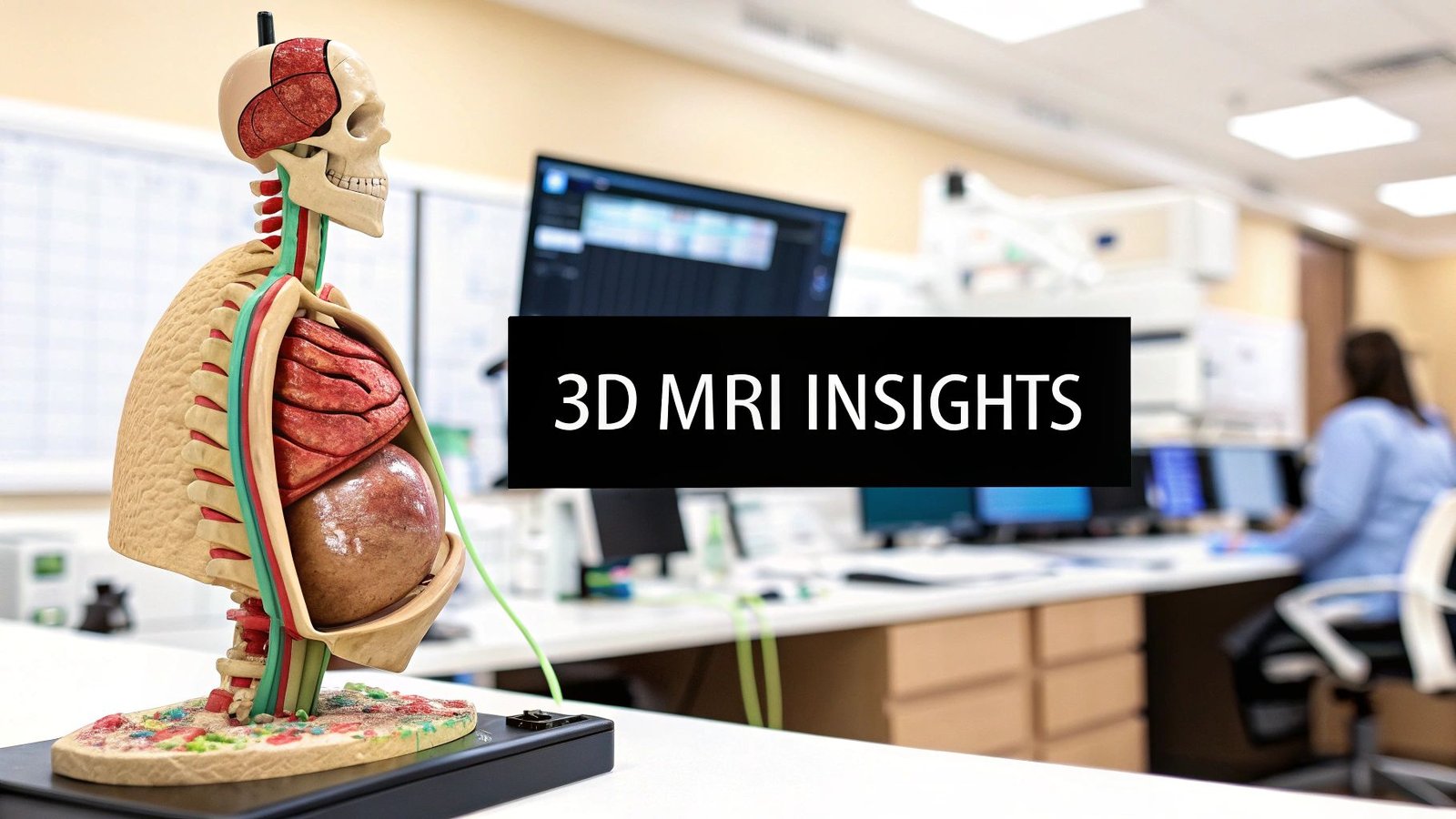
Picture this: a medical image that isn't just a flat photograph, but a fully explorable, three-dimensional model of the human body. That’s the incredible reality of a 3D MRI image. It fundamentally changes how we approach diagnostics, taking stacks of flat, 2D scans and weaving them into interactive, volumetric data that showcases our anatomy in breathtaking detail.
How We Build a 3D MRI Image

Crafting a 3D MRI image is a beautiful dance between physics and powerful computation. It's not like snapping a single 3D photo. Instead, it’s more like building a highly detailed sculpture, one incredibly thin layer at a time. The MRI machine captures a series of high-resolution 2D images, often called "slices."
These slices are captured from multiple angles—axial (like looking down from the top), sagittal (from the side), and coronal (from the front). This gives us a complete cross-sectional map of the area being examined. While a single slice offers just one perspective, something amazing happens when you bring hundreds, or even thousands, of them together.
The Art of Digital Stacking
Imagine you have a deck of cards, where each card is a detailed 2D MRI slice. When you stack them up perfectly, they no longer represent individual cards but a solid block of information. This is what we call a volumetric dataset.
Specialized software then steps in to perform what’s known as 3D reconstruction, digitally stitching every single slice together. The result is a seamless, cohesive 3D model that clinicians can rotate, zoom into, and explore from any direction.
This digital model opens up a new world of possibilities for clinicians, allowing them to:
- View anatomy from any angle: A surgeon can rotate the model to understand exactly how a tumor is interacting with nearby blood vessels.
- Measure volumes with precision: An oncologist can measure a tumor’s true size to see if a treatment is working.
- Plan complex interventions: An orthopedic specialist can map out a tricky surgery with total confidence before ever making an incision.
The real power of a 3D MRI image is its volumetric nature. It provides a complete anatomical context that flat images simply can't match, leading to smarter clinical decisions and better patient outcomes.
At PYCAD, we live and breathe this kind of complex data visualization. We at PYCAD, build custom web DICOM viewers and integrate them into medical imaging web platforms. This empowers healthcare professionals to interact with these intricate 3D models right in their web browser, making the technology both accessible and collaborative.
The ability to create and interact with a detailed 3D MRI image is a pillar of modern medicine. It gives us a window into the human body that was once the stuff of science fiction, pushing forward fields from neurology to cardiology. To see how these advanced viewers work in the real world, take a look at our portfolio. It's all about turning raw data into life-changing clinical insight.
The Journey to Seeing Inside the Body
To really appreciate the stunning detail of a modern MRI 3D image, it helps to look back at where it all started. This technology wasn't a sudden invention; it grew out of our deep-seated curiosity to see inside the human body without ever making an incision. The story begins not in a hospital, but in the world of physics, with a concept called Nuclear Magnetic Resonance (NMR).
For a long time, NMR was a niche tool for chemists, a clever way to figure out the molecular makeup of different substances. It was a fascinating scientific principle, but the idea of using it to peer inside a living person felt like science fiction—a problem just waiting for the right minds to tackle it.
The real leap forward happened when a few pioneers started to ask a different kind of question. Instead of just identifying materials with NMR, they wondered, "What if we could map their precise location in space?" That single question sparked one of the biggest revolutions in medical history.
From Physics Concept to Medical Marvel
The 1970s was the decade where theory slammed into reality. The journey toward MRI 3D imaging is rooted in that foundational NMR research, but it was in the 70s that the critical pieces fell into place. In 1973, a chemist named Paul Lauterbur figured out how to use gradients in the magnetic field to create spatial maps, which allowed him to produce the very first 2D and, later, 3D images.
Around the same time, physicist Sir Peter Mansfield was developing methods to drastically speed up the imaging process, making it a viable tool for actual clinical settings. Their combined brilliance, which earned them the 2003 Nobel Prize, built the foundation for every single MRI scan that happens today. If you're interested in the full story, you can read more about the development of MRI technology.
This pivot from a lab curiosity to a diagnostic game-changer was nothing short of monumental. The first-ever MRI scan on a human took place in 1977. It lasted nearly five hours and produced just one, very fuzzy image. But that blurry picture was everything—it was proof that we could finally make the invisible, visible.
This journey from a physics principle to a life-saving tool is a testament to human ingenuity. It highlights a persistent quest to overcome limitations and see the body in ways previously thought impossible.
The Evolution of Visualization
Those early MRI scans gave us simple 2D slices, like looking at single pages from a book. But everyone knew the potential was there to see the whole story. As computers became more powerful, so did our ability to handle the massive amounts of data the scanners could collect. This progress paved the way for the first volumetric models, turning those flat slices into a rich, interactive MRI 3D image.
At PYCAD, we see ourselves as part of this ongoing story. We at PYCAD, build custom web DICOM viewers and integrate them into medical imaging web platforms, putting these powerful 3D models into the hands of clinicians, no matter where they are. Our work allows doctors to explore and interact with complex anatomy in a way the original MRI pioneers could only dream of.
The history of MRI is really a story about turning abstract science into tangible hope. Every detailed 3D model we interact with today stands on the shoulders of giants who dared to look deeper. To get a feel for how modern platforms manage this complex data, take a look at our portfolio of advanced imaging solutions.
Building A 3D Image From 2D Slices
Creating a 3D MRI image is a little like building a detailed, layered sculpture out of a stack of ultra-thin photographs. It all starts with the MRI scanner capturing a series of individual 2D images, often called "slices."
Think of each slice—whether taken from the front (coronal), side (sagittal), or top-down (axial)—as a single, high-resolution snapshot. On its own, one slice offers just a sliver of the full picture. But when you bring them all together, they form the foundation for something truly remarkable: a complete, three-dimensional model of the patient's anatomy.
From Slices To A Seamless Volume
The real magic unfolds during the reconstruction phase. This is where sophisticated software algorithms take over, meticulously aligning and stacking these 2D slices. This isn't just a simple stacking process, though. The system uses a powerful technique called interpolation to intelligently "fill in the gaps" between each slice.
This step is crucial. It ensures the final result is a smooth, continuous 3D volume, not a clunky, pixelated model with visible steps between each layer. What you get is a rich, fully explorable digital twin of the patient's internal structures. Understanding the nuts and bolts of this process is easier when you see how modern tools are pushing the boundaries of AI-powered 2D to 3D model conversion.
This infographic gives you a sense of the incredible journey from the discovery of the underlying physics to the powerful imaging we have today.
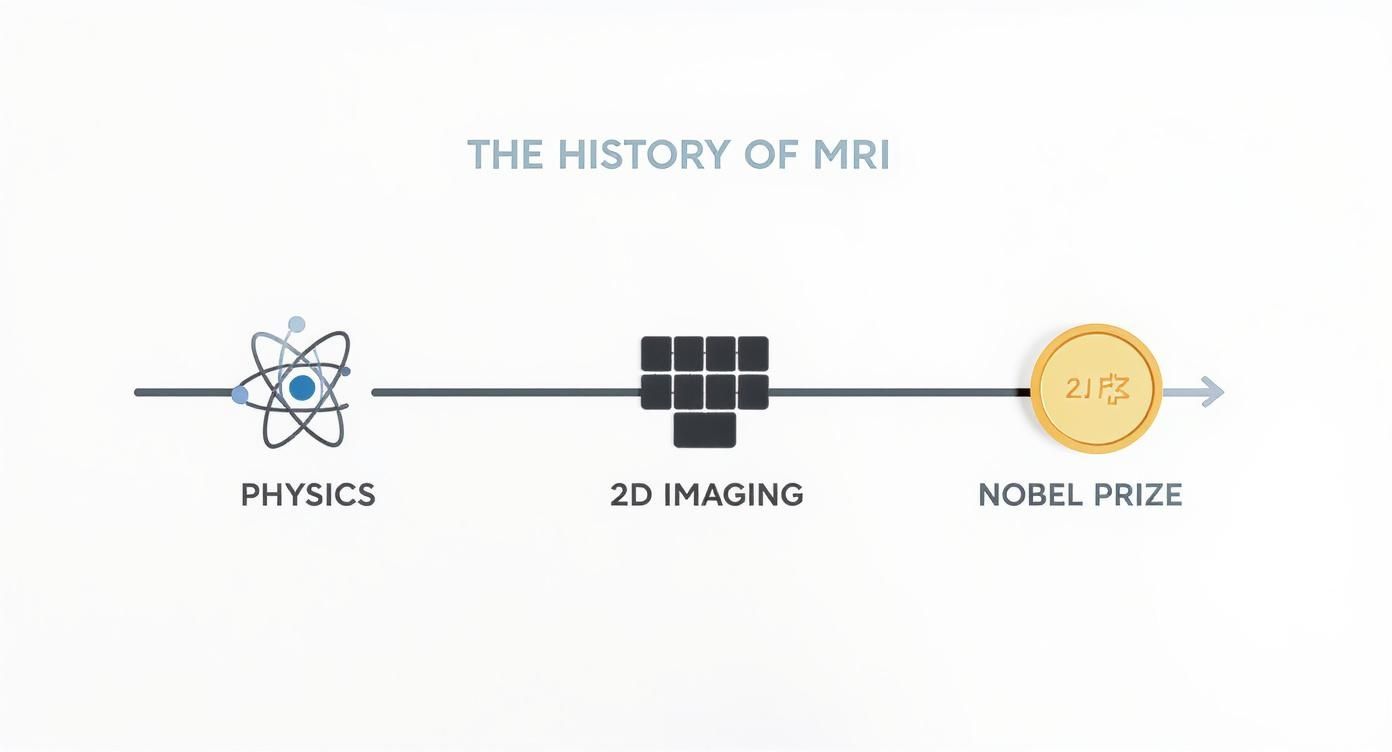
It’s a great reminder of how decades of scientific breakthroughs have paved the way for the incredible diagnostic tools we rely on.
Advanced Visualization Techniques
Once this 3D volume is built, the real fun begins. Clinicians can use specialized tools to interact with the model in ways that bring the data to life. Two of the most common and powerful techniques are Multi-Planar Reformation (MPR) and Volume Rendering (VR).
-
Multi-Planar Reformation (MPR): This lets a doctor virtually "re-slice" the 3D model from any angle they can imagine. They aren't limited to the original three planes. A surgeon, for instance, could create a custom view that perfectly matches their planned path of incision, giving them a roadmap before the procedure even starts.
-
Volume Rendering (VR): This is where the 3D MRI image becomes truly intuitive. VR algorithms assign different colors and opacities to different types of tissue based on their signal strength. The result? Vivid, color-coded models where bone, muscle, blood vessels, and tumors are all clearly distinguishable at a glance.
The ability to move beyond static, 2D images and interact with a dynamic 3D model is what gives modern diagnostics its power. It provides an anatomical understanding that is simply not possible with flat images alone.
At PYCAD, our mission is to make these advanced visualizations readily available to clinicians. We at PYCAD, build custom web DICOM viewers and integrate them into medical imaging web platforms. This allows medical professionals to perform MPR and VR right from their browser, eliminating the need for clunky, desktop-bound software.
From 2D Slices To A Full 3D View
The leap from flat, 2D slices to an interactive 3D model represents a fundamental shift in diagnostic capability. The table below breaks down exactly what that looks like in practice.
Comparing 2D Slices vs 3D Volumetric Imaging
| Feature | Traditional 2D MRI Slices | Modern MRI 3D Image |
|---|---|---|
| Data Representation | A sequence of separate, flat images. | A single, unified volumetric dataset. |
| Anatomical Context | Limited to the specific plane of the slice. | Complete spatial relationships between structures. |
| Lesion Detection | Small lesions between slices can be missed. | Provides a gap-free view, improving detection. |
| Surgical Planning | Offers a limited, 2D perspective for planning. | Allows for interactive, 3D surgical simulation. |
| Diagnostic Confidence | Can be ambiguous, requiring mental reconstruction. | Provides clear, intuitive visualization of anatomy. |
| Flexibility | Viewing is restricted to acquired planes. | Can be "re-sliced" from any angle (MPR). |
This comparison makes it clear: while 2D images are the building blocks, the complete 3D volume is where true clinical insight is found.
The Impact Of Stronger Magnets
The quality of the final 3D MRI image is deeply connected to the power of the MRI scanner’s magnet, which is measured in Tesla (T). The move from the standard 1.5T magnets to more powerful 3T scanners was a huge step forward for 3D imaging.
A 3T machine generates a much stronger magnetic field, which dramatically boosts the signal-to-noise ratio (SNR). A higher SNR means the initial 2D slices are sharper, clearer, and packed with more detail. Better ingredients make a better final product, and in this case, higher-quality slices build a far more precise and reliable 3D model.
If you're interested in going deeper, our guide on creating 3D models from 2D images explores the reconstruction process in greater detail.
How 3D Imaging Transforms Patient Care
It's one thing to talk about technology, but how does a detailed MRI 3D image actually translate into better, more hopeful outcomes for patients? The magic happens when this technology moves from a computer screen into the hands of clinicians who are changing lives every single day. Across the medical world, volumetric imaging delivers a level of clarity that was once the stuff of science fiction, making complex procedures safer and diagnoses more certain.
This isn't some brand-new trend. The clinical use of 3D MRI has been reshaping medicine for decades. Believe it or not, the very first clinically useful 3D MRI images were captured way back in 1980 by a team at the University of Aberdeen. This breakthrough finally gave doctors the power to examine complex anatomy from any angle and measure tissue volumes with incredible precision.
This was a game-changer for cardiology. Early studies in the 1980s used 3D cardiac MRI scans on volunteers, laying the essential groundwork for how we assess the heart today.
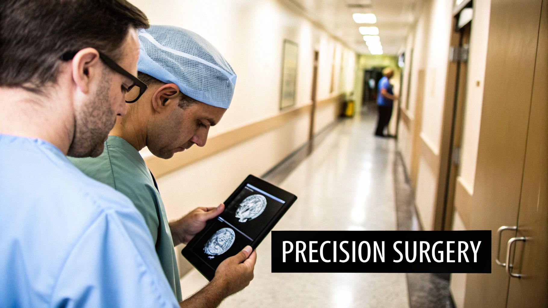
Precision in Neurosurgery
Imagine being a neurosurgeon. The brain is an incredibly delicate landscape where a single millimeter can be the difference between a full recovery and a life-altering complication. Before 3D imaging, surgeons had to rely on flat, 2D scans and their own mental gymnastics to navigate this intricate terrain.
Today, everything is different. A surgeon can load a patient's MRI 3D image and create a detailed, interactive map of their brain. They can pinpoint a tumor, see its exact size and shape, and—most importantly—understand how it relates to critical blood vessels and brain tissue. This allows them to plan the safest possible path for removal, digitally rehearse the entire surgery, and walk into the operating room with a level of confidence that simply wasn't possible before.
Advancements in Orthopedic Surgery
Orthopedic surgeons face a similar need for precision, especially when planning something like a complex joint replacement. Getting the alignment and fit of an implant just right is everything. An MRI 3D image gives them the perfect blueprint.
- Surgical Rehearsal: Surgeons use the 3D model to select the perfect implant size and shape, tailored to the patient’s unique anatomy.
- Custom Guides: In many cases, they can even 3D-print custom surgical guides that snap perfectly onto the patient's bone, ensuring every cut is made with absolute accuracy.
- Better Outcomes: This meticulous planning leads directly to better implant function, faster recovery, and a much lower risk of complications down the road.
The true gift of a 3D MRI image is its ability to remove guesswork. It turns abstract anatomical challenges into clear, solvable geometric problems, directly improving the safety and effectiveness of medical interventions.
Transforming Cardiology and Oncology
In cardiology, the heart is a dynamic, three-dimensional structure that's notoriously difficult to see clearly. A 3D model lets a cardiologist see the chambers, valves, and vessels in one cohesive view. This is invaluable for diagnosing congenital heart defects or planning a complex valve replacement with far greater accuracy.
For oncologists, tracking how a tumor responds to treatment is the core of patient care. A 3D MRI image lets them calculate the tumor's exact volume, offering a far more meaningful measure of progress than a simple 2D measurement. Seeing a 15% reduction in tumor volume gives them concrete data that a treatment is working.
At PYCAD, our passion is making this powerful data accessible. We at PYCAD, build custom web DICOM viewers and integrate them into medical imaging web platforms, empowering clinicians to collaborate and plan using these detailed 3D models from any device.
This is just the beginning. The way patients and doctors interact with this data is also evolving, with new tools like Virtual Reality as a Gateway to Accessible Healthcare promising an even more intuitive future. Ultimately, every step forward in 3D imaging is a step toward more personalized and effective medicine. You can see incredible examples of these technologies in action in our PYCAD portfolio.
The Role of AI in 3D MRI Analysis
The next great leap for the MRI 3D image is its powerful fusion with Artificial Intelligence. AI is taking this technology far beyond simple visualization, turning it into a tool for intelligent, automated analysis. This is where the future of diagnostics is heading—a world where AI acts as an expert collaborator, working alongside radiologists to uncover insights hidden deep within the data.
This partnership moves us from just seeing anatomy to truly understanding it on a scale that was previously impossible. AI algorithms are built to recognize patterns, anomalies, and structures with incredible speed and consistency.
Automating Complex Tasks
One of AI's most immediate impacts is in automatic segmentation. Imagine a radiologist needing to manually outline a complex tumor or trace the exact boundaries of an organ across hundreds of individual MRI slices. This is an incredibly precise but painfully time-consuming task, often taking hours of meticulous work.
AI changes the game entirely. Machine learning models, trained on thousands of scans, can instantly and accurately segment organs, lesions, and other anatomical structures from a 3D volume. This not only frees up a massive amount of a clinician's time but also introduces a level of standardization that is difficult to achieve manually. For those interested in the technical details, our overview of medical image segmentation offers a deeper dive into these methods.
This automation allows radiologists to focus their expertise on the most critical part of their job: interpreting the findings and making a diagnosis.
Unlocking Predictive Diagnostics
Beyond just saving time, AI is opening the door to predictive diagnostics. This is where the technology truly feels like a glimpse into the future. Machine learning algorithms can analyze subtle textures and patterns within an MRI 3D image that are completely invisible to the human eye.
By identifying these microscopic signatures, AI models can begin to predict how a disease might progress. For example, an algorithm could analyze the internal structure of a brain tumor to forecast its potential growth rate or its likely response to a specific type of chemotherapy.
AI isn't just about making current processes faster; it's about asking entirely new questions. It allows us to shift from reactive diagnosis to proactive, predictive medicine, paving the way for truly personalized patient care.
This ability to forecast disease is a monumental step forward, offering the potential to tailor treatments to an individual’s unique biology. It moves medicine closer to an era of prevention and early intervention.
A New Era of Collaboration
The ultimate goal is not for AI to replace radiologists, but to augment their abilities. Think of AI as an incredibly perceptive assistant, one that can process vast amounts of data, highlight areas of concern, and provide quantitative measurements in seconds. This allows the human expert to bring their full clinical experience and intuition to bear on the most complex cases.
At PYCAD, we are at the forefront of this integration. We at PYCAD, build custom web DICOM viewers and integrate them into medical imaging web platforms, creating the perfect environment for these AI tools to thrive. By embedding AI analysis directly into the viewing workflow, we empower clinicians with deeper insights right at their fingertips.
This powerful collaboration between human expertise and machine intelligence is redefining what's possible in medical imaging. To see how we bring these advanced solutions to life, explore our work in the PYCAD portfolio.
Bringing 3D MRI to the Web
A stunningly detailed MRI 3D image is only as good as our ability to access, share, and interact with it. The real magic happens when these rich, volumetric datasets break free from isolated hospital workstations and move into a connected, web-based world. This is how advanced medical imaging becomes a natural, fluid part of modern digital health.
The universal language for this is DICOM (Digital Imaging and Communications in Medicine). It’s the standard that ensures every medical image, including MRI scans, can be stored and shared consistently. The catch? These files are often enormous and traditionally required powerful, dedicated software just to open them.
That’s where web-based DICOM viewers completely change the game. These tools are built to render massive 3D volumes right inside a standard web browser. Suddenly, clinicians are free from installing and maintaining clunky desktop applications, making critical patient data accessible to anyone who needs it, wherever they are.
Enabling Collaboration from Anywhere
Picture this: a neurosurgeon in New York needs a second opinion on a complex brain scan. With a web-based platform, they can securely share the interactive 3D model with a specialist in London. Both of them can see and manipulate the exact same model in real-time—rotating it, highlighting areas of concern, and mapping out a surgical plan together. Not long ago, that kind of seamless teamwork was a logistical nightmare.
The goal is to bring the data to the clinician, not the other way around. Web-based platforms transform the MRI 3D image from a static file into a living, collaborative tool that can be accessed securely from any device with an internet connection.
This is exactly what we do at PYCAD. We at PYCAD, build custom web DICOM viewers and integrate them into medical imaging web platforms. Our solutions are engineered from the ground up to handle large, complex datasets with grace, giving healthcare providers the tools they need to securely view and share 3D MRI data from anywhere in the world. For a closer look at our projects, feel free to explore our work in the PYCAD portfolio.
The Future of Medical Imaging Platforms
When these viewers are woven into a larger medical platform, they create a powerful, unified ecosystem. Clinicians can pull up patient records, lab results, and intricate 3D scans all from one secure portal. This doesn't just simplify workflows; it provides a complete clinical picture that enriches the entire diagnostic process. To see how these powerful tools function in a browser, our guide to 3D Slicer online explores very similar visualization concepts.
Ultimately, this is about connecting the brilliance of advanced imaging with the practical demands of modern healthcare. It’s about ensuring that every incredible MRI 3D image is put to its best possible use—driving collaboration, accelerating diagnoses, and delivering better outcomes for patients.
Answering Your Questions About 3D MRI
As we dive into the incredible world of 3D MRI, it's natural to have questions. This technology sits at the fascinating crossroads of physics and powerful computing, so curiosity is a great sign! Let's clear up some of the most common questions and give you a deeper appreciation for this amazing diagnostic tool.
These answers are designed to give you straightforward insights into how 3D MRI works and why it’s become so essential in modern medicine.
Is a 3D MRI Scan Safe?
Absolutely. An MRI scan is widely considered to be very safe. The key difference between an MRI and a CT scan or X-ray is that it doesn't use any ionizing radiation. Instead, it works its magic with a strong magnetic field and radio waves.
The only real safety consideration is that powerful magnet. This is why you're asked to remove all metal and tell the technologist about any implants you might have. For the vast majority of people, it's an incredibly safe and non-invasive procedure.
How Long Does a 3D MRI Scan Take?
The time it takes can really vary, mostly depending on what part of the body is being scanned and just how much detail the doctors need to see. A scan could be as short as 30 minutes or last for more than an hour.
Think of it like building a very detailed map, slice by slice. Acquiring all the thin layers needed for a high-quality 3D MRI image simply takes time. Even though new techniques are helping to speed things up, you should still plan on lying still for a while to get the clearest possible images.
While the scan itself requires a bit of patience, the comprehensive data it produces is priceless. It delivers a complete, three-dimensional view that can dramatically speed up diagnosis and treatment planning, making the time spent a very worthwhile investment in your health.
What Is the Difference Between a 1.5T and 3T MRI?
Great question. The "T" stands for Tesla, which is the unit measuring the strength of the magnetic field. A 3T MRI scanner has a magnet that is literally twice as powerful as a 1.5T machine.
Why does that matter? A stronger magnet gives you a much better signal-to-noise ratio. In simple terms, it means a 3T scanner can produce images with far greater detail and higher resolution. This is a game-changer when you're trying to create a crisp and precise 3D MRI image, especially for tiny structures in the brain, joints, or blood vessels.
This is where our work at PYCAD comes in. We specialize in taking the complex, high-resolution data from advanced scanners like these and making it useful. We at PYCAD, build custom web DICOM viewers and integrate them into medical imaging web platforms, so clinicians can pull up and analyze these incredibly detailed 3D models from anywhere. It's all about turning that raw data into clear, actionable intelligence that helps doctors make better decisions.
PYCAD empowers healthcare innovators by building secure, web-based platforms with advanced DICOM viewers and integrated AI solutions. Discover how we can bring your medical imaging data to life by exploring our portfolio.


