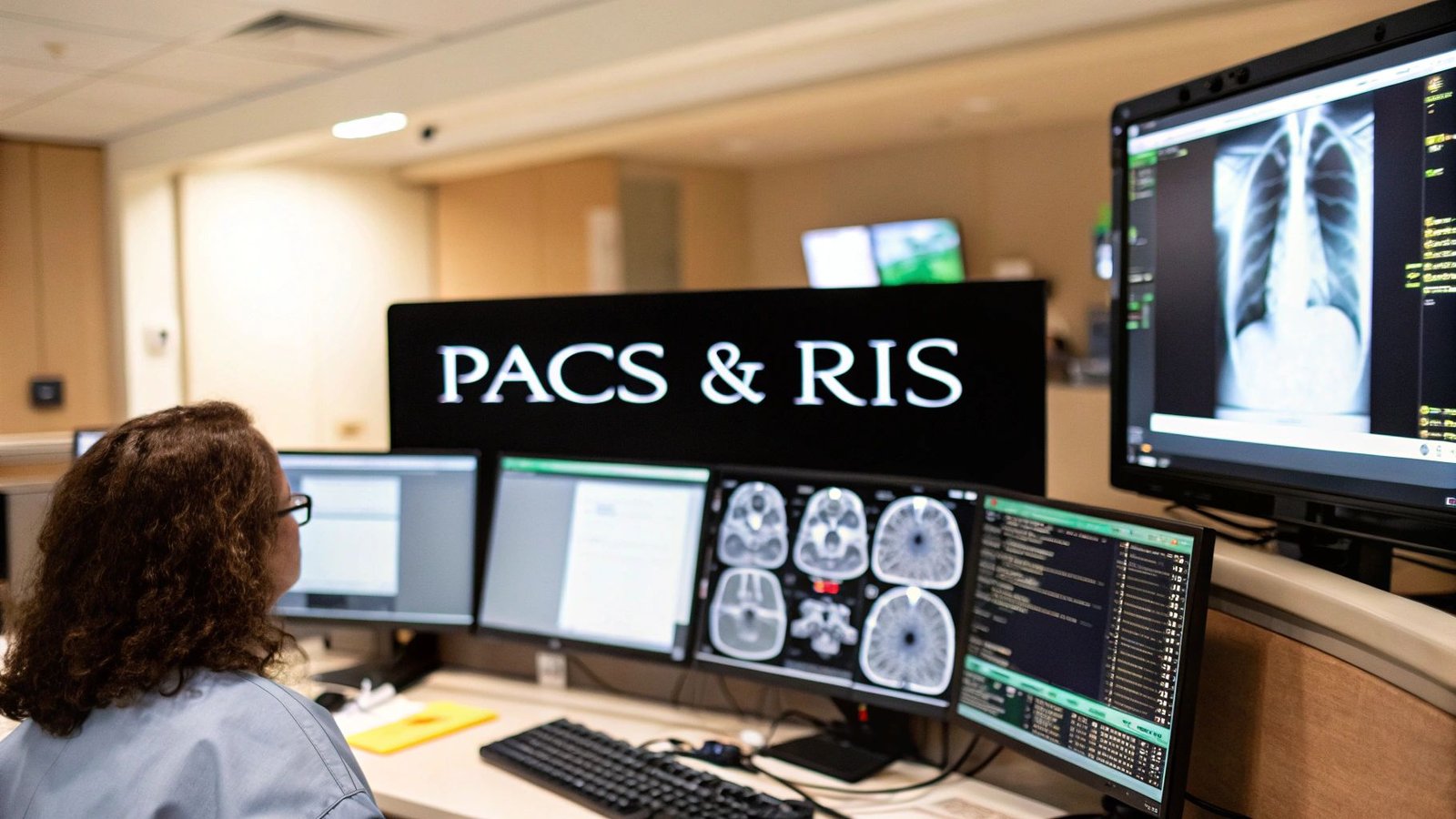
In the world of medical imaging, two systems stand as the pillars of a modern radiology department: PACS and RIS. Think of them as the brain and the heart of the operation. A Radiology Information System (RIS) is the department's command center, managing patient information and the entire workflow, while a Picture Archiving and Communication System (PACS) is the high-tech library where all the medical images live.
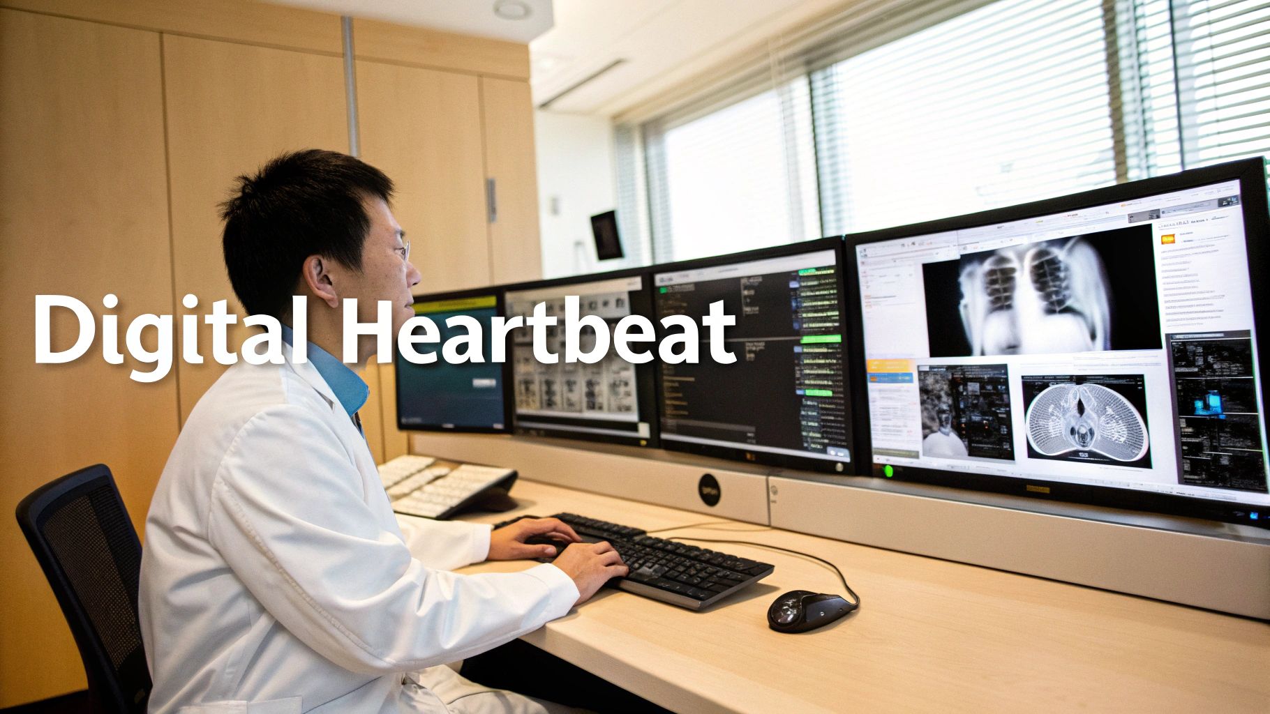
The Digital Heartbeat of Modern Radiology
Walk into any hospital's imaging department today, and you'll see a symphony of technology at work. This entire environment, from scheduling a scan to delivering a diagnosis, relies on the seamless partnership between PACS and RIS. Grasping what each one does is the first step to understanding just how powerful modern radiology has become.
Let's use an analogy. The RIS is like the air traffic controller for a patient's entire imaging journey. It handles all the logistics—scheduling the appointment, registering the patient, tracking the exam's progress, and even managing billing. It's a text-based system, focused entirely on keeping the administrative and operational side of things running smoothly.
The PACS, on the other hand, is the secure, digital archive where every single medical image is stored, protected, and made available in an instant. It’s specifically built to manage the massive visual files produced by MRI machines, CT scanners, and X-ray equipment. It's all about the pictures.
PACS vs RIS At a Glance
To make the distinction crystal clear, here’s a quick side-by-side comparison of their core responsibilities.
| Aspect | Radiology Information System (RIS) | Picture Archiving and Communication System (PACS) |
|---|---|---|
| Primary Function | Workflow and data management | Image storage and viewing |
| Data Type | Text-based (patient info, reports) | Image-based (DICOM files) |
| Core Tasks | Scheduling, billing, reporting | Archiving, retrieving, displaying images |
| Key User | Administrative staff, technicians | Radiologists, clinicians |
| Analogy | The "Air Traffic Controller" | The "Digital Image Library" |
While their jobs are different, they are completely codependent. A PACS without a RIS would be a library with no catalog, and a RIS without a PACS would be a schedule with no results to show for it.
Defining the Roles of Each System
Even though they work in tandem, their functions are clearly defined. This separation of duties is precisely what makes the whole system so incredibly efficient.
- Radiology Information System (RIS): This is the workflow engine. Its job is to manage patient scheduling, handle registration, track exams, generate reports, and process billing. Essentially, it keeps the entire administrative machine humming.
- Picture Archiving and Communication System (PACS): This is the image archive. Its sole purpose is to handle the acquisition, storage, retrieval, and display of medical images, ensuring clinicians get the high-quality diagnostic visuals they need, right when they need them.
Together, PACS and RIS turn a flood of complex administrative text and visual data into clear, life-saving insights. They are the essential foundations supporting every timely and accurate diagnosis made today.
The Power of Specialized Tools
The images stored in a PACS are more than just pictures; they are complex data sets that require powerful tools for a radiologist to interpret. This is where the universal imaging standard, DICOM, comes into play. You can dive deeper into the technical backbone of medical imaging by exploring our complete guide on DICOM standards.
This is also where specialized development makes a huge difference. At PYCAD, we build custom web DICOM viewers and integrate them directly into medical imaging platforms. These advanced viewers are the sophisticated interfaces that radiologists use to transform raw image data into actionable diagnoses, allowing them to manipulate, annotate, and compare scans with incredible precision. This guide will continue to demystify how these systems work in harmony, setting the stage for a more efficient and intelligent future in radiology.
How RIS Orchestrates the Patient Journey
If PACS is the library holding all the medical images, think of the Radiology Information System (RIS) as the expert conductor leading the entire symphony of a patient's visit. It's the operational brain that steers everything from the initial appointment request all the way to the delivery of a finished diagnostic report. The RIS is what brings calm, control, and clarity to a process that could otherwise be quite chaotic.
To really get a feel for what a RIS does, let's walk through a patient's experience. Imagine their primary care doctor orders a CT scan. That request, packed with patient details and the doctor's notes, lands in the radiology department’s system. The moment the RIS logs it, the journey officially begins.
From Scheduling to Check-In
The first job for the RIS is scheduling. This isn't just about finding an open time slot; it’s about managing the availability of every scanner, every room, and every technologist to prevent conflicts. The system helps staff find the perfect time that works for the patient and the clinic, then automatically sends out confirmations and any special preparation instructions.
When the patient arrives, they check in. The RIS instantly verifies their demographic info, confirms insurance details, and flips their status from "scheduled" to "arrived." That one simple action signals the entire team—the front desk, the technologists, the radiologists—that their patient is here and ready. No more guesswork.
Guiding the Clinical Workflow
Once checked in, the RIS shifts from an administrative tool to a clinical command center. It generates a worklist for the technologist, detailing exactly what exam to perform on which patient. This is a perfect example of data interoperability in healthcare in action, as the patient data from the RIS flows directly to the imaging modality itself.
The technologist confirms the patient’s identity against the RIS record, performs the scan, and marks the exam as "complete." This click sets off a chain reaction:
- The radiologist is notified that new images are available for review.
- The use of any contrast agents is logged for inventory and billing.
- The system preps the necessary information for the billing department.
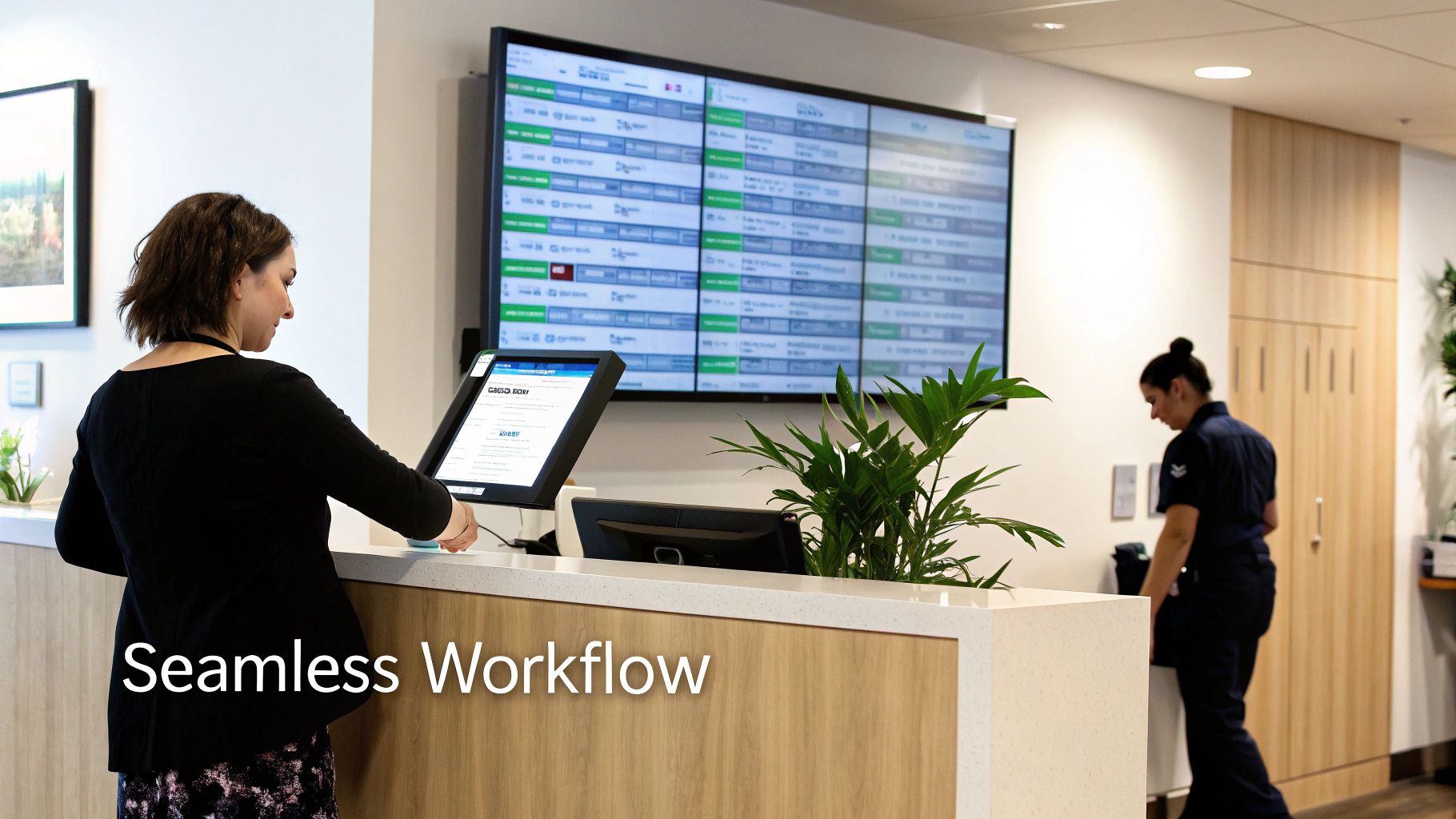
A RIS transforms a series of separate, disjointed tasks into one smooth, trackable workflow. It gets rid of clumsy manual hand-offs and ensures everyone has the exact information they need, right when they need it.
Reporting and Beyond
After the images are safely stored in the PACS, the radiologist gets an alert. They pull up the patient’s file in the RIS, which gives them the full clinical context—the reason for the exam, prior history, and any relevant notes. This background is absolutely essential for an accurate diagnosis. The radiologist then dictates their findings, and the report is transcribed right back into the RIS.
Once the report is signed off, the RIS handles the final, crucial step: report distribution. It automatically and securely sends the final report to the ordering physician. The loop is officially closed. Behind the scenes, the system is also taking care of the billing by capturing all the procedural codes and even helps manage inventory so supplies never run low.
By masterfully coordinating this entire journey, the RIS frees up the clinical team to do what they do best: focus on patient care. They can work with confidence, knowing the administrative details are being handled with precision and efficiency. The result is a faster, safer, and better experience for everyone.
The Power of PACS in Medical Imaging
If the RIS is the operational conductor, then the Picture Archiving and Communication System (PACS) is the digital vault—the secure library where the real treasures of radiology are kept and made instantly available. But don't think of it as just a simple storage drive. A PACS is a living, breathing ecosystem built for four critical functions: acquisition, archiving, access, and analysis. It's what took us from shuffling around clunky film jackets to a world of immediate, digital insight.
The magic starts with acquisition. The second a CT, MRI, or X-ray scan is finished, the images are whisked away to the PACS using the universal DICOM standard. This direct digital pipeline ensures every pixel is captured securely and automatically tied to the correct patient, wiping out the old-world problems of lost films and manual mix-ups.
From Immediate Access to Long-Term Integrity
Once the images arrive, they're archived using a clever, tiered storage system that balances speed and cost. The newest, most relevant scans stay in high-speed, online storage so radiologists can pull them up in a flash. Older images, which are needed less often, are moved to more economical, long-term archives. This keeps the system snappy for daily work while guaranteeing the data is safe and sound for years, which is crucial for both legal and medical history.
The sheer scale of this technology is staggering. The global market for pacs and ris systems, valued at USD 2.29 billion, is expected to more than double to USD 4.62 billion by 2034. The PACS component alone dominates with an 83% market share, a figure that speaks volumes about its essential role in handling the daily flood of imaging data. You can dig into more market insights on Precedence Research. This isn't just a trend; it's the backbone of modern medicine.
The most game-changing feature of a PACS is universal access. It gives clinicians the power to view high-resolution medical images from anywhere—a hospital workstation, a home office, or a colleague's screen across the country.
Elevating Diagnostics with Advanced Viewing Tools
This is where basic, off-the-shelf tools just don't cut it, and specialized solutions become mission-critical. At PYCAD, we don't just provide viewers; we build custom web DICOM viewers and integrate them directly into medical imaging web platforms. These are not simple image displays; they are sophisticated analytical instruments.
We equip radiologists with the advanced features they need to make precise, confident diagnoses. This includes:
- Intuitive manipulation tools to zoom, pan, and adjust contrast with fluid precision.
- Side-by-side comparison capabilities to easily track a patient's progress over time.
- Advanced annotation features for marking up and measuring areas of concern.
- Seamless integration that delivers lightning-fast performance right inside a web browser.
This tailored approach turns passive image viewing into an active, diagnostic exploration. For a closer look at how these systems are built, check out our comprehensive article on what a PACS system is. By building superior viewing environments, we help radiologists do what they do best: make faster, more accurate decisions. Take a look at our portfolio to see how our custom solutions are making a real-world impact.
After all, a great PACS isn't just about storing images. It's about unlocking the life-saving stories they tell.
Unlocking Real Synergy with PACS and RIS Integration
Think of a RIS as the brain of a radiology department, managing all the logistical and textual information. A PACS, on the other hand, is the eyes—handling all the visual data. They're powerful on their own, but the real magic happens when they work together in perfect harmony.
When integrated, they create a single, intelligent nervous system for your entire imaging operation. All the friction of manual data entry and disjointed steps disappears. What was once a series of separate tasks becomes one fluid, automated journey from patient scheduling to final diagnosis.
The secret to making this happen is getting them to speak the same language. Your RIS is a master of text and logistics, communicating through a standard called HL7 (Health Level Seven) to manage patient data, orders, and reports. Your PACS is a visual specialist, using DICOM (Digital Imaging and Communications in Medicine) to handle massive, complex image files. A proper integration acts as a universal translator, letting these two essential systems share information without missing a beat.
The Integrated Workflow in Action
To really grasp this, let's walk through a patient's imaging exam from start to finish. It’s a beautifully choreographed dance where data is handed off automatically, virtually eliminating errors and saving an incredible amount of time.
- The Order Begins in RIS: A referring physician schedules a CT scan. The order is created in the RIS, complete with the patient's demographics and clinical history.
- Data Flows to the Modality: The RIS instantly sends this information directly to the CT scanner's worklist. This is a crucial step—it ensures the technologist selects the right patient for the right exam, every time.
- Images are Captured and Sent to PACS: The scan is performed. Those images are immediately sent to the PACS, already embedded with the correct patient data that came from the RIS. No manual tagging needed.
- PACS Notifies RIS: The PACS then sends an automatic alert back to the RIS, confirming the images have been safely archived and are ready for the radiologist to review.
- The Loop Closes with a Report: The radiologist opens the images in the PACS viewer, dictates their findings, and the final report is generated and stored in the RIS. The RIS then automatically distributes that report back to the ordering physician.
This constant digital conversation ensures every piece of information is accurate and accounted for from beginning to end. This flow diagram breaks down the core PACS functions—acquisition, archiving, and access—that are at the heart of this integrated process.
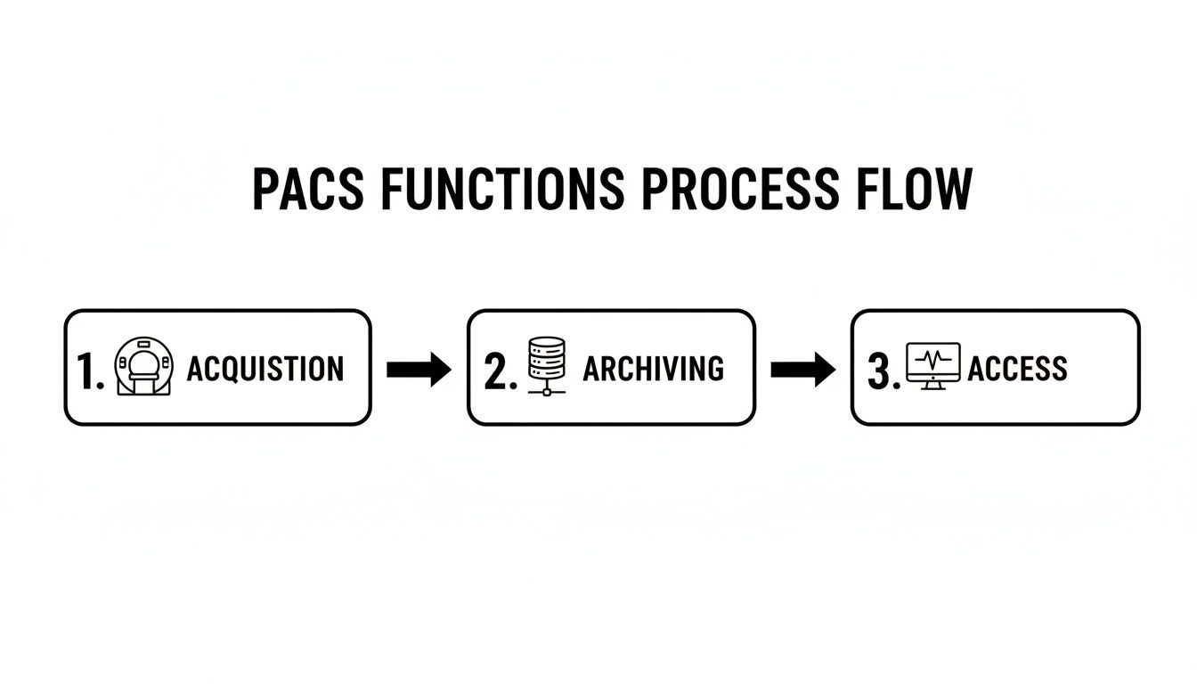
This shows just how smoothly data can move from the imaging machine to secure storage and, finally, to the radiologist's screen for a confident diagnosis.
The Financial and Operational Impact
The benefits here go far beyond just convenience; they hit the bottom line. To truly appreciate how these systems collaborate, it helps to understand the broader concept of application integration.
By automating tasks like scheduling, reporting, and even billing, an integrated system gives a massive boost to operational efficiency. For high-volume imaging centers, this is a game-changer. Just moving from old-school film to a digital system can slash storage and processing costs by 30-50% alone. It’s a huge driver for why so many practices have adopted integrated PACS and RIS solutions.
At its heart, PACS and RIS integration is about creating a single source of truth. It ensures that the right image is always linked to the right patient, the right report, and the right billing code—every single time.
This is the bedrock of modern medical imaging. At PYCAD, we take this experience to the next level. We specialize in building custom web DICOM viewers and integrating them into medical imaging platforms, giving radiologists exceptional tools that fit perfectly within these finely tuned ecosystems. See how our solutions create a more intuitive and powerful diagnostic environment by checking out our portfolio.
The Future of Radiology with AI-Enhanced Platforms
For years, we've thought of PACS and RIS as two separate, if connected, worlds. One handles the images, the other handles the information. But that distinction is starting to blur, and the force behind this change is artificial intelligence. AI isn't some far-off concept anymore; it's right here, right now, and it's fundamentally changing how we approach medical imaging.
We're moving toward a future where our tools are more than just digital filing cabinets. They're becoming active partners in the diagnostic process. Think about it: AI algorithms working quietly within the PACS, scanning every single image for subtle anomalies a human might miss on the first pass. This isn't about replacing radiologists—it's about giving them a powerful second set of eyes, boosting their confidence, and finding problems earlier than ever before.
AI Supercharging the RIS Workflow
But AI's reach goes far beyond just looking at pixels. It's also injecting a new level of intelligence into the day-to-day operations managed by the RIS. It can turn the administrative side of a radiology department from a reactive process into a smart, predictive engine.
Here are a few ways this is already happening:
- Predictive Scheduling: Instead of just booking appointments, AI can analyze patterns to forecast no-shows. This allows staff to proactively fill those gaps, keeping expensive machinery and skilled technicians busy.
- Intelligent Worklists: A radiologist's to-do list becomes dynamic. AI can intelligently reorder the queue, pushing exams with suspected critical findings to the very top. It’s about making sure the most urgent cases get attention first.
- Automated Reporting: Using Natural Language Processing (NLP), AI can help draft preliminary reports by pulling out key findings. This lets radiologists focus their brainpower on interpretation and nuance, not on repetitive typing.
When you weave AI into both pacs and ris, you create a powerful synergy. You're not just improving one piece of the puzzle; you're amplifying both clinical insight and operational efficiency at the same time.
Building the Integrated Platforms of Tomorrow
The massive growth we're seeing in the pacs and ris market isn't a coincidence. It’s being driven by the real-world adoption of AI for image analysis and a huge push to connect everything with electronic health records. The fastest-growing part of this is cloud-based platforms, which offer the flexibility and remote access that modern tele-radiology absolutely depends on.
This is exactly where we at PYCAD live and breathe. We don't just build off-the-shelf software. We build custom web DICOM viewers and integrate them into medical imaging web platforms that are designed from the ground up to make the most of AI. Our goal is to create one cohesive environment where a radiologist can view an image, see the report, and get AI insights—all in a single window. That kind of seamless workflow is how you unlock the next level of diagnostic speed and accuracy.
Of course, as these systems become more powerful and connected, we have to think about security. It's crucial to understand the pivotal role of AI in enhancing cybersecurity to protect this sensitive data.
Ultimately, this is all about empowering clinicians in a way that simply wasn't possible before, leading to smarter diagnostics and better outcomes for patients. To see how we’re making this a reality with our advanced viewer solutions, take a look at our portfolio.
Your Top PACS and RIS Questions, Answered
Even after getting the basics down, you might still have questions about how PACS and RIS really work together in the day-to-day grind of a busy radiology department. Let's tackle some of the most common questions I hear from clinicians and administrators to give you a rock-solid understanding of these critical systems.
What Is the Main Difference Between PACS and RIS?
Think of it this way: the RIS is the traffic controller, and the PACS is the photo library.
A Radiology Information System (RIS) is all about the workflow and the words. It handles text-based data—scheduling patients, registering them when they arrive, tracking their exam progress, and managing billing. It's the administrative backbone of the whole operation.
A Picture Archiving and Communication System (PACS), on the other hand, is built for the images. Its entire purpose is to store, retrieve, and display the actual medical image files (like DICOMs from an MRI or CT scan).
So, in a nutshell, the RIS manages the patient's entire journey through the department, while the PACS takes care of the images created along the way.
Can a Clinic Use PACS Without a RIS?
Technically, yes, but it would be like trying to run a busy restaurant without an order management system. It's incredibly inefficient. Without a dedicated RIS, all the crucial workflow steps like scheduling and reporting get pushed onto a generic Electronic Health Record (EHR) or, worse, rely on manual spreadsheets and paper. This creates disconnected data, slows everything down, and opens the door wide for human error.
The gold standard for a reason is a seamless integration between a RIS and a PACS. This connection ensures the right images are always tied to the right patient file and the right report, which is the foundation of efficient, accurate, and high-volume patient care.
This synergy is exactly why the market for these systems is booming. North America currently leads the charge, holding a 37% market share thanks to its advanced healthcare infrastructure. The U.S. market alone, valued at USD 590 million, is expected to skyrocket to USD 1.22 billion by 2034. These numbers speak volumes about the real-world value of getting this integration right. For a deeper dive into these trends, check out the full research on Precedence Research.
How Do These Systems Connect to an EHR?
Connecting to an Electronic Health Record (EHR) is non-negotiable for creating a complete, unified patient story. This link is usually forged using a standard messaging protocol called HL7. The process is beautifully simple: a physician orders an MRI in the EHR, and that order zips over to the RIS to get the patient scheduled and the exam started.
Once the radiologist has analyzed the images and finalized their report in the RIS, that report is sent securely back to the EHR. It becomes a permanent part of the patient's medical history. Often, the EHR will even include a link that lets the ordering physician click to open a viewer and see the actual images from the PACS, giving them the full diagnostic picture instantly.
Why Is a Custom Web DICOM Viewer So Important?
A web-based DICOM viewer is a game-changer. It lets authorized users view high-resolution medical images from any web browser, on any device, without needing to install clunky, specialized software. This untethers radiologists from their workstations and opens up huge possibilities for remote diagnosis and collaboration.
While off-the-shelf viewers are available, a custom-built solution is where the real magic happens. It allows for advanced features and workflows tailored to a radiologist's specific needs—something a generic viewer just can't offer.
This is precisely our expertise at PYCAD. We build custom web DICOM viewers and integrate them into medical imaging web platforms. By doing this, we give radiologists superior tools, blazing-fast performance, and intuitive workflows that help them make faster, more accurate diagnoses. You can see what these advanced viewers look like in action on our portfolio page.
At PYCAD, we're passionate about building the next generation of medical imaging platforms. Our deep expertise in creating custom, secure web platforms with powerful DICOM viewers is helping healthcare organizations unlock new levels of efficiency and diagnostic precision.


