A 3D ultrasound takes us from the flat, two-dimensional world of traditional scans into a fully realized, three-dimensional model of what’s happening inside the body. Instead of just seeing a single "slice," this technology pieces together a whole series of images to build a detailed, lifelike view. This gives us a much richer understanding of anatomy and is making a real difference in everything from prenatal care to planning intricate surgeries.
Seeing Beyond the Surface with 3D Ultrasound
Think about trying to understand a complex sculpture by looking at a single, flat photograph. You'd get the basic outline, maybe a few features, but you'd miss its depth, its texture—its true form. That’s the core limitation of a standard 2D ultrasound, which gives us just one grainy cross-section of the body. It's incredibly useful, but the perspective is naturally limited.
A 3D ultrasound is a whole different ballgame. It moves past that single snapshot and acts more like a digital sculptor, building a complete, explorable model. Instead of just one flat picture, it captures a whole volume of 2D images and then intelligently stitches them all together.
This process, called volume rendering, takes hundreds of individual data points and weaves them into a single, cohesive three-dimensional structure. It’s a model you can actually rotate, examine, and analyze from any angle you can imagine.
This leap forward has brought about a major shift in both medical diagnostics and the patient experience. The applications are clinically powerful and, at the same time, deeply personal, touching lives across many different fields of medicine.
From Diagnosis to Connection
The ability to see a detailed, three-dimensional image is huge. For clinicians, it offers a much clearer picture of anatomical structures, which is absolutely critical for making accurate diagnoses and planning effective treatments. For example, surgeons can use these models to walk through a complex operation beforehand, planning their approach with far greater precision.
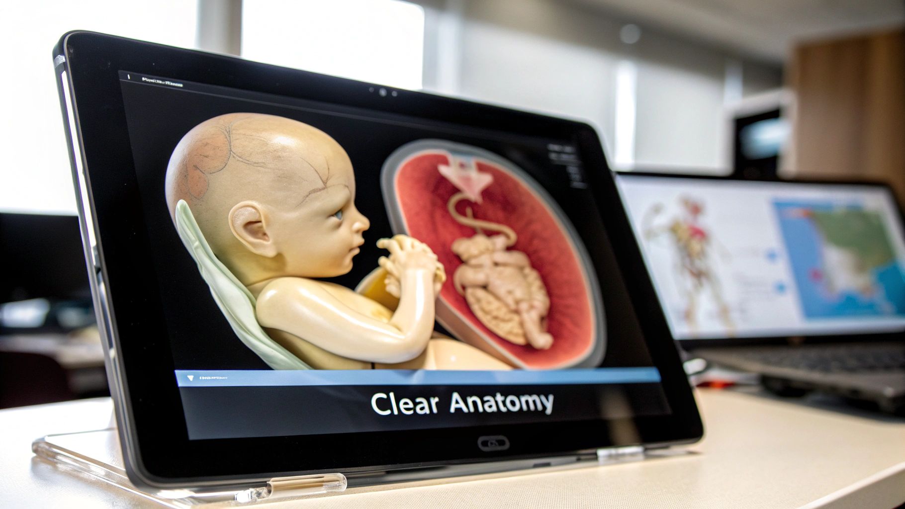
But it’s not just about the clinical side. This technology creates a powerful human connection. Its most famous use is in obstetrics, where it gives expectant parents their first realistic look at their child. Seeing the baby’s distinct facial features or watching their tiny movements forges a strong parental bond long before birth. It turns an abstract idea into a tangible, moving reality.
This is the real value of a 3D ultrasound. It’s not just about getting better pictures; it’s about gaining a deeper understanding, improving medical outcomes, and fostering a more profound connection to the life developing within. Moving from flat slices to dynamic models is fundamentally changing how we see inside the human body.
How 3D Ultrasound Technology Actually Works
To really get what makes a 3D ultrasound so special, it helps to first think about a regular 2D ultrasound. A standard ultrasound probe sends out high-frequency sound waves and then "listens" for the echoes that bounce back. This creates a flat, black-and-white picture—kind of like looking at a single, thin slice of a loaf of bread.
That one slice is incredibly useful, but it's still just a single, flat perspective. 3D ultrasound takes that basic idea and builds on it in a big way. Instead of capturing just one slice, the machine quickly grabs hundreds, or even thousands, of them from slightly different angles.
Then, powerful software stitches all those individual 2D slices together. This is a process called volume rendering, and it essentially reconstructs all that data into a complete, three-dimensional model. You can then rotate it, look at it from any angle, and truly understand the structure.
The Art of Capturing Slices
To build that detailed 3D image, the sonographer needs a way to collect all those 2D slices in an organized fashion. There are a few different ways they can capture this "volume" of data, and the best technique often depends on what they’re looking for.
-
The Freehand Technique: This is the most straightforward method. The sonographer simply sweeps the ultrasound probe (the transducer) over the area in a smooth, steady motion. A position sensor keeps track of the probe's movement, which tells the software exactly how to stack each image correctly.
-
The Mechanical Transducer: Some probes have a small motor inside that automatically sweeps the sound wave crystals back and forth. This captures all the necessary slices in a consistent, automated sequence, taking the guesswork out of a manual sweep.
-
The Matrix Array Transducer: This is the most advanced approach. These probes have thousands of tiny, individual ultrasound elements that can be controlled electronically. They can "steer" the ultrasound beam to capture a whole pyramid-shaped volume of data almost instantly, all without the probe needing to physically move.
From Sound Waves to Lifelike Images
Once all that raw data is collected, the real magic happens inside the computer. The ultrasound machine’s software gets to work, processing the massive dataset to build the final 3D model. It intelligently assigns different levels of shading and transparency to tissues based on how strongly they echo, which is what creates the depth and realistic look.
This isn't just about creating a cool picture; it's a game-changer for diagnostics. Being able to see anatomy in three dimensions helps doctors spot problems and make more accurate diagnoses for everything from heart conditions to cancer.
This clinical impact is driving huge growth. The 3D ultrasound market was valued at around USD 3.7 billion and is expected to hit USD 5.84 billion by 2030. A big reason for this is its effectiveness in diagnosing noncommunicable diseases, which are responsible for roughly 74% of all deaths globally. You can find more insights about the growing 3D ultrasound market on Polaris Market Research.
The end result is a dynamic, interactive image that tells a much richer story than a flat photo ever could. Doctors can digitally slice through the model, peer behind organs, and measure volumes with incredible accuracy. It’s a remarkable process that turns simple sound waves into a clear window inside the human body.
Comparing 2D, 3D, and 4D Ultrasound Technology
To really get a feel for what 3D ultrasound brings to the table, it helps to see how it stacks up against its technological siblings. While 2D, 3D, and 4D ultrasounds all run on the same basic principle—using sound waves to see inside the body—the view they provide is worlds apart. It's a bit like the evolution of photography.
A standard 2D ultrasound is your classic black-and-white photo. It gives you a flat, single-slice image of what’s going on inside. This is the trusted workhorse of medical imaging, perfect for taking routine measurements of a baby or checking the basic shape of an organ. It's a real-time, cross-sectional view that doctors rely on every day for essential diagnostics.
Stepping Up to 3D Imaging
Now, a 3D ultrasound is where things get interesting. It takes a whole series of those 2D slices and digitally stitches them together to create a static, three-dimensional model. Think of it like taking a stack of those flat photos and assembling them into a complete sculpture. This process gives clinicians a much more holistic view, letting them rotate and examine organs or a developing fetus from virtually any angle.
The jump in detail from 2D is substantial. This graphic breaks down some of the key improvements that come with these more advanced imaging techniques.
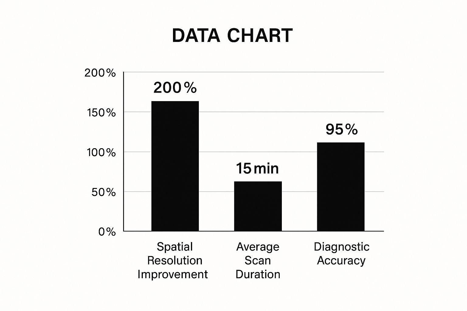
As you can see, the newer technology delivers a huge boost in resolution and diagnostic accuracy without making scan times impossibly long. Finding that sweet spot is exactly why it’s becoming so common in clinics everywhere.
Adding the Dimension of Time with 4D
And then there's 4D ultrasound. This takes that detailed 3D model and adds the fourth dimension: time. Suddenly, your sculpture isn't just a static object—it's a live video of that sculpture moving. The machine captures 3D images one after another in quick succession, creating a real-time movie.
In short, 4D is just "3D in motion." This is an incredibly powerful tool for watching processes as they happen, like seeing a baby yawn in the womb or checking how well a heart valve is working.
Being able to see actual movement gives doctors diagnostic clues that a still image just can't provide. It lets them assess not just the structure of an organ, but its function, which is a game-changer in specialties like cardiology and fetal medicine.
To put it all into perspective, this table lays out the key differences in a straightforward way.
Ultrasound Technology Comparison: 2D vs. 3D vs. 4D
Here’s a side-by-side look at how these three imaging modalities compare, from what they produce to how they are used.
| Feature | 2D Ultrasound | 3D Ultrasound | 4D Ultrasound |
|---|---|---|---|
| Image Output | A flat, cross-sectional image, like a single slice. | A static, three-dimensional model you can view from all angles. | A live, moving 3D video, showing action in real-time. |
| Primary Use | Routine check-ups, standard measurements, and basic structural reviews. | Detailed anatomical assessments and diagnosing complex conditions. | Observing movement and evaluating how organs are functioning. |
| Patient Experience | Provides a basic, outlined view of internal anatomy. | Offers a clear, lifelike still image—great for seeing a baby's face. | Delivers a "live stream" video, often creating a strong bonding moment. |
Each type of ultrasound has its own important job. While 2D is still the go-to for many routine exams, 3D offers critical depth for complex anatomical questions, and 4D adds that vital layer of functional analysis. The right choice always comes down to what the doctor needs to see, with each technology offering its own unique and powerful window into the body.
Clinical Applications Transforming Patient Care
The theory behind ultrasound in 3D is fascinating, but its real story unfolds in the exam room. When this technology moves from the lab to the clinic, its ability to build detailed, volumetric models opens up entirely new ways for doctors to diagnose problems and map out treatments.
Obstetrics is where most people have seen 3D ultrasound in action. It gives physicians an astonishingly clear view of a developing fetus, letting them evaluate anatomical structures with a level of precision that was once unimaginable. This is a game-changer for spotting potential issues like cleft lip, spina bifida, or skeletal problems much earlier and more reliably than 2D imaging ever could.
But it’s not just about diagnostics. These incredibly lifelike images forge a powerful emotional connection. For expectant parents, seeing their baby’s face and watching them move turns an abstract idea into a real, living person, strengthening that parental bond long before birth.
Enhancing Cardiac Diagnostics
In cardiology, 3D echocardiography has become indispensable for looking at the heart's intricate machinery. A standard 2D echo gives you flat, disconnected slices. Trying to understand the whole heart from those is like trying to grasp how a car engine works by looking at separate, flat blueprints.
A 3D echo, on the other hand, constructs a dynamic, moving model of the entire organ. This gives cardiologists the whole picture, allowing them to:
- Measure heart chamber volume with far greater accuracy, which is vital for judging how well the heart is functioning.
- Inspect the delicate leaflets of heart valves to find leaks or blockages that are easy to miss on a 2D scan.
- Guide surgeons in real-time during tricky procedures like valve repairs, providing a live anatomical roadmap.
This deeper view gives doctors the confidence to diagnose conditions like congenital heart defects and valvular disease with much more certainty.
The ability to see the heart in three dimensions removes much of the guesswork. It allows for precise measurements and a clear understanding of spatial relationships, leading directly to better-informed treatment decisions and improved patient outcomes.
The rapid adoption of this technology is fueling major market growth. The global 3D ultrasound market was valued at around USD 4.25 billion and is expected to hit USD 11.43 billion by 2034, growing at a compound annual rate of 10.40%. You can dig deeper into the numbers on the 3D ultrasound market's impressive growth over at Precedence Research.
Expanding into New Medical Frontiers
The reach of ultrasound in 3D goes well beyond babies and hearts. Its applications are constantly growing, offering critical insights across a whole host of medical fields.
Take gynecology, for example. It provides a much better way to evaluate the uterus, letting doctors accurately spot and measure fibroids, check for congenital abnormalities, and get to the bottom of infertility issues with a clarity 2D imaging just can't provide.
And it’s making a real difference in other key areas, too:
- Oncology: Doctors use it to get a precise measurement of a tumor's volume and see how it’s responding to treatments like chemotherapy. This helps tailor cancer care to the individual.
- Musculoskeletal Imaging: It gives sharp, detailed views of joints, tendons, and ligaments, making it easier to diagnose complex injuries like rotator cuff tears.
- Regional Anesthesia: Anesthesiologists rely on 3D imaging to guide needles with pinpoint accuracy when performing nerve blocks, making the procedure safer and more effective.
The story is the same across the board. By offering a more complete and intuitive look at our anatomy, 3D ultrasound helps medical professionals see beyond flat pictures, make smarter diagnoses, and ultimately, provide a higher standard of care.
The Rise of Portable and Handheld 3D Ultrasound
For decades, medical imaging meant being tethered to large, stationary machines tucked away in specialized hospital rooms. That entire model is being turned on its head by one of the biggest moves in modern medicine: the shrinking of ultrasound in 3D technology. We're seeing a massive shift from bulky, cart-based systems to powerful devices that can literally fit in a pocket, bringing diagnostics right to the patient, no matter where they are.
This push for portability is knocking down the physical walls that once kept advanced imaging out of reach. It gives clinicians the power to make critical assessments at the point of care—whether that’s in a chaotic ER, a remote rural clinic, or right at a patient's bedside.
Putting Imaging Power in Every Pocket
The latest handheld 3D ultrasound scanners are a product of incredible jumps in processing power and wireless tech. These new devices connect right to a smartphone or tablet, which acts as the high-resolution screen. This not only makes the hardware tiny but also slashes the cost, opening the door for more people to access advanced imaging.
This new accessibility is completely changing who can perform scans. It’s no longer just the domain of radiologists and sonographers. Now, paramedics, general practitioners, and nurses can grab a device and get the quick, essential images they need to make life-saving decisions on the spot.
Think of it this way: we're essentially putting the diagnostic capability of an entire hospital department into the hands of frontline medical staff. That kind of speed can be the deciding factor in emergencies like trauma care or a sudden cardiac event.
This practical advantage is driving major market trends. Handheld 3D ultrasound devices are carving out a serious space in the global market because they’re portable, user-friendly, and affordable. These small scanners let medical pros perform point-of-care scans everywhere—in hospitals, ambulances, during home visits, and even in remote areas with poor infrastructure. You can learn more about this trend by exploring the growth of the 3D ultrasound market.
The Real-World Impact of Portability
The benefits of portable ultrasound in 3D are showing up in countless real-world medical situations, changing how healthcare is delivered.
- In the Emergency Room: A doctor can instantly check for internal bleeding in a trauma patient, no waiting for a trip down to radiology. This gets a diagnosis faster and treatment started sooner.
- In Remote Villages: A community health worker can perform prenatal scans on expectant mothers who can't get to a hospital, flagging high-risk pregnancies that need immediate attention.
- At the Bedside: A physician can do a quick scan to check for fluid around a patient's heart or lungs, getting an answer in minutes for something that might have otherwise taken hours to diagnose.
This technology is more than just a convenience; it's a fundamental shift in the medical workflow. By bringing top-tier imaging directly to the patient, portable 3D ultrasound cuts down on dangerous delays, lowers healthcare costs, and gives clinicians the tools to provide faster, better care exactly when and where it’s needed most. It’s a small device making a huge difference in global health.
The Future of Imaging AI and Robotics

Looking ahead, the combination of ultrasound in 3D, artificial intelligence (AI), and robotics isn't just about small tweaks to how we see inside the body. It’s about building a fundamentally smarter, more precise, and automated way to diagnose disease. We're moving toward diagnostic systems that can learn on their own, assist clinicians in real-time, and perform certain tasks with a consistency that’s frankly beyond human capability.
At the center of this evolution are AI algorithms. These powerful systems are being fed enormous datasets of medical scans, teaching them to spot subtle patterns, flag anomalies, and take measurements with incredible accuracy and speed. This doesn't make sonographers obsolete; it frees them from tedious, repetitive work so they can focus their expertise on the toughest parts of the diagnosis.
Intelligent Image Enhancement and Automation
One of the first things you'll notice with AI integration is how much better the ultrasound in 3D images look. AI models can instantly clean up the "speckle," that grainy noise inherent in ultrasound, and sharpen the edges of anatomical structures. The result is a much clearer, more confident view for the clinician.
But AI’s role goes far beyond just cleaning up pictures. It’s starting to automate tasks that are critical but often time-consuming. Think of it as a brilliant co-pilot for the sonographer.
- Automated Measurements: Instead of manually clicking to measure a fetal head or a heart chamber, the AI can find the landmarks on its own and calculate the dimensions instantly and accurately.
- Anomaly Detection: Having analyzed thousands of "normal" scans, the AI can highlight areas that look unusual, acting as a second pair of eyes to catch things a human operator might miss during a long session.
- Workflow Optimization: Smart systems can even guide a newer technician through a complex scan, prompting them to capture the right views and angles, which ensures a high-quality study every time.
This automation isn’t about replacing the expert in the room. It’s about augmenting their skills. By offloading the routine work, AI minimizes the chance of human error and helps standardize a higher level of care for every patient.
The role of artificial intelligence is expanding rapidly, especially when it comes to diagnostics. To get a better sense of where this is all heading, it's worth exploring the different agentic AI use cases in healthcare for diagnostics.
The Rise of Robotic Precision
The next leap forward is connecting this powerful AI to robotic hardware. Robotic ultrasound systems use an automated arm to position and move the probe with perfect stability. This directly addresses one of the biggest variables in ultrasound: the skill and steadiness of the operator. A robotic arm can hold a probe perfectly still for long periods or execute a sweep with a smoothness that even the most experienced sonographer can't replicate every single time.
This opens up some incredible new possibilities, especially for remote and underserved areas. Imagine a top specialist in New York City remotely controlling a robotic ultrasound scanner to examine a patient in a small rural clinic hundreds of miles away. It's a way to bring world-class expertise to anyone, anywhere.
Fusion Imaging and Immersive Realities
The future isn't just about one technology; it's about blending them. Fusion imaging is a great example, where a live 3D ultrasound feed is overlaid directly onto a patient's pre-op MRI or CT scan. For a surgeon, this is like having a real-time GPS, combining the live, dynamic view of ultrasound with the detailed anatomical map from another scan.
And it gets even more interesting. Technologies like virtual and augmented reality (VR/AR) are changing how doctors interact with this data. Soon, a surgeon could put on a headset and virtually "step inside" a 3D model of a patient's heart to plan their approach. Or, an AR display could project that same 3D ultrasound image right onto the patient's body during a procedure, guiding the surgeon's hands with pinpoint accuracy. This convergence of AI, robotics, and immersive tech is pushing medical imaging into a new era—one that is smarter and more connected than we ever thought possible.
Common Questions About 3D Ultrasound
It's one thing to understand the tech, but it's another to know what it means for you or a patient. Let's dive into some of the questions we hear all the time about getting an ultrasound in 3D.
Is 3D Ultrasound Safe?
This is always the first and most important question, and the answer is a confident yes. A 3D ultrasound is just as safe as the standard 2D scans that have been used for decades. It works on the exact same principle: high-frequency sound waves, not radiation.
Unlike an X-ray or CT scan, there's no exposure to ionizing radiation. Major medical bodies worldwide have studied ultrasound extensively and found no evidence of harm to a mother or her developing baby. It remains one of the most trusted and safest tools in medical imaging.
What Is the Main Purpose of a 3D Scan?
Those amazing baby pictures are a fantastic bonus, but they aren't the primary reason doctors order these scans. The real power of 3D ultrasound is its diagnostic capability. It gives doctors a volumetric, multi-dimensional view that can reveal issues a flat, 2D image might hide.
Clinically, these scans are ordered to:
- Diagnose Fetal Abnormalities: Getting a clear look at conditions like a cleft lip or spina bifida is much easier with a three-dimensional view.
- Examine Complex Organs: Doctors can better assess the structure of the heart, uterus, or other organs for potential problems.
- Assess Tumor Volume: In oncology, a 3D scan helps accurately measure a tumor and track how it's responding to treatment.
The main event is always a more precise diagnosis; the lifelike images are just a happy side effect.
When Is the Best Time for a 3D Ultrasound in Pregnancy?
If you’re an expecting parent hoping for those clear, detailed pictures of your baby’s face, timing is everything. For the best keepsake photos, the sweet spot is usually between 26 and 32 weeks into the pregnancy.
Why then? By this stage, your baby has developed a bit of fat under their skin, filling out their facial features. There’s also still plenty of amniotic fluid around them, which helps the sound waves create a crisp, clear image.
Go too early, and they might be too small to see clearly. Wait too late, and they might be too snuggled into the womb to get an unobstructed view.
Will My Baby Look Exactly Like the 3D Ultrasound Image?
The images you get from a 3D ultrasound are an incredible preview, but think of them as a very realistic sketch rather than a perfect photograph. The final image quality really depends on the baby’s cooperation! Their position, the amount of fluid, and even where the placenta is can all affect the final picture.
You'll definitely see your baby's features, but their true appearance at birth will still be a wonderful surprise.
At PYCAD, we're focused on what's next for medical imaging. We build advanced AI that enhances diagnostic accuracy and simplifies workflows for medical device companies and healthcare innovators. See how our expertise can advance your technology at https://pycad.co.


