For years, doctors had to piece together a mental picture of what was happening inside a patient using flat, two-dimensional images. Think of it like trying to understand the intricate design of a complex machine by only looking at its blueprint. 3D medical imaging changes everything, taking those flat slices and building them into a complete, interactive model you can turn, explore, and analyze from any angle.
It’s the difference between looking at a photograph of a mountain and being able to walk around a detailed sculpture of it. This leap gives clinicians a level of depth and clarity that was once unimaginable.
A New Dimension in Healthcare
Traditional tools like X-rays are incredibly valuable, but they have inherent limitations. On a flat image, organs and tissues are layered on top of each other, which can obscure critical details or create a confusing picture of how structures relate to one another. It's like trying to navigate a city using only its shadow.
3D medical imaging overcomes this challenge by taking hundreds, sometimes thousands, of individual 2D "slices" and computationally stacking them. The result is a complete, volumetric model that allows a surgeon to virtually fly through a patient's arteries or a radiologist to precisely measure a tumor without any guesswork. This enhanced view directly translates to better, safer patient care.
The Foundation of Modern Diagnostics
This powerful visualization is made possible by several core imaging technologies. Each one offers a unique perspective on the human body, acting as the engine that captures the raw data needed to build these sophisticated digital models.
- Computed Tomography (CT): The go-to for seeing dense structures. It provides incredible detail of bones, organs, and blood vessels.
- Magnetic Resonance Imaging (MRI): The gold standard for soft tissues. It gives an unparalleled look at the brain, muscles, ligaments, and tendons.
- 3D Ultrasound: Offers the magic of real-time imaging without any radiation, most famously used to watch a fetus grow and move.
- Positron Emission Tomography (PET): Goes beyond structure to show function. It highlights metabolic activity, making it essential for finding active diseases like cancer.
The medical community's growing reliance on these tools is driving massive market growth. The global 3D imaging market was valued at roughly $41.96 billion in 2024 and is expected to climb to around $115.98 billion by 2030. This boom, detailed in a 3D imaging market forecast from Grand View Research, shows just how integral this technology has become.
By turning flat data into a dynamic, explorable map of the human body, 3D medical imaging gives clinicians the context they need. This perspective is vital for making the right diagnosis, planning a complex surgery, and creating treatment plans that are truly tailored to the individual.
In this guide, we'll dive deeper into each of these technologies, see how artificial intelligence is making them even smarter, and explore the real-world applications that are changing the face of medicine.
The Core Technologies Behind 3D Medical Imaging
At the heart of any 3D medical image is a sophisticated technology designed to capture specific information about the body. The best way to think about these technologies is to see them as different kinds of cameras, each built for a unique job. One might be perfect for capturing fast motion, while another excels at seeing in the dark.
In the same way, the core tools of 3D medical imaging—CT, MRI, 3D Ultrasound, and PET—each give us a completely different window into the human body. Understanding what makes each of them tick is the first step to seeing how these incredible three-dimensional models are built and why they’ve become so vital in medicine.
Comparison of Key 3D Medical Imaging Modalities
To get a clearer picture, it helps to see these technologies side-by-side. The table below breaks down the four main imaging types, explaining in simple terms how they work and where they shine in a clinical setting.
| Modality | How It Works (Simplified) | Best For Visualizing | Common Applications |
|---|---|---|---|
| CT | Stacks hundreds of thin X-ray "slices" to build a 3D model. | Dense structures like bone, organs, and blood vessels. | Trauma, complex fractures, tumor detection, stroke diagnosis. |
| MRI | Uses a strong magnet and radio waves to "listen" to water atoms in the body. | Soft tissues with incredible detail: brain, nerves, muscles, ligaments. | Brain tumors, spinal cord injuries, torn ligaments, joint damage. |
| 3D Ultrasound | Captures a volume of data using high-frequency sound waves. | Real-time movement and surface structures. | Fetal imaging (obstetrics), heart function (cardiology), guiding procedures. |
| PET | A radioactive tracer highlights areas of high metabolic activity. | Biological function, not just structure. | Cancer detection and staging, assessing treatment response, brain disorders. |
As you can see, these methods aren't in competition with one another; they're complementary. Each provides a unique piece of the diagnostic puzzle that the others can't see.
Computed Tomography (CT): Slicing the Body Like Bread
A Computed Tomography (CT) scan is one of the most widely used methods for creating detailed 3D models. The easiest way to picture how it works is to imagine a loaf of bread. The scanner takes hundreds of thin, cross-sectional X-ray images, almost as if it's slicing the loaf into paper-thin pieces.
Specialized software then digitally stacks these 2D slices back together, reconstructing the entire "loaf" into a complete 3D object. This process gives doctors a remarkably detailed view of hard, dense structures inside the body.
CT scans are the gold standard for visualizing:
- Bone: Spotting complex fractures, checking on joint damage, and meticulously planning orthopedic surgeries.
- Organs: Detecting tumors, abscesses, or internal injuries in organs like the liver, kidneys, and lungs.
- Blood Vessels: Identifying blockages or dangerous aneurysms in arteries, often with the help of a contrast dye.
Because a CT scan is incredibly fast—often taking just a few minutes—it’s indispensable in emergency rooms for things like evaluating trauma patients or diagnosing a stroke.
The infographic below shows how each of these imaging techniques is suited for looking at different parts of the body.
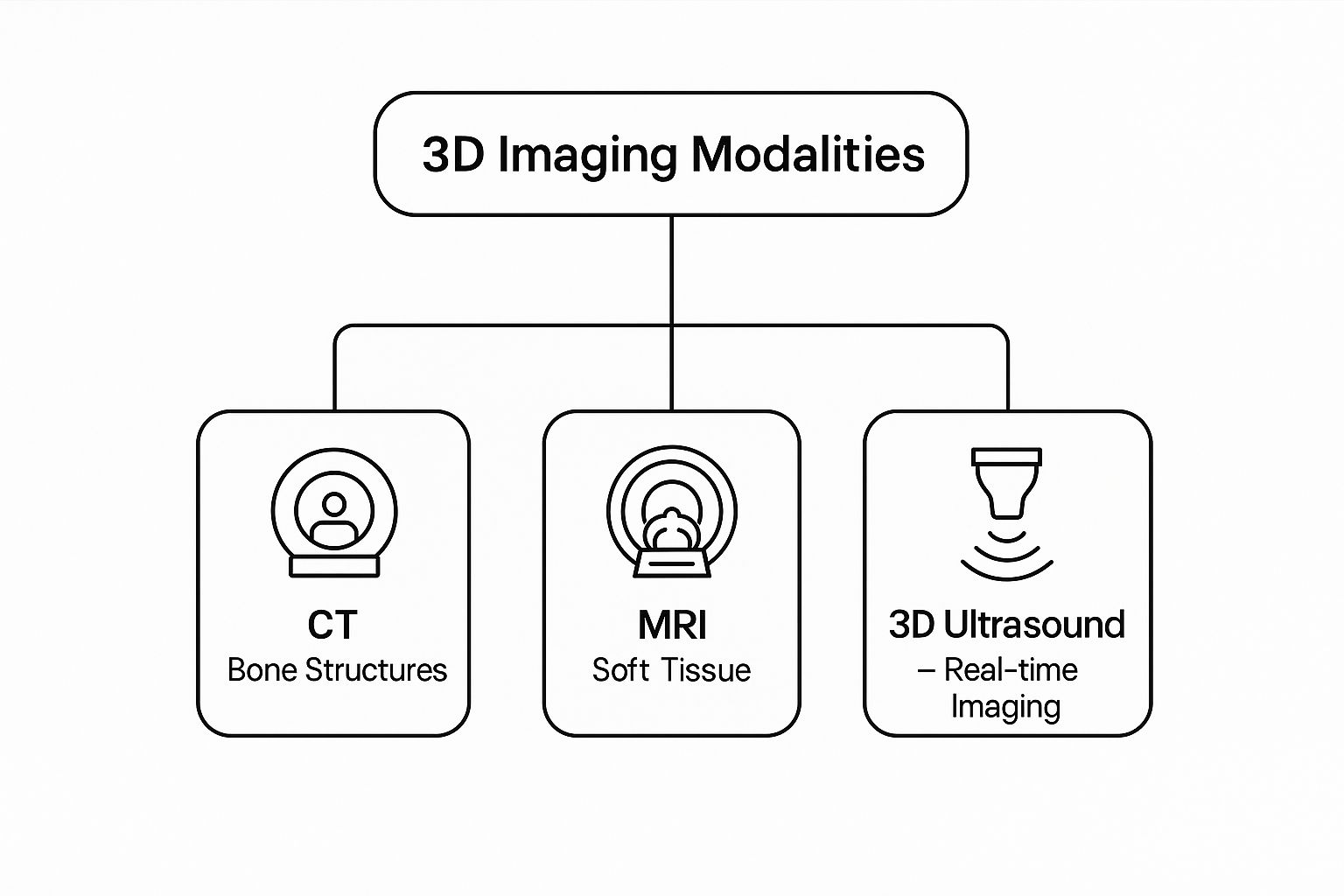
This really drives home the complementary nature of these technologies. Each one has its own special strength, giving doctors the right tool for the right diagnostic job.
Magnetic Resonance Imaging (MRI): Listening to the Body’s Atoms
If CT is like slicing bread, then Magnetic Resonance Imaging (MRI) is more like listening to the unique song sung by the body’s atoms. Instead of X-rays, an MRI uses a powerful magnetic field and radio waves to get the hydrogen atoms in your body's water molecules to "talk."
Different tissues—muscle, fat, cartilage, nerves—all have different amounts of water and respond to the magnetic field in their own distinct way. The MRI scanner picks up on these subtle signals and translates them into images with stunning detail.
An MRI provides exceptional soft-tissue contrast, revealing anatomical details that are often invisible on a CT scan. This makes it an indispensable tool for neurology, sports medicine, and oncology.
This incredible ability makes MRI the top choice for examining:
- The Brain and Spinal Cord: Finding tumors, evidence of a stroke, multiple sclerosis, or herniated discs.
- Joints and Muscles: Visualizing torn ligaments, cartilage damage, and other soft-tissue injuries with amazing clarity.
- Internal Organs: Assessing organs like the heart and liver without using any ionizing radiation.
3D Ultrasound: Capturing Motion in Real Time
3D Ultrasound takes the familiar 2D ultrasound we all know and literally adds another dimension. It doesn't use radiation or magnets; instead, it relies on high-frequency sound waves that bounce off internal structures to create images.
Think of a standard ultrasound as a single snapshot. A 3D ultrasound, on the other hand, captures an entire volume of data, which can then be displayed as a still 3D image. When you take it a step further, you get 4D ultrasound, which adds time to the mix to create a moving, real-time 3D video.
This technology is famous for producing those lifelike images of a fetus in the womb. But it's also incredibly valuable in cardiology for watching the heart and its valves beat, and for guiding surgeons during minimally invasive procedures.
Positron Emission Tomography (PET): Visualizing How the Body Works
While CT and MRI are masters at showing anatomical structure, Positron Emission Tomography (PET) is all about revealing function. It uncovers the metabolic activity humming away inside the body, highlighting areas where cells are burning through energy faster than normal.
Before a PET scan, a patient is injected with a safe, radioactive tracer. This tracer moves through the bloodstream and gathers in areas with high metabolic rates, like rapidly dividing cancer cells. The PET scanner detects the energy given off by this tracer, creating a map of biological activity.
PET scans are often combined with CT or MRI scans (in what's called a PET-CT or PET-MRI) to overlay the functional data onto a detailed anatomical map. This fusion gives doctors the full story, showing not just where a tumor is, but also how active it is. This is crucial for diagnosing cancer, planning treatments, and checking to see if those treatments are actually working.
How AI Is Supercharging 3D Medical Imaging
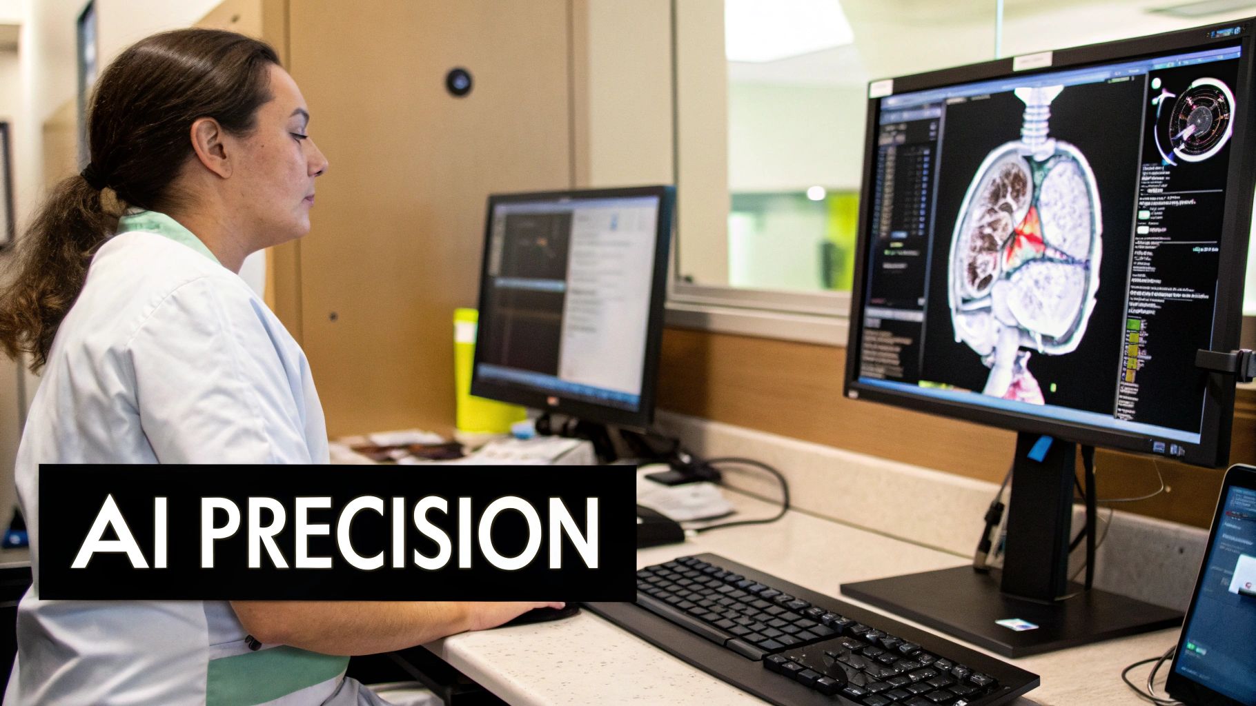
If you think of 3D medical imaging as a powerful new camera, then Artificial Intelligence (AI) is the genius photo editor that works at superhuman speed. It sifts through the massive datasets from these scans, finding patterns that are either too small or too complex for the human eye to catch on its own.
This isn’t about replacing doctors. Far from it. The goal is to give clinicians a set of incredibly powerful tools. Think of it as an expert assistant that can review an entire 3D scan in seconds, highlighting the exact spots that need a closer look from a human expert. This frees up clinicians to focus on what they do best: patient diagnosis and treatment planning.
Automated Segmentation and Analysis
One of the most practical ways AI helps today is with automated segmentation. This is just a technical term for outlining specific organs, tumors, or other structures inside a 3D scan. Done by hand, it’s a slow, meticulous process that can take a radiologist hours.
AI models, however, can be trained on thousands of existing scans to do this job in minutes, and with impressive accuracy. This is a game-changer for things like radiation therapy, where a treatment's success depends on precisely targeting a tumor while leaving healthy tissue untouched. The practical deployment of AI in these critical areas often involves complex frameworks like Agentic AI and Human-in-the-Loop systems to ensure every step is safe and reliable.
By automating segmentation, AI not only frees up countless clinical hours but also brings a new level of consistency to the process. This leads to more standardized, reliable, and effective treatment plans.
Enhancing Image Reconstruction and Clarity
AI is also making the raw 3D images themselves much better. Scans can sometimes have visual "noise" or artifacts that make it hard to see fine details, especially when using faster scan times or lower radiation doses. AI algorithms are fantastic at cleaning up this noise, producing images that are sharper and clearer.
This process, called enhanced image reconstruction, has some huge real-world benefits. It lets clinicians:
- Get clear images from lower radiation doses, which is a big win for patient safety, particularly with CT scans.
- Reduce scan times for patients, since the AI can build a detailed model from less raw data.
- Sharpen the edges between different types of tissue, making it easier to spot even tiny abnormalities.
Essentially, doctors get a much better look inside the body, while the patient often gets a faster and safer experience. This is a field that’s moving fast, with companies like GE Healthcare and Siemens Healthineers pushing the technology forward all the time. While the entire medical imaging market is huge, it’s 3D medical imaging that is really fueling this progress, as recent market reports show. You can explore more insights on the 3D medical imaging market here to see the trends.
Predictive Diagnostics From Hidden Patterns
This is where things get really exciting. AI is starting to move beyond just seeing what's there and into the realm of predictive diagnostics. Algorithms can be trained to find subtle textures and patterns in 3D scans that are invisible to us but actually correlate with future disease.
For example, an AI could analyze a 3D scan of a small lung nodule and, based on its unique texture and shape features, predict how likely it is to become cancerous. In another case, it might analyze a brain scan to forecast the probable progression of a neurodegenerative disease.
This fundamentally shifts medicine from being reactive to proactive. By using 3d medical imaging to identify high-risk patients long before symptoms appear, AI gives clinicians the chance to step in sooner, potentially changing the course of a disease and leading to much better outcomes.
Putting 3D Imaging to Work in the Clinic

Technology is only as good as its real-world impact. When we talk about 3D medical imaging, its true power isn't in the pixels or processing speeds—it’s measured by how it helps doctors save and improve lives. In the hands of a skilled clinician, these detailed models become a roadmap to the human body, fundamentally changing how they tackle the toughest medical challenges.
From the operating theater to the cancer ward, these dynamic, three-dimensional views provide a level of clarity that flat, 2D scans just can't match. It’s like switching from a paper map to a fully interactive GPS, giving medical teams the confidence to plan, practice, and perform with incredible precision.
Precision in Surgical Planning
Think about a neurosurgeon tasked with removing a tumor deep inside a patient's brain. With old-school 2D scans, they have to mentally build a 3D picture of the tumor's location, trying to guess its relationship to critical blood vessels and delicate tissue. It’s an incredibly high-stakes process that leaves a lot to the imagination.
Now, imagine that same surgeon with a detailed 3D model spun from the patient’s MRI data. They can virtually fly through the brain, rotating the model to inspect the tumor from every possible angle. This digital dress rehearsal lets them map out the safest surgical path and spot potential roadblocks long before making the first cut.
This ability to practice a complex surgery on a digital twin of the patient is a massive leap forward for safety. It drastically reduces surprises in the operating room, which often leads to shorter procedures, less blood loss, and a smoother recovery.
And this isn't just for neurosurgery. This approach is quickly becoming the standard of care across many different surgical fields.
Enhancing Cancer Treatment in Oncology
When fighting cancer, precision is everything. The entire goal is to obliterate cancerous cells while doing as little harm as possible to healthy tissue. This is where the functional detail from 3D PET scans becomes a game-changer.
A 3D PET scan doesn't just show a tumor's size and shape; it reveals its metabolic activity, basically lighting up a "heat map" of the most aggressive cancer cells. When doctors fuse this functional data with a detailed anatomical 3D CT scan, they get the complete story.
This comprehensive view allows for incredibly precise radiation therapy. Oncologists can literally shape the radiation beams to match the tumor's exact 3D contours, delivering a powerful, targeted dose right where it's needed while steering clear of healthy organs. This focused approach is key to reducing the grueling side effects that often come with cancer treatment.
Innovations in Cardiology and Orthopedics
The influence of 3D imaging is rippling through countless other specialties, reshaping how doctors treat some of the most common and challenging health problems.
Cardiology: When a patient needs a heart valve replacement, a 3D model of their heart built from a CT scan helps the surgical team pick the perfect-sized prosthetic valve before the procedure even begins. They can also map out the best insertion route—a huge advantage in minimally invasive surgeries where the surgeon's view is limited.
Orthopedics: This field has been completely energized by the rise of custom, 3D-printed implants. Surgeons start with high-resolution 3D CT scans to create a perfect digital blueprint of a patient’s damaged joint. That data then fuels the design and printing of a replacement that’s a one-of-a-kind match for that person's anatomy.
This custom-fit approach has major benefits over the old one-size-fits-all implants:
- A much better and more natural fit for the patient.
- Improved joint function and a greater range of motion.
- The implant itself often lasts longer.
Whether it’s crafting a personalized knee replacement or navigating the brain's complex network, these examples make it clear: 3D medical imaging is more than just a fancy visualization tool. It’s a cornerstone of modern, personalized medicine, giving doctors what they need to deliver safer, more effective care for every single patient.
Challenges and the Future of 3D Visualization in Medicine
While the potential of 3D medical imaging is immense, its path into everyday clinical practice isn't always a straight line. Like any powerful technology, there's a gap between what's possible in a lab and what's practical in a busy hospital. A number of real-world hurdles involving cost, training, and infrastructure still need to be cleared.
One of the biggest roadblocks is the sheer cost. MRI and PET/CT scanners are multimillion-dollar machines, an investment that’s simply out of reach for many smaller hospitals and clinics. And the spending doesn't stop there. Specialized software for rendering 3D models adds to the bill, as does the robust digital plumbing needed to store and manage the massive files these scans produce.
Beyond the price tag, there’s a critical human element. Reading a complex 3D model isn't the same as looking at a flat X-ray. It demands a different skillset and specialized training. Radiologists, surgeons, and techs need to get comfortable with new software and visualization techniques to truly tap into the power of this data, which means healthcare systems must invest in ongoing education.
Getting Past the Bottlenecks
These challenges aren't stopping progress, but they do require smart solutions. From wrestling with enormous datasets to ensuring patients everywhere can benefit, the medical community is actively working to smooth the road for broader 3D adoption.
A huge logistical headache is just managing the data. A single 3D scan can easily generate gigabytes of information. For a large medical center, that adds up to petabytes of data in no time, creating a serious bottleneck for storage, security, and sharing. Smart, cloud-based systems are becoming essential to make this data manageable and accessible across different departments or even different hospitals.
There's also the issue of global health disparities. While wealthy countries are installing the latest imaging systems, many low- and middle-income regions lack the funding, infrastructure, and trained staff to do the same. This creates a technology gap. Thankfully, work is underway to develop more affordable and rugged systems built for diverse healthcare environments. You can read the full research on the 3D medical imaging market to see how these dynamics are playing out globally.
"The ultimate goal is to make advanced visualization tools as accessible and user-friendly as a stethoscope. Overcoming the current barriers of cost, training, and data management is the critical next step in that journey."
This push for accessibility is key. It's about ensuring these powerful diagnostic tools become a standard part of care everywhere, not just in major research hubs.
The Next Frontier for 3D Medical Imaging
Looking ahead, the evolution of 3D medical imaging is starting to look like something out of science fiction. The focus is shifting from just creating clearer pictures to interacting with patient data in entirely new ways.
Augmented Reality (AR) integration is one of the most exciting developments on the horizon. Picture a surgeon wearing AR glasses that overlay a patient's 3D organ models directly onto their body during a procedure. This kind of "X-ray vision" would offer unprecedented precision, helping the surgeon navigate tricky anatomy and steer clear of critical blood vessels or nerves with incredible confidence.
Another major trend is the push for smaller, more portable imaging devices. Researchers are working on lower-cost scanners that could bring 3D imaging capabilities into ambulances, local clinics, or remote villages, making high-end diagnostics available where they're needed most.
Finally, the concept of the "digital twin" is set to completely change the game. This involves creating a dynamic, virtual replica of a patient from their 3D imaging data and other health records. With a digital twin, doctors could:
- Simulate a complex surgery multiple times to find the safest and most effective approach.
- Test how a specific patient might react to a new drug before ever prescribing it.
- Predict the likely progression of a disease, allowing for earlier and more proactive intervention.
This isn't just about personalized medicine; it's about building a predictive, proactive model of healthcare, all driven by the ever-growing power of 3D visualization.
A Few Common Questions About 3D Medical Imaging
As we dive into the world of 3D medical imaging, a few practical questions always come up. How does this really compare to what we're used to? Is it safe? What's it actually like to get one of these scans? Let's clear up some of the most common questions.
Is 3D Medical Imaging Safe?
The short answer is: it depends entirely on the type of scan. Each technology works differently, and their safety profiles reflect that.
For instance, MRI and 3D Ultrasound are considered exceptionally safe. Why? Because they don't use any ionizing radiation. An MRI uses a powerful magnet and radio waves, while an ultrasound uses simple sound waves—neither of which harms the body. This makes them perfect for situations that require extra care, like imaging an unborn baby or when a patient needs frequent follow-up scans.
On the other hand, CT and PET scans do rely on a small, controlled amount of ionizing radiation to create their images. This is where a critical safety principle called ALARA comes in, which stands for "As Low As Reasonably Achievable." Every technician and radiologist is trained to use the absolute minimum dose of radiation needed to get a clear, diagnostic-quality image. In almost every case, the benefit of getting a definitive, often life-saving, diagnosis far outweighs the minimal risk from the radiation.
How Is a 3D Scan Different from a Regular X-Ray?
Think of a standard X-ray as a flat shadow puppet. It gives you a single, two-dimensional image where all your internal structures are layered on top of each other. A CT scan is a big step up from that, capturing a series of 2D "slice" images from different angles—like flipping through the individual pages of a book.
3D medical imaging takes all those "pages" and does something remarkable. It uses sophisticated software to digitally stack the 2D slices from a CT or MRI scan and reconstruct them into a complete, interactive three-dimensional model.
It's the difference between looking at a single photo of a car and being able to walk around a full 3D model of it. A doctor can digitally rotate, slice into, and view the anatomy from any angle, revealing crucial spatial relationships between organs, blood vessels, and tumors that are simply invisible in flat images.
What Is the Patient Experience Like During a 3D Imaging Procedure?
Here's the good news: for the patient, getting a 3D scan feels exactly the same as getting a traditional 2D scan. All the "3D" magic happens behind the scenes, in the computer, long after you've left the room.
If you’re having a CT or MRI, you'll lie on a table that slides into a machine shaped like a doughnut or a short tunnel. Your main job is to stay as still as possible to avoid blurring the images. The scan itself is painless, though the tight space of an MRI machine can feel a bit confining, and it makes a lot of loud thumping noises (which is why they almost always offer you headphones).
A 3D ultrasound is identical to a standard one: a technician puts a clear gel on your skin and moves a small wand, called a transducer, over the area. The bottom line is your physical experience doesn't change—the only difference is the incredible depth and detail of the final images produced from the data.
How Long Does It Take to Get Results from a 3D Scan?
Getting your results involves a few steps. First is the scan itself, which can be as quick as 15 minutes for a CT or last over an hour for a complex MRI.
Once the scan is done, the raw data is sent to a powerful computer where specialized software builds the 3D model. With modern equipment, this reconstruction is practically instant.
The final and most important step is the interpretation. A radiologist—a doctor who specializes in reading medical images—carefully reviews everything. They look at both the original 2D slices and the interactive 3D model, searching for any abnormalities. They then write up a detailed report for your doctor. Usually, your physician will have the final report and can discuss the findings with you within a few business days. In an emergency, of course, a preliminary reading can be ready much sooner.
At PYCAD, we’re focused on pushing medical diagnostics into the future. We develop advanced AI solutions that help medical device companies and healthcare providers unlock the full story hidden within their imaging data. The goal is always more accurate diagnoses and better patient outcomes. To see how our expertise in AI and computer vision can support your medical imaging projects, visit us at https://pycad.co.


