
If you sell dental implants, your dentists already plan cases somewhere. Often it is a desktop planning suite bought from another vendor, with another vendor’s implant library and logo on the splash screen. Sometimes it is whatever viewer came on the disc from the imaging center, with no implant library at all.
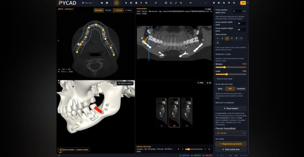
At PYCAD we have a web-based dental DICOM viewer that opens CBCT scans, rebuilds the panoramic from the dental arch, and lets the dentist place implants from a manufacturer’s own STL library. Implant companies can hand that viewer to their dentist network under their own brand.
Who this is for
This page is for implant manufacturers and dental OEM teams who want three things:
- Dentists in their network open a patient’s CBCT and plan with the company’s implants, in a browser.
- The company’s logo and colours sit on the tool the dentist plans in.
- Cases move between the dentist, a colleague, and the company’s clinical support team by share link.
If you are a dentist who wants to look at one scan today, go straight to the dental viewer demo.
What the dentist does in the viewer
Open the CBCT
The dentist opens the scan in the browser, with nothing to install. Axial, coronal, and sagittal views load together, with distance, angle, and density measurements read from the DICOM geometry in millimetres. Large scans are served at server-derived resolution levels, so the viewer stays usable on a tablet or a slow clinic connection.
See the anatomy as 3D meshes
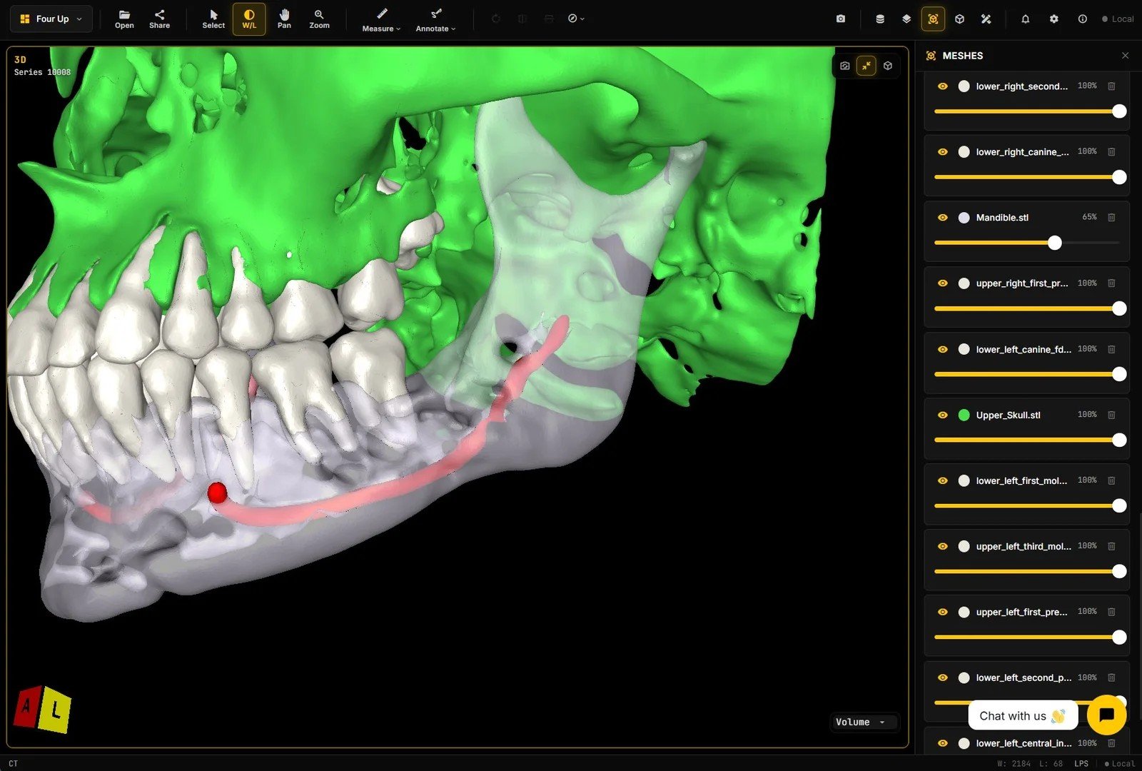
Meshes of the mandible, maxilla, individual teeth, and mandibular canal are generated by applying a segmentation algorithm to the CBCT. They load as STL files on top of the scan. Segmentation masks can also be shown as colour overlays on every plane, with per-structure visibility and opacity.
The canal gets the most attention. On a raw scan it has to be traced slice by slice. As a mesh, the dentist sees its full path through the jaw before choosing a site. We cover the canal overlay in more detail in mandibular canal CBCT to STL in the browser.
Draw the arch and rebuild the panoramic
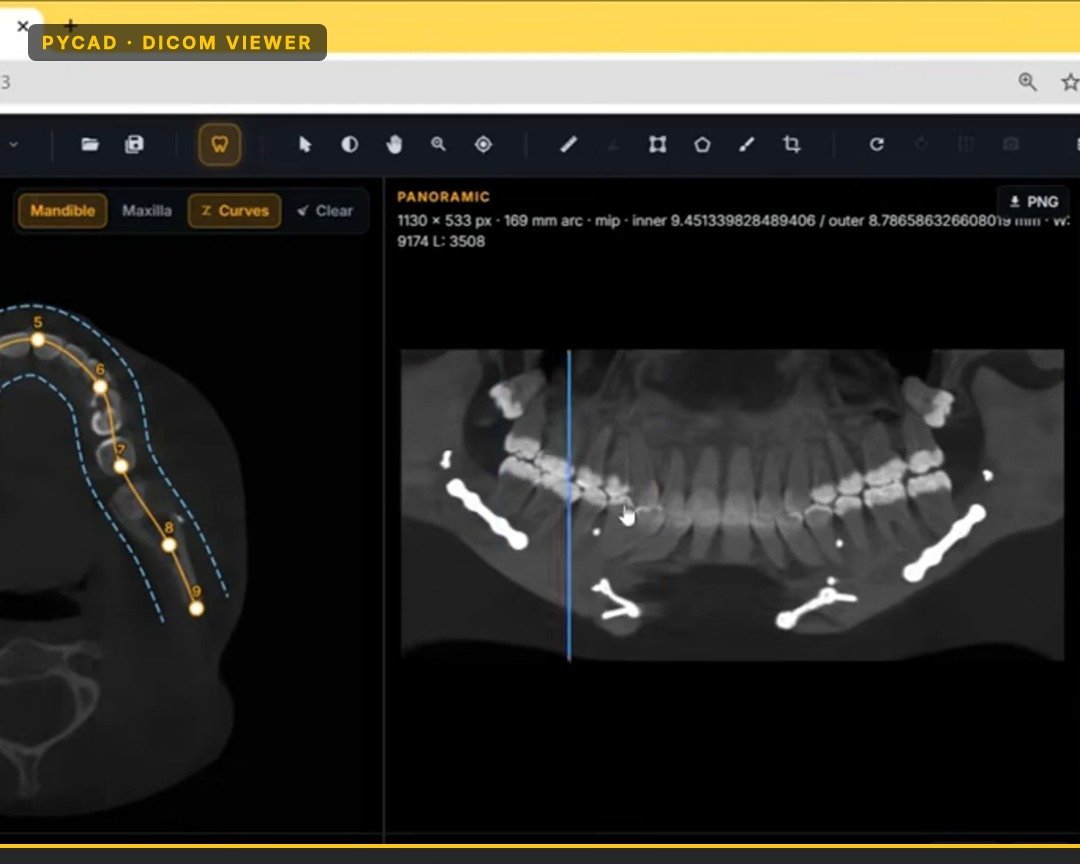
In the panoramic workspace, a curve is drawn along the upper or lower arch. This step can be automated, and the dentist keeps manual control of the spline points and the spacing between the inner and outer curves.
From that curve the viewer rebuilds a panoramic image and shows it next to the axial view and a set of cross-sections. A slider moves the cross-sections along the arch, so the dentist can stop at a tooth site and check bone height and width. The arch, cross-section, and panoramic rebuild post shows this workspace step by step.
Place your implants
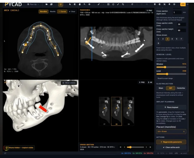
The dentist clicks on the panoramic to drop an implant at the chosen site. The implant comes from your STL library, so the geometry on screen is the geometry you sell. Each SKU (Stock Keeping Unit) in your range, meaning each diameter and length, can appear as its own entry in the dentist’s implant list.
Once placed, the implant shows in 3D and can be moved and angled. As the dentist moves it, or moves the slider, the cross-sections update so the implant can be checked against bone and canal in every plane. Distance measurements show how far the implant sits from the canal.
Share the case by link
When the dentist wants a second opinion, they share a link from a saved session. The second clinician opens the same case in their own browser, reviews the plan, and adds notes. Nobody buys a second desktop seat or copies the scan onto a USB stick for that review.
The same link works for your clinical support team. When a dentist calls about a case, your specialist opens it on their own laptop and sees the plan the dentist made.
Why a branded web viewer works better for your dentist network
Look at how a planning tool reaches your dentists today.
With desktop seats, each dentist needs an install, a licence key, and a computer that meets the specs. Your team fields support tickets about operating system versions, graphics drivers, and Mac users who cannot run the software. Every new implant line means a library update pushed to every seat. When a dentist wants a colleague to look at a case, the colleague needs a seat too.
With disc, USB, and ZIP handoffs, the imaging center burns the CBCT to a disc or sends a ZIP. The dentist hunts for a viewer that opens it. The case is then copied again to reach a colleague or your support team. Your brand appears nowhere in that chain.
A web viewer changes the rollout:
- One URL for your whole network. Dentists open it on the computer or tablet they already have.
- One implant library on the server. When you add a new diameter, every dentist sees it in their next session.
- Your logo and colours on the screen where the dentist plans.
- Share links replace discs for second opinions and support calls.
- Your sales reps can open a demo case on a tablet during a clinic visit.
Film-style animation or an interactive CBCT embed
Some implant companies already pay for film-style medical animation, with a fixed camera path showing an implant going into a generic jaw. That works for a launch video or a congress screen. A dentist watching it cannot turn the jaw, swap the implant size, or load their own patient’s scan.
An interactive CBCT embed lets them do all three. Keep the film for launches and give dentists the viewer for daily cases. We compared the two formats for device marketing in medical device animation vs interactive implant 3D.
Try the viewer
Open a sample CBCT in our dental DICOM viewer demo. It has the MPR views, the curved panoramic, implant placement, measurements, and segmentation overlays. A free account lets you save a session and share a link with a colleague.
Talk to us about a branded viewer for your dentists
If you are an implant manufacturer and want a branded web CBCT viewer for your dentist network, Contact us. Send these three details with your message so the first call covers your setup:
- The implant lines you want in the library, and the STL files you have for them.
- How many dentists you plan to onboard first, and in which countries.
- Who should receive share links: dentist to dentist, dentist to your support team, or both.
We will reply with how the branded viewer, your implant library, and share links would be set up for your network.