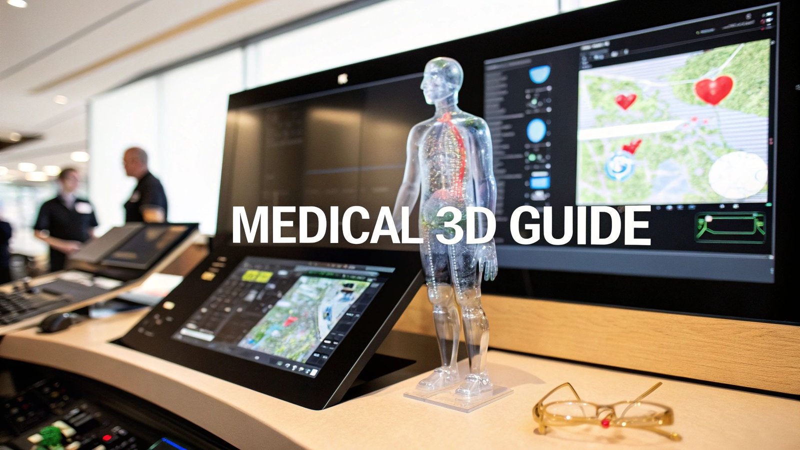
Picture this: a surgeon meticulously rehearsing a complex heart operation on a patient's perfect digital twin. Or an oncologist watching a tumor shrink in three dimensions, tracking its response to treatment in real-time. This isn't a glimpse into the future—it's what 3D visualization software makes possible today. Think of it as a powerful translator, turning flat, grayscale medical scans into vibrant, interactive maps of the human body.
Seeing Medicine in a New Dimension
For decades, clinicians have been masters at interpreting 2D images from CT and MRI scans. They mentally pieced together anatomical structures from a series of flat slices, a skill that takes immense experience—almost like building an intricate sculpture from a stack of blueprints. 3D visualization software does all that heavy lifting for them, instantly creating detailed models that you can understand at a glance.
This leap forward changes everything. It moves diagnosis and treatment planning from a process of interpretation to one of direct interaction. Instead of just looking at images, medical professionals can now grab, rotate, and even virtually dissect anatomical models. It provides a deep, spatial awareness that was simply out of reach before.
The Power of Perspective
The real magic of this technology is how it provides a completely new perspective. By turning raw medical data into something you can see and touch, it unlocks deeper insights and gives clinicians the confidence to make better decisions. This shift makes a huge difference in several key areas:
- Sharper Diagnostic Clarity: Doctors can see the exact size, shape, and location of an abnormality and how it relates to the tissues around it.
- Smarter Surgical Planning: Surgeons can map out entire procedures, anticipate challenges, and choose the perfect tools before ever making an incision.
- Clearer Patient Communication: Imagine explaining a complex condition to a patient using a simple, clear 3D model. It helps them understand their own health, fostering better engagement and informed consent.
At its core, 3D visualization software closes the gap between complex medical data and human understanding. It creates a common visual language for doctors, surgeons, and patients alike.
A Growing Field of Opportunity
This technology is taking off, and not just in medicine. The global visualization and 3D rendering software market was valued at around USD 3.4 billion in 2024. It’s expected to grow at a staggering rate of 30.55% annually through 2030, as industries like architecture, design, and entertainment also find new ways to use it. You can read the full research about this market growth to see just how widespread its impact is.
In this guide, we'll go beyond the technical jargon to show you how 3D visualization is fundamentally reshaping healthcare. We’ll look at how it turns pixels into patients, see how AI is supercharging its capabilities, and explore what it all means for the future of medicine.
Here at PYCAD, we're obsessed with making this advanced technology both accessible and powerful. We at PYCAD, build custom web DICOM viewers and integrate them into medical imaging web platforms. Our work is all about giving healthcare professionals the tools they need to see medicine in a whole new dimension—you can see it for yourself on our portfolio page.
How Software Turns Pixels Into Patients
Have you ever wondered how a series of flat, black-and-white scan images can become a vibrant, interactive 3D model of a patient's anatomy? It’s a bit like a digital sculptor at work. Instead of clay, 3D visualization software takes hundreds, sometimes thousands, of individual 2D image slices from a CT or MRI scan and meticulously pieces them together.
This isn't just about stacking pictures on top of each other. It's a sophisticated process of reconstruction, intelligently assembling raw data into a living digital twin of the patient. This transformation turns abstract pixels into profound clinical insight, giving doctors a perspective on the human body that was once the stuff of science fiction.
This infographic breaks down that core journey, from a simple 2D scan to a dynamic, fully interactive 3D model.
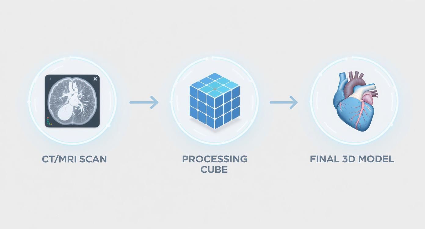
As you can see, the software acts as the critical bridge between the raw scan data and a valuable clinical asset. The real magic, though, happens in the various ways this software can present the final 3D model.
Different Lenses for Deeper Understanding
Once the 3D model is built, the software provides clinicians with a set of "lenses" to view it through. These aren't just for show; they are powerful diagnostic tools designed to highlight specific aspects of the anatomy. Each technique offers a unique perspective, allowing doctors to zero in on the exact tissues or structures they need to see.
Think of it like a photographer using different lighting setups. One light might reveal the fine texture on a surface, while another emphasizes the object's overall shape. In the same way, these rendering techniques illuminate different clinical details inside the body.
Let’s look at a few of the most important ones:
- Multi-Planar Reconstruction (MPR): This is the workhorse technique. It frees clinicians from the standard axial, coronal, and sagittal views, allowing them to slice through the 3D model from any angle imaginable. This provides a complete, custom view that can uncover pathologies hidden from a traditional perspective.
- Maximum Intensity Projection (MIP): This lens is designed to make dense structures pop. It essentially peers through the entire 3D dataset and projects only the brightest pixels forward. This is incredibly valuable in angiography, where it makes contrast-filled blood vessels shine with brilliant clarity against the surrounding tissue.
- Volume Rendering (VR): This is perhaps the most visually stunning technique. Volume Rendering creates a semi-transparent, almost glass-like model of an organ or region. By assigning different colors and opacities to various tissue densities, it allows a doctor to see through skin and bone to view the intricate interplay between organs, vessels, and tumors simultaneously.
These rendering methods are more than just features; they represent different ways of asking questions of the data. They empower clinicians to tailor their view to the specific diagnostic challenge at hand, leading to more confident and accurate conclusions. You can see these techniques in action in our detailed overview of Computed Tomography 3D reconstruction.
Interactivity: The Key to Insight
The real leap forward isn't just seeing a beautiful 3D model—it's being able to interact with it. A static picture is helpful, but an interactive one is a different beast entirely.
Clinicians can grab the model and rotate, pan, and zoom in real time, gaining an unparalleled understanding of spatial relationships. They can digitally peel away layers of tissue to reveal what lies beneath or isolate a single blood vessel to trace its entire path.
This interactivity is what elevates a diagnostic tool into a surgical planning powerhouse. Surgeons can measure precise distances, map out incision points, and even simulate procedures, all within the safety of the software. This direct engagement with patient-specific anatomy is how pixels truly translate into better patient outcomes.
Supercharging Diagnosis with AI
If 3D visualization software is the incredible vehicle letting clinicians navigate the human body, then Artificial Intelligence is its high-performance engine. AI takes an already powerful tool and makes it faster, smarter, and more insightful. It’s a partnership that is truly pushing the boundaries of what’s possible in medical imaging.
This fusion of AI and 3D modeling creates a synergy that amplifies a clinician's abilities. It automates the tedious, time-sapping tasks and, more importantly, uncovers subtle patterns that might otherwise go unnoticed. This isn't about replacing human expertise; it's about augmenting it in the most profound way.
From Manual Effort to Automatic Insight
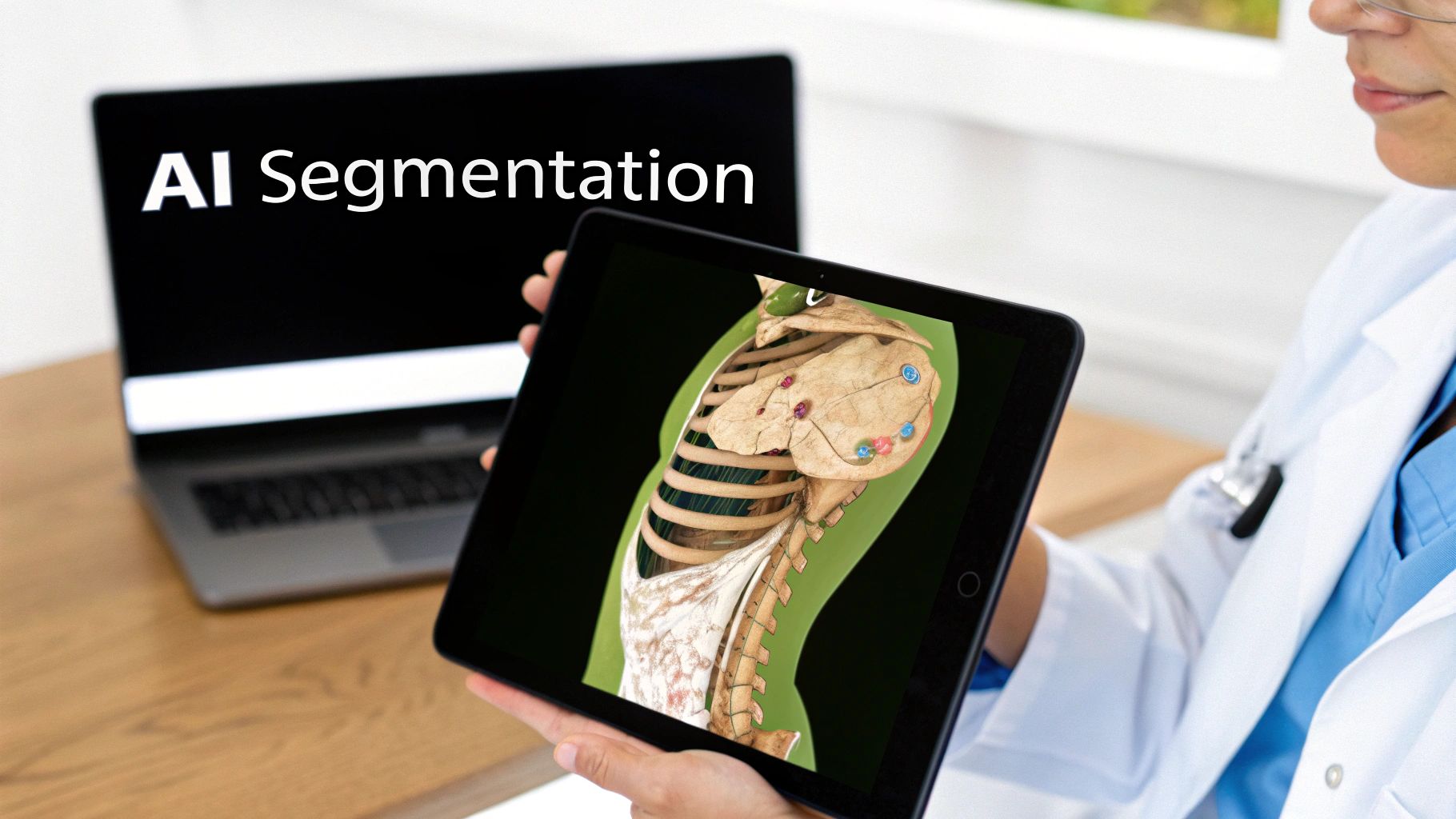
One of the most immediate impacts of AI is in segmentation—the process of outlining specific organs or structures within a 3D scan. Traditionally, this was a painstaking manual job. A radiologist would have to meticulously trace boundaries, slice by tedious slice. It was time-consuming and, naturally, could have minor variations between specialists.
Now, imagine an artist carefully tracing an intricate image by hand versus a smart tool that perfectly outlines the subject with a single click. That’s the leap AI provides. Sophisticated algorithms can now identify and segment organs, tumors, and blood vessels in a fraction of the time, with incredible consistency.
This automation frees up clinicians to spend less time on manual prep and more time on what matters most: accurate diagnosis and effective treatment planning.
Key Applications of AI in 3D Imaging
The influence of AI extends far beyond simple segmentation. It introduces a whole new layer of intelligence to the diagnostic workflow. Here are some of the most inspiring applications:
- Automated Anomaly Detection: AI models can be trained on thousands of scans to recognize the subtle signs of disease. They act as a vigilant second pair of eyes, flagging potential tumors, lesions, or other abnormalities for the clinician's review.
- Predictive Analytics: By analyzing the 3D structure and characteristics of a tumor, AI can help predict its potential growth patterns or its likely response to certain therapies. This opens the door to truly personalized medicine.
- Surgical Pathway Optimization: During surgical planning, AI can analyze a 3D model to suggest the safest and most efficient path for an instrument, helping to minimize risk to surrounding healthy tissue.
The goal of AI isn't to give answers, but to help us ask better questions. It directs a clinician's attention, quantifies complex data, and provides a deeper context for every decision.
The impact of this technology is also driving incredible growth in related fields. The 3D visualization software market for design disciplines like architecture and product design, for instance, was projected to hit USD 15 billion in 2025. With a steady growth rate, it could expand to nearly USD 35 billion by 2033, a trend fueled by the same AI and VR integration that makes realistic simulations possible. You can explore the full research behind this market expansion to understand its broader influence.
At PYCAD, we live at the forefront of this integration. We at PYCAD, build custom web DICOM viewers and integrate them into medical imaging web platforms, infusing them with the power of AI to create smarter diagnostic tools. You can learn more about how AI is transforming the field of medical imaging in our guide on artificial intelligence for radiology. Our work, which you can see on our portfolio page, shows just how this powerful combination can create a brighter future for patient care.
Transforming Patient Outcomes with Real-World Wins
The true test of any medical technology isn't found in a lab—it's measured by its impact on people's lives. While the technical specs of 3D visualization software are impressive, its real magic happens in the clinic. This is where it helps doctors save lives, dramatically reduce surgical risks, and deliver diagnoses with newfound clarity.
It’s about moving medicine from abstract data on a screen to something tangible and real, giving clinicians a profound connection to their patient's unique anatomy. These aren't just theoretical benefits; they are happening in hospitals worldwide, every single day. The technology acts as a clear, interactive roadmap, empowering medical professionals to navigate the complexities of the human body with more confidence than ever before.

Precision in the Operating Room
Picture a surgical planning session for a complex spinal fusion. The surgeon's task is to place several tiny screws into a patient's vertebrae—a procedure where a single millimeter can be the difference between a successful outcome and severe nerve damage. It’s incredibly high-stakes.
Now, imagine that surgeon rotating a perfect 3D model of that patient's spine, rebuilt from a CT scan. They can zoom in, examine it from any angle, and virtually place each screw, testing different trajectories to find the perfect path. This isn't some generic textbook diagram; it's the patient's actual bone structure, complete with all its unique curves and imperfections.
This digital dress rehearsal allows the surgeon to anticipate challenges and refine their strategy long before the first incision is made. The result? A more precise, faster, and safer procedure with a much lower risk of complications.
Vigilance in Early Detection
Let’s shift gears to a radiologist tracking a tiny, suspicious nodule in a patient's lung over several months. On a traditional flat, 2D scan, measuring subtle changes in size is often difficult and can be subjective.
With 3D visualization, the entire process changes. The software can instantly isolate the nodule from the surrounding tissue and calculate its exact volume down to the cubic millimeter. When the patient returns for a follow-up, the new model can be compared directly against the old one, providing objective, quantifiable data on its growth.
This level of precision is a game-changer. It allows doctors to spot even minuscule changes that could signal a turn toward malignancy, enabling them to intervene at the earliest possible stage when treatment is most effective. It strips away the guesswork and replaces it with data-driven certainty.
"In medicine, we often deal with ambiguity. 3D visualization cuts through the noise, giving us a clear, undeniable view of the problem. It’s like turning on the lights in a dark room."
This clarity is also a powerful tool for patient communication. A doctor can pull up a 3D model of a patient's heart and point to the exact valve that needs repair. Seeing it for themselves demystifies the condition, helping patients truly understand their own health and feel like active partners in their care. The work we do at PYCAD is all about making this level of insight accessible. We at PYCAD, build custom web DICOM viewers and integrate them into medical imaging web platforms, putting these powerful tools right where clinicians need them.
Guiding Complex Cardiac Procedures
In cardiology, there's zero room for error. Think about a surgeon preparing to replace a failing aortic valve. The heart’s anatomy is a labyrinth of critical structures, and success hinges on choosing a perfectly sized replacement valve and placing it with absolute accuracy.
Using 3D models built from the patient's scans, the surgical team can take precise measurements and even simulate the deployment of different valve sizes. This lets them select the ideal device and map out the entire procedure, minimizing risk and paving the way for a better outcome. It’s a beautiful example of how digital planning leads to superior physical results.
A Snapshot of Clinical Applications
The impact of 3D visualization echoes across nearly every medical specialty, with each field discovering unique ways to solve its biggest challenges. You can see how this plays out in some of the real-world projects on our portfolio page.
Here’s a quick look at how different specialties are putting this technology to work.
3D Visualization Applications Across Medical Fields
| Medical Specialty | Primary Use Case | Key Benefit |
|---|---|---|
| Orthopedics | Planning joint replacements and trauma surgery | Improved implant sizing and surgical accuracy |
| Oncology | Tumor volume tracking and treatment planning | Precise monitoring of treatment response |
| Cardiology | Guiding valve replacements and interventions | Reduced procedural risk and better outcomes |
| Neurosurgery | Mapping brain tumors relative to critical areas | Enhanced safety by avoiding delicate structures |
| Dentistry | Planning dental implants and maxillofacial surgery | Accurate placement and predictable results |
From the operating room to the radiologist’s reading room, 3D visualization software is so much more than a tool for seeing—it’s a tool for understanding. It delivers the deep insight and confidence clinicians need to achieve the best possible outcomes for their patients, one procedure at a time.
How to Choose Your 3D Visualization Solution
Picking the right 3D visualization software is one of those big decisions that will shape how your entire organization sees and uses medical data. It’s so much more than just ticking boxes on a feature list. You're really looking for a partner that fits seamlessly into your clinical workflows, your technical setup, and your vision for the future. Get it right, and you empower your team. Get it wrong, and you introduce friction that no one has time for.
This isn't a choice to be rushed. It requires a thoughtful look at your immediate needs while also keeping an eye on where you're headed. You need to consider how the software handles sensitive patient data, how it will play with your existing systems, and whether it can grow with you. It’s a decision that will ripple through every department that touches medical imaging.
On-Premise Stability or Cloud Agility
One of the first forks in the road is deciding where your software will live. You can go the traditional route with an on-premise installation on your own servers, or you can embrace a more modern, cloud-based solution. Each path has its own appeal and serves different priorities.
-
On-Premise Solutions: These put you in the driver's seat, giving you total control over your data and infrastructure. This is often the best fit for institutions with iron-clad data governance rules or very specific hardware needs. The trade-off? Your IT team is on the hook for all the maintenance, updates, and security.
-
Cloud-Based Solutions: This is where the real flexibility and scalability lie. You can forget about massive upfront hardware costs and give your team secure access to their tools from practically anywhere. It’s no surprise this model is gaining so much traction.
The industry trend is clearly pointing towards the cloud. The visualization and 3D rendering software market was valued at USD 3.7 billion in 2024, but IMARC Group projects it will rocket to USD 14.5 billion by 2033. Much of that explosive growth is fueled by cloud-based rendering that delivers incredible speed and scale. You can discover more insights about these market trends to get a feel for where things are going.
The Build vs. Buy Dilemma
Now for the biggest question of all: should you buy an off-the-shelf product or build a custom solution from the ground up? "Buying" gets you up and running quickly with a standard set of tools designed for a broad audience. The catch is that you often end up forcing your team to bend their workflows to fit the software's rigid structure.
"Building," on the other hand, is a strategic move. A custom-built solution is molded around your specific clinical needs, ensuring the tool works for the user, not the other way around. This approach means every single feature has a purpose, directly supporting your unique diagnostic processes or surgical planning. As you weigh your options, it's helpful to understand the general landscape of the best 3D rendering software solutions on the market to see what's possible, no matter which path you take.
A custom solution isn't just about features; it's about creating a frictionless workflow that feels like a natural extension of your clinical expertise.
Why a Custom Solution Delivers Deeper Value
At PYCAD, we believe deeply in the "build" philosophy because we've seen firsthand the incredible difference it makes. We specialize in crafting bespoke solutions that solve the exact problems that generic software just can't touch. We at PYCAD, build custom web DICOM viewers and integrate them into medical imaging web platforms, creating something that’s a perfect fit for your institution.
This tailored approach guarantees a smooth connection with your existing Picture Archiving and Communication System (PACS) and other essential systems. It completely sidesteps the compatibility headaches and workflow chaos that often come with one-size-fits-all products. By designing a solution from the ground up, we can build an intuitive experience that cuts down on training time and gets your team on board faster.
To get a better idea of what this level of custom development looks like in the real world, we invite you to explore our work on our portfolio page. You can also dive into our guide on the various 3D visualization programs out there to get a broader perspective.
The Future of Medical Imaging Is 3D
https://www.youtube.com/embed/8bZ0z4xL2zY
We're standing at the edge of a new era in medicine, a point where the digital and physical worlds of patient care are starting to merge in truly remarkable ways. The story of 3D visualization software isn't ending; it's just getting started. It's growing beyond a powerful diagnostic tool to become the very backbone of tomorrow's healthcare.
This isn't some far-off dream—it's happening right now. We are moving past simply seeing anatomy on a screen and stepping into a future where that same data guides every action with astonishing precision.
Into New Dimensions of Care
Picture this: a surgeon, mid-operation, wearing augmented reality (AR) glasses. They aren't just looking at the patient on the table; they're seeing through them. A live, transparent 3D model of the patient's organs is projected directly onto their body, giving the surgeon a kind of superpower—an intuitive map that guides every single incision with pinpoint accuracy.
The next leap is already taking shape with 4D visualization, which adds the crucial element of time. Instead of a static 3D model of a heart, imagine watching it beat, seeing it as a dynamic, rhythmic process. This allows cardiologists to study function, not just structure, and watch blood flow and valve movements as they happen.
This is where medical imaging is headed: a world where we don't just see data, we experience it, letting it guide every decision and action in real time.
This powerful vision makes one thing clear: 3D visualization is no longer a niche tool for specialists. It’s becoming a core technology that will define the future of medicine. It's an invitation for all of us to embrace this shift and help lead the way.
Here at PYCAD, we're passionate about building the platforms that will shape this new era. We at PYCAD, build custom web DICOM viewers and integrate them into medical imaging web platforms, creating the exact tools you need to innovate.
Take a look at our portfolio of innovative projects to see what's possible, and let's build the future of patient care together.
Frequently Asked Questions
As medical imaging technology leaps forward, it’s only natural for clinicians and administrators to wonder where 3D visualization software fits into their daily work. Getting a handle on what this technology can do—and what it needs to run—is the first step toward unlocking a whole new dimension of patient care. Let's walk through some of the most common questions we hear.
These tools are built to be incredibly flexible, capable of transforming a wide range of standard medical scans into rich, interactive 3D models.
What Types of Medical Scans Work with This Software?
At its core, this software can work with almost any imaging method that creates a series of cross-sectional pictures, what we in the field call a DICOM series. Think of it like a digital flipbook of the human body. The most common sources are the ones you’re already using every day:
- Computed Tomography (CT): Your go-to for incredible detail on bone, blood vessels, and soft tissues.
- Magnetic Resonance Imaging (MRI): Unbeatable for getting a clear, detailed look at soft tissues like the brain, muscles, and organs.
- Cone Beam Computed Tomography (CBCT): A favorite in dentistry and maxillofacial surgery for its super-sharp images of hard tissues.
Basically, if the scan gives you a stack of image "slices," the software can stitch them together into a fully explorable 3D model.
Is Special Hardware Required to Run These Tools?
In the past, absolutely. The sheer amount of data needed for 3D rendering required beastly, dedicated workstations with top-of-the-line graphics cards. This often meant the technology was stuck in a specific reading room, creating frustrating delays and bottlenecks. Thankfully, that’s no longer the case.
Modern web-based solutions have completely changed the game by moving all the heavy lifting to powerful servers or the cloud. This means you and your team can now pull up and interact with complex 3D models on a standard computer, a laptop, or even a tablet, all through a simple web browser. This has made 3D visualization software more accessible and practical for everyone.
How Does a Custom Viewer Improve Workflow?
Off-the-shelf software gives you a set of pre-packaged tools, and it's up to you to figure out how to fit them into your workflow. A custom-built viewer flips that script entirely. It's designed from day one to match your specific clinical needs and plug right into the systems you already use.
A custom solution removes friction. It’s not just about adding features—it’s about creating a workflow that feels intuitive, efficient, and like a natural extension of a clinician's expertise.
This tailored approach makes a world of difference. At PYCAD, we specialize in building custom web DICOM viewers and integrating them into medical imaging web platforms. We make the tool fit your team, not the other way around. The result is a seamless, user-focused experience that standard software just can't offer, leading to quicker diagnoses and more confident planning.
At PYCAD, we build the integrated, custom solutions that put the power of 3D visualization right where it belongs: in your hands. To see what a truly bespoke medical imaging platform can do, we invite you to explore our work.


