
Imagine trying to navigate a new city. For decades, you’d rely on a folded paper map—static, cumbersome, and totally unaware of the traffic jam just around the corner. Now, think about using a real-time GPS on your phone. It doesn't just show you the map; it analyzes traffic, predicts your arrival, and reroutes you around obstacles instantly.
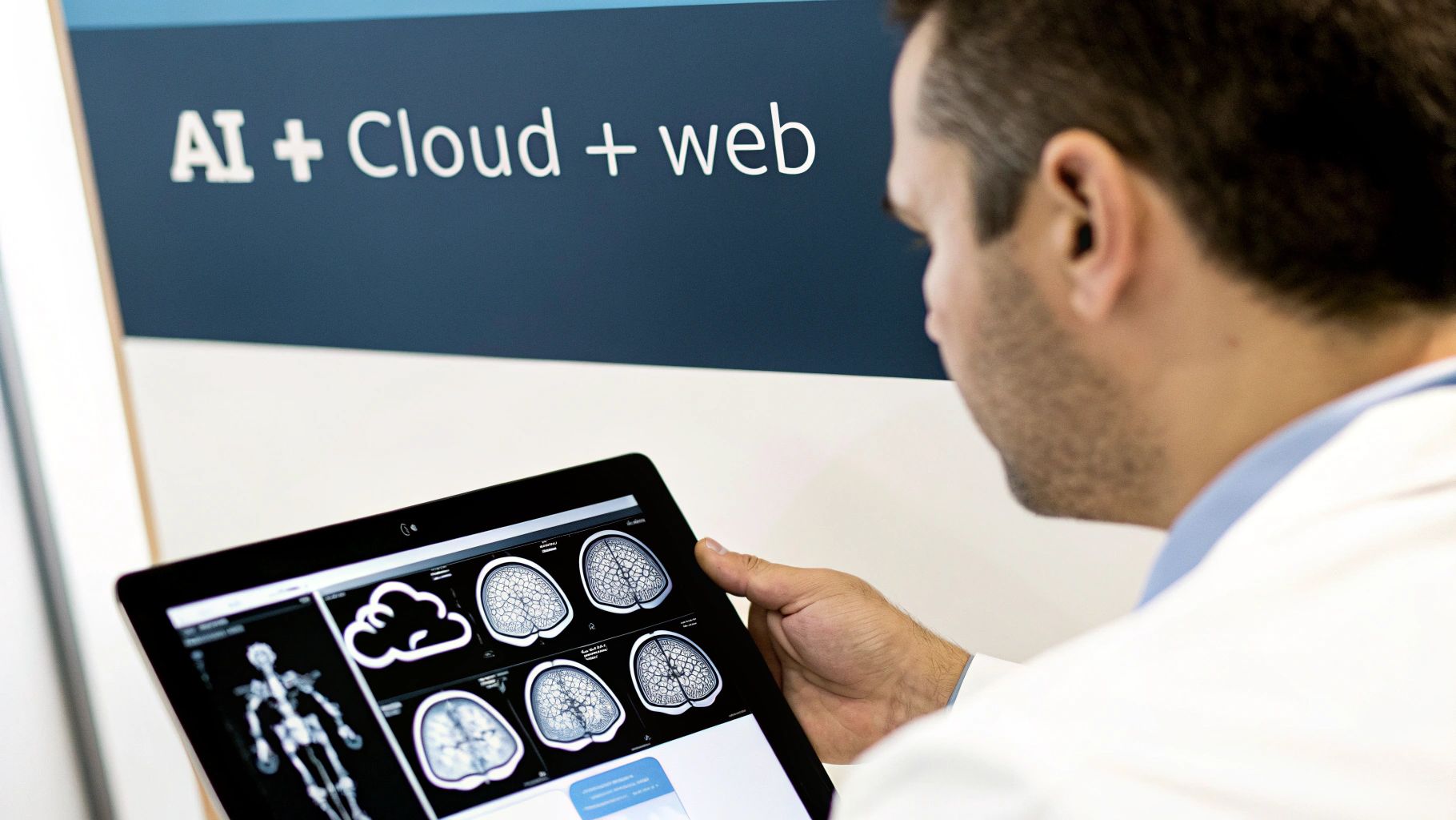
That’s the exact leap we’re making from traditional radiology to next gen radiology. This isn't some futuristic fantasy. It's a fundamental change happening right now, transforming medical images from flat, static pictures into dynamic, intelligent data that can predict, inform, and guide care like never before.
The Future of Medical Imaging Is Already Here
The days of clunky, on-premise imaging servers and siloed departments are numbered. We're moving into an era defined by a smart, interconnected ecosystem where technology acts as a trusted co-pilot for radiologists and clinicians. This new approach is about more than just looking at scans; it’s about building an intelligent workflow that delivers faster, more precise diagnostics and improves the entire patient journey.
This evolution is built on a few core pillars that are completely changing the game for how we acquire, analyze, and share medical imaging data. These aren't just minor tweaks; they're foundational shifts creating a more responsive and insightful diagnostic environment.
-
Intelligent Automation: Think of Artificial Intelligence (AI) as a second set of eyes. These algorithms can spot subtle anomalies the human eye might miss, automate time-consuming measurements, and triage critical cases to the top of the worklist.
-
Seamless Cloud Access: Bulky on-site servers are being replaced by secure, cloud-native platforms. This gives clinicians the power to access imaging studies instantly, from anywhere, on any device—no more being tethered to a specific workstation in a dark room.
-
Universal Visualization: Modern, web-based DICOM viewers are the new universal translator for medical images. They provide sophisticated visualization tools right in a web browser, eliminating the need for clunky software installations and compatibility headaches.
A New Diagnostic Paradigm
At its heart, next gen radiology is about demolishing the walls that have historically fragmented the imaging workflow. The old process is painfully familiar: a patient gets a scan, the images are locked away on a local PACS server, a radiologist reads them on a dedicated machine, and the report is slowly routed back to the referring doctor. Every single step introduces friction and potential delays.
This new model connects all the dots. A scan performed in a rural clinic can be read by a sub-specialist across the country in seconds, with an AI algorithm providing a preliminary analysis before the file is even fully loaded. This isn't just about speed; it's about unlocking a level of collaboration and expertise that was simply out of reach before.
This shift empowers healthcare providers to stop reacting to disease and start proactively managing patient health.
To give you a clearer picture, here’s a simple breakdown of how things are changing.
Traditional vs Next Gen Radiology at a Glance
This table contrasts the old and new paradigms in radiology, highlighting the key technological and operational shifts that define the next generation.
| Aspect | Traditional Radiology | Next Gen Radiology |
|---|---|---|
| Data Storage | On-premise PACS servers | Cloud-native, scalable storage |
| Image Access | Dedicated, on-site workstations | Any device, anywhere, via web browser |
| Analysis | Manual human interpretation | AI-assisted detection & quantification |
| Workflow | Linear, siloed, and manual | Integrated, automated, and collaborative |
| Interoperability | Proprietary formats, difficult sharing | Standardized APIs (DICOMweb, FHIR) |
| Infrastructure | High upfront hardware costs | Flexible, subscription-based (SaaS) |
As you can see, the move to next gen isn't just about better software—it’s a complete reimagining of the infrastructure and philosophy behind medical imaging.
At PYCAD, we live and breathe this transformation. We specialize in building custom web DICOM viewers and integrate them into medical imaging web platforms, creating the precise tools our partners need to make this future a reality. You can explore some of these powerful solutions in our portfolio.
The Core Technologies Driving the Revolution
This incredible leap forward in radiology isn't powered by a single breakthrough, but by four distinct technologies working together. Think of them as the pillars supporting a new era of medical imaging. First, Artificial Intelligence (AI) serves as an expert partner to radiologists, finding subtle patterns and automating the tedious parts of the job.
Next up is cloud-native PACS and DICOMweb. This is all about moving away from clunky, on-site servers and giving medical teams instant, secure access to images from literally anywhere.
Then we have web-based DICOM viewers, which finally free us from the chains of specialized desktop software. Now, powerful visualization tools are available on any device with a web browser.
And finally, integrated platforms tie everything together. They connect imaging workflows directly to EHRs, scheduling tools, and patient management systems, creating a seamless flow of information that eliminates frustrating manual work.
Together, these technologies give clinicians sharper insights, dramatically faster results, and lower overheads. More importantly, they clear the path for better patient outcomes and deliver a real, measurable return on investment by simplifying IT management.
Here at PYCAD, we live and breathe this stuff. We build custom web DICOM viewers and integrate them into medical imaging web platforms, unifying these four pillars for hospitals and medtech companies. You can explore some of these powerful solutions in our portfolio.
Artificial Intelligence as a Co-pilot
Imagine having a tireless assistant who can scan thousands of images and never miss a single detail. That's the power of AI in radiology. For instance, an AI algorithm can flag tiny microcalcifications in a mammogram that a human eye might overlook after a long and demanding shift.
This allows radiologists to focus their attention on the most critical cases first, which can shave precious time off a diagnosis. AI also brings a new level of consistency to reporting by standardizing measurements and terminology study after study.
The growth here is simply astonishing. The AI in radiology market, currently valued at USD 10.57 billion, is expected to explode to USD 193.02 billion by 2033. That’s a compound annual growth rate of 38.12%. This boom is fueled by rising demand for imaging, a greater focus on early detection, and supportive regulations. You can get more insights on the AI in radiology market from Grand View Research.
“AI integration can cut review time by up to 40%, freeing radiologists to focus on complex interpretation and patient care.”
In practice, AI-driven workflows often look like this:
- Automated lesion detection that highlights suspicious areas in seconds.
- Predictive models that can forecast how a disease might progress over time.
By treating AI as a trusted collaborator, not a replacement, radiology departments are unlocking incredible new levels of efficiency and diagnostic accuracy.
Cloud-Native Storage With DICOMweb
Moving from local PACS servers to the cloud is like swapping a filing cabinet for an infinitely expandable, always-accessible digital library. It’s a game-changer, but it absolutely has to be done with an ironclad focus on security.
Modern cloud platforms use DICOMweb APIs to make sure that images, metadata, and reports flow between systems without any friction. Plus, cloud providers typically offer SLA-backed uptime guarantees that are far more reliable than what most on-premise data centers can promise.
You can learn more in our deep dive on PACS and DICOMweb integration.
- End-to-end encryption protects data whether it's stored or being transferred.
- Role-based access controls ensure only authorized personnel can view sensitive information.
Universal Web-Based Image Viewers
Remember the switch from DVDs to Netflix? No more discs, no more downloads, no more compatibility issues—just click and watch. Web DICOM viewers bring that same beautiful simplicity to medical imaging.
Now, clinicians get access to sophisticated tools directly through a simple web link.
- Open a study in any modern browser on any device.
- Run complex AI analysis with a single click.
- Annotate images in real-time and share your findings instantly with colleagues.
These modern viewers are packed with powerful features, including:
- Multi-planar reformats (MPR) and volume rendering for deep 3D insights.
- A responsive interface that works perfectly on desktops, tablets, and even smartphones.
Connected Platforms and Workflows
Working with disconnected systems is a recipe for delays and mistakes, forcing staff into a clumsy dance of manual data exports and endless email chains. Integrated platforms fix this by linking everything—EHRs, CRMs, and imaging viewers—into one smooth, automated pipeline.
Picture this: a radiology order placed in the scheduling system automatically triggers a barcode check at the scanner, sends the resulting images through an AI triage model, and pushes the preliminary findings directly to the referring physician’s dashboard. No manual steps needed.
At PYCAD, this is what we build. We build custom web DICOM viewers and integrate them into medical imaging web platforms to tear down information silos and help clinical teams make faster, better-informed decisions.
- Real-time alerts notify clinicians of critical findings the moment they appear.
- Task management boards assign follow-up actions and track them to completion.
- Audit logs record every interaction, ensuring full compliance and quality control.
As next-gen radiology begins to incorporate more smart devices and real-time data, having a solid IoT infrastructure becomes essential. To see how these technologies are being applied across the medical field, check out this excellent resource on A Practical Guide to IoT Healthcare Solutions.
Our portfolio is full of examples showing how we bring these technologies together in the real world. Take a look at our success stories at our portfolio.
Where the Rubber Meets the Road: The Real-World Impact of Next-Gen Radiology
All this amazing technology is just an academic exercise if it doesn’t deliver real-world value. The shift to next-gen radiology isn’t about chasing trends; it's about delivering clear, measurable wins for hospitals, medtech innovators, and—most critically—the patients they serve. This is where the true power of this new era really shines.
The most immediate and profound impact is a huge leap in diagnostic accuracy. Think of AI tools as a tireless, expert second opinion for every radiologist, trained to spot subtle patterns the human eye might overlook. We're talking about algorithms that can pinpoint early-stage lung nodules or faint microcalcifications in mammograms with incredible precision, acting as a perfect complement to a radiologist's expertise.
This directly translates into earlier disease detection. Finding something like cancer sooner is often the single most important factor in a patient's outcome. When you can identify problems at their earliest stages, you open the door for treatments that are more effective and less invasive, completely altering a patient's journey.
Breaking Free from Inefficiency
Beyond just being more accurate, next-gen radiology is about reclaiming time. The traditional reporting workflow can be a frustrating bottleneck, bogged down by clunky systems, manual dictation, and endless cycles of edits. This administrative grind eats up hours that radiologists could be dedicating to challenging cases or collaborating with clinical teams.
Now, imagine a radiologist simply speaking their findings aloud as an AI system instantly organizes them into a perfectly structured, guideline-compliant report. This isn't science fiction; it’s happening today, and it’s shrinking report turnaround times from several minutes down to just a few seconds.
By taking over the repetitive, time-consuming tasks, next-gen platforms give radiologists their most valuable resource back: the mental space to focus their expertise on complex diagnostics and patient care.
This speed doesn't just benefit the radiologist. It creates a domino effect across the entire hospital, speeding up clinical decisions and dramatically cutting down that agonizing wait time for patients and their referring doctors.
The Financial and Clinical Payoff
The return on investment here is undeniable, both clinically and financially. Moving to the cloud immediately gets rid of the headache and high cost of maintaining on-premise servers. This slashes IT overhead and frees up capital, making top-tier imaging technology a realistic option for facilities of all sizes.
The benefits stack up quickly in a few key areas:
- Slashed IT Costs: Migrating to a cloud-native platform means saying goodbye to expensive hardware maintenance, high energy bills, and the need for a dedicated team to babysit local servers.
- Reduced Downstream Treatment Costs: Catching diseases earlier and more accurately often means patients can undergo less invasive, less expensive treatments. This lowers the long-term cost of care for everyone involved.
- Higher Throughput: When you can analyze scans and generate reports faster, your radiology department can handle more volume without burning out your staff. This directly boosts the department's financial health.
These aren't just line items on a budget. Saving money in one area means you can reinvest it where it truly counts—in direct patient care.
Crafting a Better Patient Experience
At the end of the day, the most important results are felt by the patient. The anxiety of waiting for test results is immense. Next-gen radiology tackles this head-on by closing the gap between the scan and the diagnosis.
What's more, the precision offered by AI-powered diagnostics helps create truly personalized care plans. Instead of a one-size-fits-all approach, treatments can be adapted to the unique characteristics of a patient's condition, as revealed by advanced imaging. This leads to more effective care with fewer side effects.
At PYCAD, our entire focus is on building the tools that make these results possible. We specialize in building custom web DICOM viewers and integrating them into medical imaging web platforms that put powerful, intuitive technology in the hands of clinicians. To see how these solutions are changing the game, we invite you to explore the projects in our portfolio.
Charting a Course Through the Implementation Challenges
Stepping into the future of radiology is exciting, but it’s a journey that comes with its own set of obstacles. Adopting next gen radiology isn't as simple as installing a new piece of software. It’s a foundational shift that forces us to rethink everything from data security and AI ethics to how our tangled web of healthcare systems finally starts talking to each other. To make this leap successfully, we need to meet these challenges with a clear, intentional strategy.
The first and most important hurdle is always data security. In a world governed by HIPAA, the very idea of moving sensitive patient data to the cloud can give anyone pause. But here’s the thing: modern cloud platforms and zero-footprint web viewers can actually offer a far more secure environment than the on-premise systems we’ve relied on for decades.
Think of a zero-footprint viewer like a secure window into the data. Instead of downloading a patient’s entire imaging file onto a personal laptop or workstation—where it could be lost, stolen, or forgotten—clinicians are simply streaming the pixels. The actual data never leaves the safety of the encrypted central server, dramatically shrinking the potential for a breach and simplifying compliance.
Making Sure AI Works for Everyone
Beyond the technical safeguards, we have to address the ethics of artificial intelligence. An AI algorithm is a product of its education, and it's only as unbiased as the data it was trained on. If that dataset isn't truly diverse and representative of the real world, the AI can inadvertently perpetuate and even amplify human biases, leading to care that isn't equitable for all.
This is why rigorous, ongoing AI model validation is absolutely essential. Before any algorithm goes live, it must be put through its paces against a broad spectrum of data covering different ages, ethnicities, and genders to prove it performs accurately and fairly for everyone. This isn't a one-and-done check; it’s a commitment to ensuring the tool is a trustworthy partner for clinicians and a genuine benefit to patients.
The real goal is to build AI that serves all of humanity, not just a subset. It requires a constant loop of testing, monitoring, and refining to ensure these models stay fair and effective out in the messy, unpredictable real world.
A big part of getting teams to embrace these new tools is earning their trust. This often comes down to the user experience itself. For a great read on this, check out this article on building trust through smart design.
Creating a Common Language for Healthcare Data
Another massive challenge has always been interoperability—or the lack thereof. Most hospital ecosystems are a patchwork of different systems from countless vendors, each speaking its own proprietary language. This creates frustrating data silos that slow down care and make true collaboration nearly impossible.
This is where next gen radiology truly shines, using modern standards like DICOMweb and FHIR as universal translators.
- DICOMweb liberates medical images, allowing them to be accessed and shared over the web without being locked inside a specific PACS.
- FHIR (Fast Healthcare Interoperability Resources) does the same for all other clinical data, from patient histories to lab results and reports.
When used together, they create a beautifully fluid environment. An imaging study can travel from the scanner to the EHR, then to an AI platform for analysis, and finally to a surgeon's tablet in the OR—all without a single manual step. This seamless connection is the very heart of a modern, efficient diagnostic workflow. If you want to get into the nuts and bolts of this, our guide on how to successfully implement AI in medical imaging is a great place to start.
At PYCAD, this is exactly what we live and breathe. We build custom web DICOM viewers and integrate them into medical imaging web platforms from scratch, with security, compliance, and interoperability woven into their DNA. We guide our partners through these complexities, making sure their transition to next gen radiology isn't just a technical upgrade, but a leap forward in patient care. You can see the results for yourself in our portfolio.
Your Roadmap for Adopting Next-Gen Radiology
Making the leap to next-gen radiology can feel like a massive undertaking, but it’s really about a thoughtful, step-by-step evolution. It’s not about flipping a switch and hoping for the best. Instead, think of it as a journey with a clear map, where each move is deliberate and builds on the last, adding real value along the way.
The entire process hinges on one simple idea: know where you are and where you want to go. This isn't just a tech upgrade; it's about aligning your clinical and patient needs with the incredible possibilities of modern imaging.
Laying the Groundwork With Discovery and Strategy
Before you can even think about new software, you have to get an honest look at your current imaging world. This means a deep dive into your existing PACS, your network's muscle, how you store data, and the day-to-day workflows of your team. The real goal here is to pinpoint the snags, security weak spots, and inefficiencies that are quietly draining time and resources.
Once you have that clear picture, you can set real, tangible goals. Forget vague statements like "let's be more efficient." Get specific.
- Aim to cut the average report turnaround time by 30% in the first year.
- Target a 20% reduction in annual IT infrastructure costs by moving to a cloud-native platform.
- Boost diagnostic confidence in key areas by rolling out specialized AI tools.
This kind of strategic clarity acts as your North Star, guiding every decision. It’s also your best tool for getting everyone—from the C-suite to the radiologists in the reading room—excited and on board.
Choosing Partners and Launching Pilot Programs
With your goals locked in, it's time to find the right people to help you get there. The market is flooded with vendors pushing one-size-fits-all boxes, but that’s not what this is about. You need a true partner, someone who listens and is ready to shape their technology to fit your unique way of working, not the other way around.
This is where pilot programs are your best friend. A pilot lets you test-drive new technology in a controlled, real-world setting without committing to a massive rollout. Pick a specific department or a single clinical challenge—like an AI for lung nodule detection—and see how it performs on a smaller scale. This "try before you buy" approach takes the risk out of the investment and gives you hard data to make your case for a wider launch.
At PYCAD, this partnership model is in our DNA. We don't just hand over software. We build custom web DICOM viewers and integrate them into medical imaging web platforms, working side-by-side with our clients to solve their most pressing challenges. You can explore some of these collaborations in our portfolio of medical imaging solutions.
This journey can be visualized as a clear, four-step process.
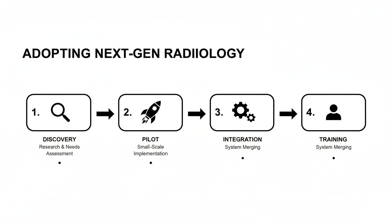
As you can see, each phase logically flows into the next, creating a solid foundation for success from start to finish.
Seamless Integration and Focused Training
With the right partner on board, the work shifts to the technical side of things: integration and moving your data. A modern, API-first approach is absolutely critical here. Think of APIs as secure bridges that let your new platforms talk effortlessly with your existing systems, like your Electronic Health Record (EHR) or patient CRM. This prevents information from getting trapped in silos and keeps everything running smoothly.
The final—and arguably most important—piece of the puzzle is training. You can have the most powerful tools in the world, but they’re worthless if your team isn’t confident using them. A solid training program, built together with your tech partner, is the key to empowering your staff and ensuring they genuinely embrace the new way of working. Once they’ve mastered the tools, you can strategically expand the solution to other departments, one success story at a time.
The global next imaging technology market, a core part of next-gen radiology, was valued at USD 7.04 billion and is expected to hit USD 14.80 billion by 2033, growing at a CAGR of 8.6%. This isn't just a trend; it's a worldwide movement toward the very technologies that make this journey possible. Learn more about the next imaging technology market trends from Straits Research.
Exploring the Advanced Hardware Powering the Shift
As much as software is changing the game, the revolution in next-gen radiology truly rests on a foundation of groundbreaking hardware. The incredible diagnostic feats we're seeing today are born from imaging equipment that captures details once thought impossible, opening up a whole new world of clinical insight.
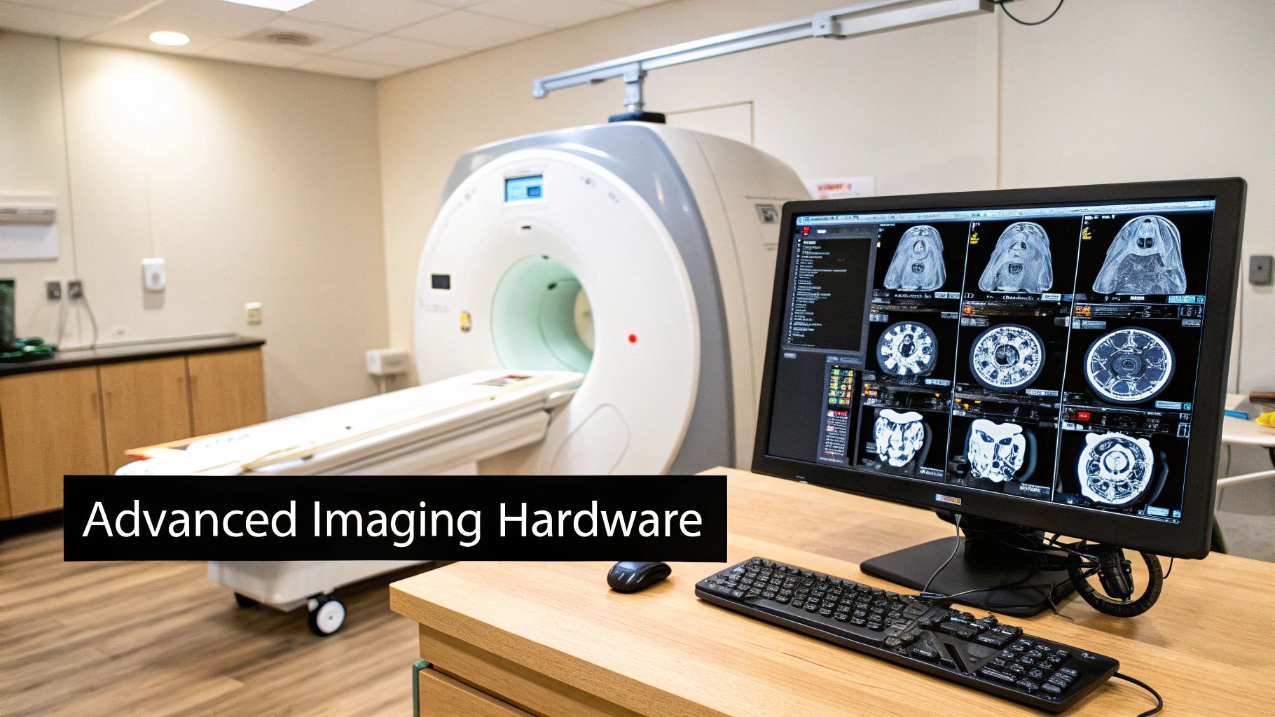
This new hardware isn't just a minor upgrade; it's a fundamental leap in how we see inside the human body. These machines are engineered for precision, designed for safety, and built for sustainability, creating a ripple effect of benefits that touches everyone from the patient to the hospital administrator.
Photon-Counting CT Scanners
Imagine taking a photograph with near-perfect resolution while using far less light. That's the core idea behind Photon-Counting CT (PCCT) scanners. While traditional CTs measure the total energy of X-rays passing through the body, PCCT systems literally count individual photons. The result? Images with extraordinary detail and clarity.
This technology brings two massive benefits to the table:
- Drastically Reduced Radiation: PCCT can significantly lower the required radiation dose. This is a huge win for patient safety, especially for children and cancer patients who need frequent scans.
- Enhanced Image Quality: The higher resolution and reduced image noise help clinicians spot subtle signs of disease much, much earlier. You can dive deeper into visualizing these complex datasets in our guide on CT volume rendering.
The momentum behind next-gen hardware is undeniable. The medical digital imaging systems market is projected to swell from USD 37.89 billion to USD 58.11 billion by 2033. Photon-counting CT is a major force here, cutting radiation doses by up to 50% in some procedures while delivering better contrast.
Helium-Free MRI Systems
Another critical hardware advancement is the arrival of Helium-Free MRI systems. For decades, MRI machines have depended on massive amounts of liquid helium—a finite and expensive resource—to keep their powerful magnets cool. New designs are changing that by drastically reducing or even eliminating this dependency.
This innovation is making advanced MRI technology more accessible and sustainable. It lowers the day-to-day operational costs and shrinks the environmental footprint of medical imaging, which is a win-win for everyone.
The key takeaway here is that all this remarkable hardware produces massive, incredibly intricate datasets. Legacy software simply wasn't built for this. To unlock the full potential of these scans, a powerful visualization platform isn't a luxury; it's essential. At PYCAD, we build custom web DICOM viewers and integrate them into medical imaging web platforms precisely for this purpose. You can see our solutions at our portfolio.
Your Top Questions About Next-Gen Radiology, Answered
As we look toward the future of medical imaging, it's natural for hospital leaders, IT teams, and medtech visionaries to have practical questions. Stepping into the world of next-gen radiology is an exciting journey, and getting clear answers is the best way to move forward with confidence. Let's tackle some of the most common things people ask.
Is This Technology Just for Big Hospital Systems?
Absolutely not. In fact, one of the best things about the shift to cloud-native platforms is how it opens up advanced radiology for everyone. Gone are the days of needing massive upfront investments in on-premise servers and the large IT teams to manage them.
This change completely levels the playing field. Smaller clinics and innovative medtech startups can now tap into the same powerful imaging tools as major institutions, all on a flexible, pay-as-you-go basis. At PYCAD, we build custom web DICOM viewers and integrate them into medical imaging web platforms designed to fit the specific scale and budget of our partners, making this incredible technology truly accessible. You can see our work in our portfolio.
What Happens to All Our Existing Data? How Do We Make Sure It Works?
This is a huge concern, and it all comes down to interoperability. A truly modern imaging platform must be built on open standards like DICOMweb and FHIR. Think of these as universal languages that allow your new systems to speak fluently with your existing picture archiving systems (PACS) and electronic health records (EHR).
A smart data migration plan is crucial here. The goal isn't just to move old files from one place to another. It's about revitalizing that data, structuring it so that new AI tools can actually use it. This process can unlock a ton of hidden value in your archives, transforming them from a dusty digital library into a living, breathing resource for new discoveries.
The best transitions don't treat legacy data as a problem to be solved, but as a priceless asset to be activated. The right platform builds a bridge connecting the past, present, and future of your imaging data.
Is AI Going to Make Radiologists Obsolete?
This is probably the question we hear most, and it comes from a common misconception. The role of AI in next-gen radiology isn’t to replace radiologists—it’s to supercharge them.
Imagine AI as the ultimate assistant, a co-pilot that never gets tired. It can handle the time-consuming, repetitive tasks like precisely measuring lesions across multiple scans or flagging subtle anomalies that need a closer look.
This frees up radiologists to focus their invaluable expertise where it matters most: solving complex diagnostic puzzles, collaborating with other physicians on care plans, and spending more time with patients. AI enhances human skill, leading to better accuracy, faster turnarounds, and ultimately, better outcomes. It lets experts do what only they can do.
At PYCAD, our passion is building the platforms that solve these challenges and make the future of medical imaging a reality. We build custom web DICOM viewers and integrate them into medical imaging web platforms, creating solutions that are secure, connected, and intelligent.
See how we're helping our partners drive this change by exploring our work at https://pycad.co/portfolio.