
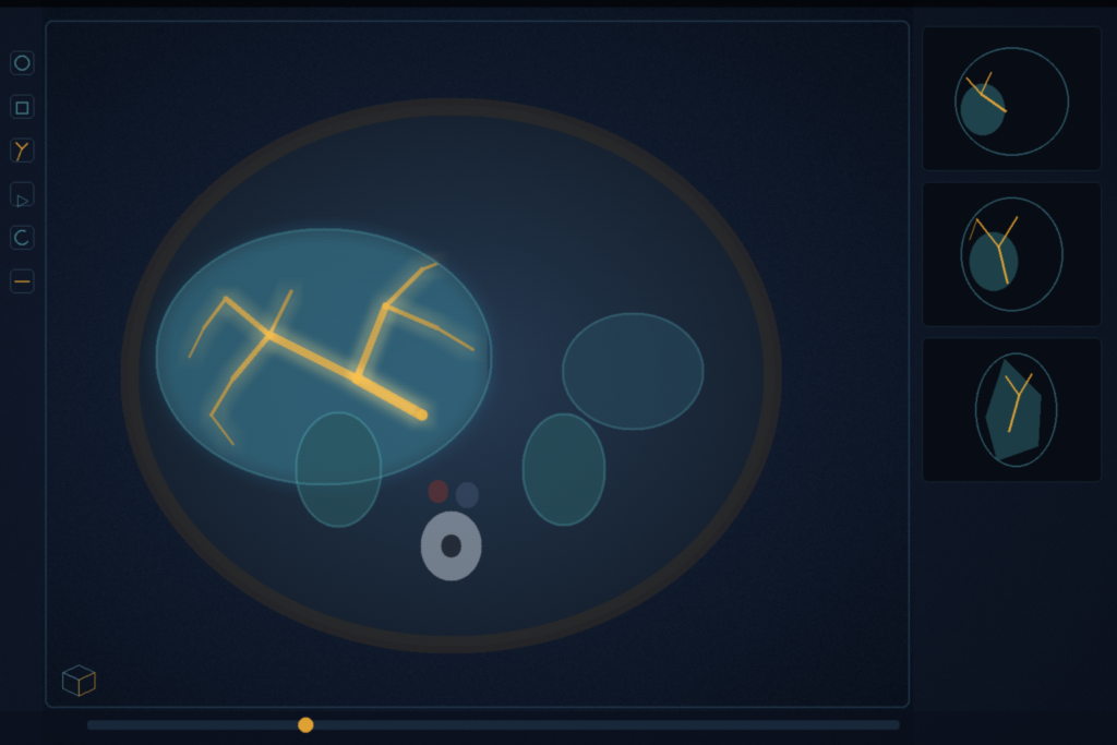
3D-CurvSegFlow is a 23.49 M flow-matching 3D U-Net that writes a vessel-tree mask in three Euler steps. It learns a velocity field from Gaussian noise to a binary mask, conditioned on the volume, then a sigmoid. Sid'El Moctar, Vitry, and Bouvrais at CNRS, University of Rennes, and the Institute of Genetics and Development of Rennes submitted arXiv:2608.19965 on 20 August 2026. The paper is Flow Matching Meets 3D Curvilinear Structure Segmentation in Medical Imaging. The same architecture and training recipe run on portal vein CT, 7 T cerebral TOF-MRA, and coronary CTA.
The preprint does not list a code URL. GPU type for the inference timings is not reported.
Time-conditioned 3D U-Net, three Euler steps
The input volume is I. The target mask is x1. Noise x0 is drawn from a standard Gaussian, independent of I. Time t sits in [0, 1]. The path is the linear interpolant xt = (1-t)x0 + t x1, with target velocity ut = x1 – x0. The network predicts a vector field νθ(xt, I, t). At test time the ODE is integrated with explicit Euler, Δt = 1/3, three updates, then a sigmoid. Diffusion-style samplers in the paper's framing use hundreds of steps; this experiment uses three.
The backbone is a 3D U-Net with encoder channels 32, 64, 128, 256, a 512-channel bottleneck, and a symmetric decoder. Blocks are 3 x 3 x 3 convolutions, GroupNorm, and SiLU. Attention gates sit on every skip. Scalar t is mapped with a 256-dimensional sinusoidal embedding, then a two-layer MLP, and added to the feature maps in each block. Parameter count is 23.49 M. Loss is flow-matching MSE plus weighted BCE (foreground weight 1.0, background 0.15) plus soft Dice. The three λ weights on that sum are not published. Optimiser is AdamW at 5 x 10⁻⁵, weight decay 1 x 10⁻⁵, cosine decay to 10-6. Augmentation is axis flips, affine scale up to 10%, rotations up to 10 degrees, gamma, and Gaussian noise. Training is up to 700 epochs on 3Dircadb and SMILE-UHURA and 200 on ImageCAS, with early stopping on an EMA of validation loss (α = 0.1). The 2D predecessor is CurvSegFlow (arXiv:2606.21608). This paper is the 3D vascular extension.
Portal CT, 7 T MRA, 1000 CTA
Three public sets share one architecture. There are no anatomy-specific heads.
- 3Dircadb portal vein (Soler et al.): 19 contrast-enhanced abdominal CTs, occupancy typically under 2% of liver volume, slice thickness 1 to 4 mm, 74 to 260 slices, 512 x 512 in-plane. Split is 15 train / 4 test, patient-wise.
- SMILE-UHURA (Chatterjee et al., arXiv:2411.09593): 7 T Siemens MAGNETOM multi-slab TOF-MRA at 300 μm isotropic. Twenty StudyForrest subjects; 14 labelled volumes were released. Split is 12 train / 2 test. Targets are fine Circle of Willis branches and aneurysms.
- ImageCAS (Zeng et al., 2023): 1000 coronary CTA volumes on a Siemens 128-slice dual-source scanner, 512 x 512 x (206 to 275) voxels, one patient per volume. Split is 800 train / 200 test. Calcified plaque and motion artefact are in the set.
nnU-Net, 3D U-Net, and CS2-Net in Table 1 were trained and tested on the same images as 3D-CurvSegFlow. D2-RD-UNet (hepatic vessels), vesselFM and SAM-Med3D (few-shot), and MSFP-Net (5-fold cross-validation) are rows copied from other papers. Those cells are not same-split numbers. If you still mix the coefficient with the training loss, read the note on Dice versus Dice loss.
Thin branches and centerline continuity
The authors put the Dice and clDice gains on missed peripheral branches and broken connectivity, not on the large trunks. Recall rises on every same-split set. Table 1 is same-split nnU-Net versus 3D-CurvSegFlow.
On 3Dircadb portal vein (4 test volumes), Dice is 0.74 against nnU-Net 0.70 and clDice is 0.70 against 0.64. HD95 is 18.54 against 20.85. 3D U-Net is 0.36 Dice. CS2-Net is 0.40. D2-RD-UNet, from a different paper, is 0.66.
On SMILE-UHURA (2 test volumes), Dice is 0.8032 against 0.7981. nnU-Net still leads precision (0.9238 against 0.8780) and HD95 (3.38 against 3.87). vesselFM few-shot is 0.5815 Dice; SAM-Med3D few-shot is 0.4659. Those two are not same-protocol full-train runs.
On ImageCAS (200 test volumes), Dice is 0.824 against 0.8059 and clDice is 0.8793 against 0.8721. HD95 is 22.62 against nnU-Net 19.58. Distal over-segmentation is the paper's account of that HD95 loss. MSFP-Net under 5-fold CV reports HD95 11.25; that split is not this 800/200 cut.
SMILE-UHURA n=2 and 3Dircadb n=4 are small test cells. ImageCAS n=200 is the large one. The paper notes that SMILE-UHURA labels look wider than the visible vessels, and that 3D-CurvSegFlow tracks the visible caliber more tightly than those labels. That is an author observation. There is no second-reader study in the paper.
Three steps beat five and ten
On SMILE-UHURA, N=3 is 22.3 s per volume at Dice 0.8032. N=5 is 36.4 s at 0.773. N=10 is 71.9 s at 0.753. More Euler steps cost time and drop overlap on this set. Alternative ODE solvers are listed as untested. Training uses fixed-size volumetric patches; the patch size is not published. Low-contrast and non-contrast protocols are untested. Hardware for the 22.3 s figure is not named.
How this lands in a viewer
If you already hang AI in a DICOM viewer, treat this as a vessel-tree overlay recipe: one 23.49 M 3D U-Net, three Euler steps, a sigmoid, then a mask you can contour or surface. Keep nnU-Net or TotalSegmentator on large organs. Use this stack when the job is thin branches and centerline continuity, portal vein on 3Dircadb being the measured example, coronary CTA on ImageCAS being the large-n check. nnU-Net still leads SMILE precision (0.9238 vs 0.8780) and SMILE HD95 (3.38 vs 3.87), and it leads ImageCAS HD95 (19.58 vs 22.62). Hang both masks if your overlay budget allows it. RadYOLO is still the faster first-pass box. SAT3D is tumour plus uncertainty in Slicer. SLIP is click latency and undo. Ten-case MedSAM3 LoRA is a site adapter on a promptable SAM. CoM3eT is a frozen Swin plus a pyramid transformer. This paper is flow matching on a 3D U-Net with three Euler steps.
The 4-volume and 2-volume test cells are small. ImageCAS is the number to quote first for coronary work. Rebuild from the protocol in arXiv:2608.19965. Do not wait on study weights. None were listed. Retrospective overlap is not a device claim. The preprint is the source of record until a venue version exists.
Sources
- Sid'El Moctar, S. M., Vitry, N., Bouvrais, H. Flow Matching Meets 3D Curvilinear Structure Segmentation in Medical Imaging. arXiv:2608.19965. Submitted 20 August 2026. https://arxiv.org/abs/2608.19965 (HTML: https://arxiv.org/html/2608.19965). No code URL listed in the preprint. No public 3D-CurvSegFlow repository found when checked 26 August 2026.
- Sid'El Moctar, S. M., Ait Laydi, A., Beber, A., Braun, M., Lansky, Z., El Mourabit, Y., Bouvrais, H. CurvSegFlow: Time-Conditioned Flow Matching for Robust Segmentation of Curvilinear Structures in Noisy Biomedical Images. arXiv:2606.21608. https://arxiv.org/abs/2606.21608 (2D predecessor)
- Isensee, F., et al. nnU-Net: a self-configuring method for deep learning-based biomedical image segmentation. Nat. Methods 2021. DOI: 10.1038/s41592-020-01008-z
- Soler, L., et al. 3D-IRCADb. https://www.ircad.fr/research/data-sets/liver-segmentation-3d-ircadb-01/
- Chatterjee, S., et al. SMILE-UHURA challenge. arXiv:2411.09593. https://arxiv.org/abs/2411.09593
- Zeng, A., et al. ImageCAS: a large-scale dataset and benchmark for coronary artery segmentation based on computed tomography angiography images. Comput. Med. Imaging Graph. 2023. DOI: 10.1016/j.compmedimag.2023.102287. Dataset: github.com/XiaoweiXu/ImageCAS-…
- Lipman, Y., Chen, R. T. Q., Ben-Hamu, H., Nickel, M., Le, M. Flow Matching for Generative Modeling. arXiv:2210.02747. https://arxiv.org/abs/2210.02747
- Wittmann, B., et al. vesselFM: A Foundation Model for Universal 3D Blood Vessel Segmentation. CVPR 2025. (few-shot row in Table 1)