
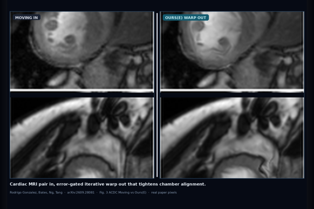
Hang a fixed and moving cardiac MRI or echo pair into a viewer and you want the warped overlays, chamber contours, and registration-based ejection fraction to sit on the fixed frame, not drift a few pixels off the blood pool. That is the clinic ask Clara Rodrigo González, Oscar Bates, Fu Siong Ng, and Meng-Xing Tang take on in arXiv:2609.28081, posted late September 2026 from Imperial College London. The paper is Recursive Uncertainty-Gated Image Registration for Learning-based Algorithms, and the authors name the method RUGI. They refine a learning-based deformation iteratively, with a spatial gate that decides where each update is allowed to move, then check whether that cleaner warp also cuts error on a registration-based EF surrogate.
Single-step registration networks are fast, but they commit to one full-field prediction. Hard regions (thin walls, large ED-ES motion, echo speckle) stay under-corrected while easy myocardium may already be fine. RUGI keeps the same base model and runs it again on the latest warped moving image, multiplying later increments by a gate so refinement concentrates where residual mismatch or predicted uncertainty is still high. The error-gated variant can wrap pretrained VoxelMorph, TransMorph, or CycleMorph at inference with no retrain.
What hangs on the viewer
Upstream is a fixed/moving pair from cine cardiac MRI (ACDC short-axis in the paper) or echocardiography (CAMUS). Downstream is a composed deformation field, a warped moving image that should match the fixed frame, optional residual or uncertainty maps, and, when you also have an ES left-ventricular mask, a registration-based EF number obtained by warping that mask into the ED domain.
For a DICOM or echo viewer rail, the hangable pieces are: the iteratively warped overlay beside the fixed series, a residual heatmap (fixed minus warp) for quick QC, and an EF panel driven by the same Φ rather than a second segmentation stack. Error-gated RUGI is the practical plug-in when you already ship VoxelMorph-family weights and do not want a training campaign. Uncertainty-gated RUGI adds a small U-Net that predicts where the base model is unreliable, which is useful if you want per-pixel trust maps and Monte Carlo intervals on EF.
How it works in plain words
Start with Φ as identity and I(0) as the moving image. At step t the registration model R predicts an incremental deformation from the fixed image and the current warped moving image. A gating map s(t) is computed from either a learned uncertainty network U or from the image residual error between fixed and current warp. The first step applies the full increment; later steps multiply the increment by the smoothed, min-max-normalised gate so well-aligned regions freeze earlier. Increments compose into Φ until an MSE-based similarity stop fires, or until a candidate update would push the Jacobian too far into folding (the paper rejects updates when the Jacobian distribution falls below −0.4).
Uncertainty gating trains U to predict per-pixel standard deviation of stochastic deformation samples from R, then uses that map as the gate at inference. Error gating skips the extra network: residual |fixed − warped| (optionally smoothed) becomes the gate. That is why error-gated RUGI drops onto pretrained IXI brain models without touching their training recipe. The authors also show that iterative refinement alone helps, but informative spatial gating beats no-gating, random gating, and a simple intensity prior in their ablation (global signed-rank effect G against those ablations stays large on both ACDC and CAMUS).
Training for their main cardiac experiments used a Bayesian registration network with image MSE (scaled by 100) plus bending-energy regularisation, and a lightweight 2-layer U-Net (~465k parameters) for uncertainty. Evaluation held out 50 ACDC patients and 100 CAMUS patients with patient-level splits. Pretrained wrap experiments used VoxelMorph, TransMorph, and CycleMorph weights from the literature on 58 paired samples.
What the numbers say
On the authors’ BNN baseline, both RUGI variants improve MSE, NCC, MI, and SSIM on ACDC and CAMUS relative to single-step inference, with patient-level Wilcoxon tests remaining significant after Benjamini-Hochberg correction. Anatomical alignment follows the same pattern when they warp segmentation masks and score Dice. On ACDC, median tissue DSC rises from 73.8% (single-step) to 83.7% with uncertainty gating and 84.6% with error gating. On CAMUS the lifts are smaller: 78.2% to 82.3% (U) and 82.0% (E), which matches how noisy ultrasound correspondence is harder to rescue with intensity residuals alone.
The headline for a shop that already owns pretrained registration weights is the error-gated wrap: median MSE fell by 30.2% on VoxelMorph, 37.1% on TransMorph, and 27.4% on CycleMorph, with NCC and MI also up across all three (rank-biserial correlation 1.00 on those comparisons). That is the abstract’s 27-37% band, cited exactly. SSIM was architecture-dependent; CycleMorph even lost SSIM while still gaining on MSE/NCC/MI, so do not treat SSIM as your sole go/no-go.
Registration-based EF is the functional check. Warp the ES LV mask to ED with Φ and compute an area-based EF from the relative chamber change. On ACDC, EF mean absolute error drops from 27.5% (BNN) to 14.9% (RUGI-Unc) and 16.0% (RUGI-Err). On CAMUS it drops from 30.9% to 23.0% and 25.2%. All methods still tend to underestimate EF when they under-capture contraction; RUGI mainly closes that gap. Uncertainty variants can also attach Monte Carlo 95% intervals on EF by perturbing Φ with the per-pixel uncertainty map.
Compute is the trade: multistep inference ran about 5× to 10× longer than one-step on the paper’s timings (median wall-clock per sample), with error gating often needing fewer steps than uncertainty gating on ACDC. Fine for offline cine batches; tight for real-time echo loops unless you cap T hard.
Where it fails and what not to trust
This is research software on 2D cardiac pairs in a simulated study setting, not a cleared medical device. The EF number is an area-based surrogate from 2D masks, not a volumetric clinical EF from a full short-axis stack. Rebuild and validate on your own cine protocol, vendor mix, and ED/ES definition before you hang EF next to a reportable value.
Uncertainty gating needs supervised samples from the base model, so the trust map is only as honest as that model’s stochastic behaviour. Error gating is simpler but leans on intensity residual as a proxy for anatomical error; echo speckle and out-of-plane motion can fool it, which is consistent with the smaller CAMUS gains. Stopping on image similarity can diverge from landmark or segmentation truth. Jacobian folding stayed under 2% of voxels in the paper’s tables, but iterative composition can still accumulate odd local warps: keep a folding and residual QC gate in the viewer.
Do not advertise cross-architecture SSIM wins after the CycleMorph result. Do not treat the IXI-pretrained wrap numbers as a promise on your cardiac domain without a local holdout. And do not hide the compute multiplier from whoever owns the batch queue.
For a viewer or clinic AI shop
Wire fixed/moving cardiac MRI or echo in; hang the composed warp, a residual QC layer, and optional uncertainty heatmaps out. Prefer error-gated RUGI as an inference wrapper around the VoxelMorph-family model you already deploy, with a max-iteration and Jacobian reject matching the paper’s spirit. Keep registration-based EF behind a research or assistive flag until you correlate it against your volumetric reference on a tagged cohort.
If you already ship a cardiac viewer, start with warped overlays and residual scrubbers for ED-ES pairs, then add EF as a secondary panel that shows the area surrogate label explicitly. Log gate type (U vs E), step count, final MSE, and percent negative Jacobian per study so support can debug bad warps. Validate on at least one internal ACDC-like MRI cohort and one echo cohort before you claim multi-modality hang. Rebuild from arXiv:2609.28081. PDF: https://arxiv.org/pdf/2609.28081.
Sources
- Rodrigo González, C., Bates, O., Ng, F. S., Tang, M.-X. Recursive Uncertainty-Gated Image Registration for Learning-based Algorithms. arXiv:2609.28081, 2026. https://arxiv.org/abs/2609.28081. PDF: https://arxiv.org/pdf/2609.28081.
- Error-gated wrap on pretrained models (paper): median MSE reductions of 30.2% (VoxelMorph), 37.1% (TransMorph), 27.4% (CycleMorph); abstract band 27-37% with no training changes.
- ACDC mask Dice (Table 3 means): single-step BNN 73.8% → RUGI-Unc 83.7% → RUGI-Err 84.6%. CAMUS: 78.2% → 82.3% / 82.0%.
- Registration-based EF MAE (Table 3): ACDC 27.5% → 14.9% (Unc) / 16.0% (Err); CAMUS 30.9% → 23.0% / 25.2%. Area-based 2D surrogate, not volumetric clinical EF.