
If your DICOM viewer only ever sees thick-slice clinical FLAIR or mixed-contrast brain MRI, FreeSurfer-style cortical surfaces are usually out of reach. Jesper Duemose Nielsen, Karthik Gopinath, Andrew Hoopes, Adrian Dalca, Axel Thielscher, Juan Eugenio Iglesias and Oula Puonti (DRCMR Copenhagen / DTU, Martinos MGH, MIT CSAIL, Harvard, UCL) posted arXiv:2610.16595 on 12 October 2026 with BrainNet, a deep model that reconstructs white-matter and gray-matter cortical surfaces, vertex-wise placement uncertainty, and a spherical registration to fsaverage from heterogeneous MRI, including non-T1w contrast and 1x1x5 mm clinical FLAIR. Inference is about 0.6 s on a GPU after affine template alignment. Code ships as a public CLI under GPL-3.0 with three pretrained TopoFit variants.
What goes in and what comes out
The input is a brain MRI volume. The networks expect a 1 mm isotropic, RAS-oriented (“conformed”) grid; the CLI can resample with --conform. Three TopoFit variants cover different training regimes: T1w-1mm (specialized on 1 mm T1w), synth-1mm (synthetic contrast at 1 mm), and synth-random (synthetic contrast with randomized resolution, the one that covers thick-slice clinical scans).
The output is a WM surface, a GM (pial) surface, a per-vertex placement uncertainty, and spherical coordinates on fsaverage for atlas overlays such as Desikan-Killiany. A viewer can use those meshes for thickness and curvature, paint uncertainty as a QC layer, and drop atlas labels without waiting hours for FreeSurfer or recon-all-clinical (RAC).
How it works
BrainNet deforms a template mesh with a UNet image feature extractor and graph-convolution blocks that predict per-vertex velocities, uncertainty, and spherical coordinates. Training uses SynthSeg-style domain randomization: random contrast, resolution augmentation for thick-slice clinical patterns, and extracerebral tissue synthesis. At inference the package first affinely aligns the scan to a template (TREGA), then runs TopoFit. The GM surface is estimated from the WM surface, and uncertainty weights the data-fidelity terms so low-confidence vertices pull less hard on the fit.
What the numbers say
On the 1 mm T1w test set (n=490), the T1w-1mm model reaches ASSD about 0.143 mm on WM and 0.197 mm on GM against FreeSurfer silver-standard surfaces, on par with DeepSurfer and V2C-Flow (Table 4). Synth models trail the T1w-specialized model on research T1w, which is expected.
On 1x1x5 mm FLAIR from ADNI GO-2 (n=199), synth-random reports ASSD about 0.70 mm WM and 0.68 mm GM, better than RAC at about 0.94 / 0.93 mm, with mesh quality metrics still close to the 1 mm T1w numbers. Subject-mean uncertainty correlates with error on that low-res FLAIR set (Pearson r about 0.67 WM, 0.69 GM). Spherical registration Desikan-Killiany region accuracy is about 0.887 for the model prediction, versus CAT12 at 0.894 and JOSA at 0.867 on the generalization T1w set.
What the pictures show
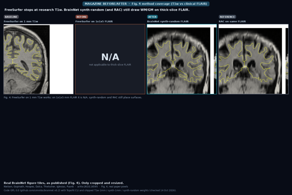
The featured image is cut from Fig. 4 of the paper. Left to right: FreeSurfer on 1 mm T1w (works), FreeSurfer on 1x1x5 mm FLAIR (N/A), BrainNet synth-random on the same FLAIR (surfaces present), and RAC on that FLAIR as the clinical reference. The yellow overlays are WM/GM contours on coronal slices. The point for a clinic shop is the middle gap: tools locked to research T1w leave clinical thick-slice series blank, while synth-random still returns a surface you can overlay in a viewer.
Where it falls short
FreeSurfer is a silver standard, not gold. Small ASSD gaps between DL methods on 1 mm T1w can be meaningless, and the authors note cases where a DL surface looks locally better around WM hypointensities than FreeSurfer. Synth models lag the T1w-specialized model when the input is research T1w.
Inputs are forced onto a 1 mm grid, so sub-millimeter acquisitions are downsampled and do not gain from finer native spacing. Major cortical pathology and strong artifacts are untested; the authors say accuracy under major cortical abnormalities remains to be evaluated. The noise model used in synthesis is Gaussian and does not match real MRI noise. There is no formal uncertainty calibration against ground truth, mainly because no ground-truth uncertainty exists. Ex vivo performance is lower than in vivo, though still ahead of RAC on the reported BigBrain and Edlow cases for most metrics.
Code, weights, and data
The package is github.com/simnibs/brainnet, GNU GPL-3.0, tagged v0.2 (checked 14 October 2026). It is not on PyPI yet; install from git with the documented cortech and brainsynth wheels. CLI entry points: brainnet trega for affine template alignment and brainnet topofit for surfaces, uncertainty and spherical registration. Contrast/resolution flags select the variant, for example brainnet topofit -c synth -r random image.nii.gz ./out.
Pretrained TopoFit weights ship inside the repo under brainnet/resources/models/topofit/: state_t1w_1mm.pt, state_synth_1mm.pt, state_synth_random.pt, plus a TREGA affine model. Imaging data does not ship with the package; the paper trains and evaluates on public cohorts (ABIDE, ADHD200, ADNI, AIBL, HCP, OASIS3, ADNI GO-2 FLAIR, BigBrain, Edlow, and others listed in Table 3).
How we would wire it into a viewer
We would run TopoFit as a server-side job that never writes back into the original DICOM pixels. Read the brain series, conform to 1 mm RAS if needed, pick synth-random for clinical mixed contrast or thick-slice FLAIR and T1w-1mm when you already have research T1w, then store WM and GM meshes plus a vertex uncertainty attribute as derived objects. In the viewer, paint uncertainty as a heat overlay for QC, and map Desikan (or another fsaverage atlas) through the predicted spherical coordinates for region tooltips. Failed or high-uncertainty patches (insula, temporal pole, occipital small folds) should stay visually distinct so a reader can correct them.
Before shipping we would time the full pipeline on our matrix sizes, compare a sample of T1w cases against FreeSurfer silver-standard surfaces, and separately check FLAIR-only studies against RAC or a reader review. Keep site-specific conform and model-variant settings in config rather than hard-coding T1w-1mm for every series. For related work on this site, see our posts on liver MRI masks under scanner shift and numbered rib centerlines from thoracic CT.
Sources
- Nielsen, J.D., Gopinath, K., Hoopes, A., Dalca, A., Thielscher, A., Iglesias, J.E., Puonti, O. Uncertainty-Aware Cortical Surface Reconstruction and Spherical Registration for Heterogeneous Brain MRI. arXiv:2610.16595, 2026. https://arxiv.org/abs/2610.16595. PDF: https://arxiv.org/pdf/2610.16595.
- Code: https://github.com/simnibs/brainnet (GPL-3.0, v0.2, TopoFit + TREGA CLI, checked 14 October 2026).
- Weights: shipped in-repo under
brainnet/resources/models/topofit/(T1w-1mm, synth-1mm, synth-random) andtrega/. - Related baselines cited in the paper: FreeSurfer, DeepSurfer, V2C-Flow, RAC (recon-all-clinical), CAT12, JOSA, SynthSeg-style domain randomization.
- Featured image: Fig. 4 of arXiv:2610.16595 (FreeSurfer T1w, FreeSurfer FLAIR N/A, synth-random FLAIR, RAC FLAIR), as published.