
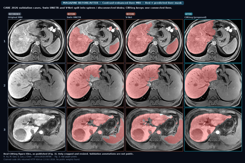
Contrast-enhanced liver MRI intensities shift across scanners, vendors and protocols. A mask that looks fine on the training scanner can grow false-positive blobs and jagged boundaries on a new site, often spilling into the spleen. Ruoshi Xu (Chinese Medicine Guangdong Laboratory, Zhuhai), Mingqi Gao (University of Sheffield), and corresponding authors Shengda Luo (Chinese Medicine Guangdong Laboratory) and Jingkun Chen (Northwestern Polytechnical University, Xi’an) posted arXiv:2610.09784 on 7 October 2026 with CIRSeg. It is a cascaded nnU-Netv2 liver segmenter for the CARE 2026 GED4 Contrast-Enhanced LiSeg subtask that trains with 3D CutMix and stochastic intensity transfer, runs a coarse-then-fine cascade, optionally adapts at test time without source data, and finishes with largest-connected-component filtering. The claimed GitHub URL returned HTTP 404 when we checked on 13 October 2026, and we found no public weights.
What goes in and what comes out
The input is a 3D contrast-enhanced liver MRI volume from the CARE 2026 official train, validation or test split. No extra public or private datasets are used. The official metrics are Dice and the 95th-percentile Hausdorff distance (HD95).
The output you hang on a series is a 3D binary liver mask. In a DICOM viewer that usually means an RTSTRUCT or a DICOM SEG overlay tied to the series, never burned into the pixels. After post-processing the mask is a single connected liver component.
How it works
The baseline is CoSSeg-TTA, the CARE Liver 2025 first-place recipe: nnU-Netv2 with mean-teacher semi-supervised learning. CIRSeg keeps that setup and adds three pieces.
During training, 3D CutMix mixes a random cuboid between two labeled volumes (and their labels) with probability 0.5. Separately, each selected volume gets a stochastic intensity transfer: histogram matching with probability 0.7 or Nyul landmark matching with probability 0.3, with the reference drawn from the full CARE 2026 training set (labeled and unlabeled). Both intensity transforms leave the segmentation label unchanged. That exposes the model to more intensity distributions than the 30 annotated training cases alone would show.
The model is a cascade. A low-resolution nnU-Netv2 predicts coarse liver extent. That prediction is upsampled and concatenated as an extra channel into a full-resolution cascade that refines boundaries. Both stages use SGD on one NVIDIA RTX 6000 24GB, learning rate 1e-2, weight decay 3e-5, with supervised Dice+CE loss and an MSE consistency term between student and EMA teacher on unlabeled data.
At inference, the low-resolution stage runs without adaptation. The full-resolution cascade can run source-free continual test-time adaptation inspired by SicTTA: a frozen anchor and an adaptive student, confidence-filtered pseudo-labels, and a probability-prior regularizer so class proportions do not drift. Adapted student weights carry forward across the test stream. Finally, a deterministic 3D largest-connected-component filter keeps only the biggest foreground component and drops isolated false-positive blobs.
What the numbers say
On five-fold cross-validation over the 30 annotated training cases (Table 1), CIRSeg reaches Dice 0.9595 and HD95 5.29 mm. The reproduced CoSSeg-TTA baseline is 0.9445 / 7.74, and plain nnU-Netv2 is 0.8973 / 26.95. The HD95 drop tracks the reduction in distant false positives and rough boundaries.
On the official CARE evaluation website (Table 2, cumulative ablation), the baseline alone already has Dice 0.9693 but HD95 24.69 mm. Adding largest-connected-component filtering cuts HD95 to 20.61. The full CIRSeg stack (Exp E: LCC + CutMix + composite intensity aug + cascade + TTA) reaches Dice 0.9713 and HD95 18.31. On the official test splits with TTA enabled, in-domain Test1 is 0.9713 / 20.18, in-domain Test2 is 0.9709 / 20.85, and the out-of-distribution test is 0.9797 / 11.36. The abstract rounds the in-domain and unseen figures to 97.13% / 20.18 mm and 97.93% / 11.30 mm; Table 2 is the more precise source.
The authors note that TTA helps the OOD subset more than in-domain cases. All of their test submissions used TTA, so there is no paired no-TTA comparison, and the sensitivity of continual TTA to case ordering was not measured.
What the pictures show
The featured image is cut from Fig. 3 of the paper: three CARE 2026 validation cases with red liver overlays. From left to right in the paper the columns are original image, nnU-Net, Attention U-Net, SegResNet, Swin UNETR, 3D U-Net, V-Net and CIRSeg. The banner keeps the original as reference, Swin UNETR and V-Net as the clear failure modes (spleen spill and disconnected blobs), and CIRSeg as the cleaned result. Validation annotations are not public, so this is a visual comparison only.
Where it falls short
The method is built for one CARE 2026 subtask on official splits only. There is no public code or weights as of 13 October 2026, despite the paper listing a GitHub URL. Continual TTA updates the student across the test stream, so results can depend on case order, and the authors could not measure that because organizers ran the official ordering. TTA did not help the mainly in-domain validation set. HD95 on the official in-domain tests is still around 20 mm, which is better than the baseline’s 24.69 but not a tight surface match. There is no reader study and no test outside the CARE GED4 contrast-enhanced liver MRI distribution.
Code, weights, and data
The paper points to https://github.com/jingkunchen/MICCAI_CARE_2026. On 13 October 2026 that URL returned HTTP 404, as did a /CIRSeg variant. We found no Hugging Face model card and no public checkpoints. The only data used is the CARE 2026 official release for the GED4 Contrast-Enhanced LiSeg subtask; validation labels are not public.
How we would wire it into a viewer
Once weights exist, we would run CIRSeg as a server-side job when a contrast-enhanced liver MRI series arrives. Write the output as a new DICOM SEG or RTSTRUCT with its own Series Instance UID, ImageType DERIVED\SECONDARY, and a derivation description that names the model, cascade stage and whether TTA ran. In the viewer the overlay opens next to the source with a clear “AI liver mask” label so a reader can hide it in one click. We would keep largest-connected-component filtering on by default, log any discarded components, and gate deployment with a local holdout across our own scanners before trusting OOD claims from the challenge set. For related work on this site, see our posts on prompt-free MRI organ boxes (A4-MR) and MedSAM2 for hip MRI anatomy.
Sources
- Xu, R., Gao, M., Luo, S., Chen, J. CIRSeg: Coarse-to-Fine Intensity-Robust Liver Segmentation with Source-Free Continual Test-Time Adaptation. arXiv:2610.09784, 2026. https://arxiv.org/abs/2610.09784. PDF: https://arxiv.org/pdf/2610.09784.
- Claimed code: https://github.com/jingkunchen/MICCAI_CARE_2026 (HTTP 404 on 13 October 2026). No public weights found.
- CARE 2026: Comprehensive Analysis of Real-world Medical Images, Liver Track, GED4 Contrast-Enhanced LiSeg subtask. https://www.zmic.org.cn/care_2026/.
- Lou, J. et al. CoSSeg-TTA (CARE Liver 2025 first place), as cited. Isensee, F. et al. nnU-Net. Wu, J. et al. SicTTA.
- Featured image: Fig. 3 of arXiv:2610.09784 (three CARE 2026 validation cases), as published.