
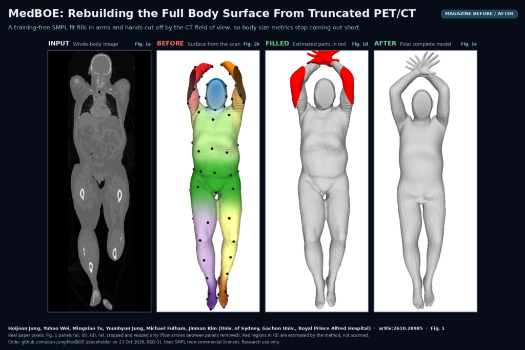
Anyone who has rendered the skin surface of a routine whole-body PET/CT has seen the problem. The arms are clipped at the elbows, the hands are missing or sit outside the reconstructed CT field of view, and sometimes the side of the torso is shaved flat where the patient touched the edge of the scan circle. In a 3D view that is only cosmetic. Anything that measures the body from that surface, such as body surface area, body volume or waist circumference, comes out too small, and nothing in the output says so. Hoijoon Jung and Yuhao Wei (equal contribution), with Mingxiao Tu, Younhyun Jung, Michael Fulham and Jinman Kim (University of Sydney, Gachon University and Royal Prince Alfred Hospital, Sydney) posted arXiv:2610.28985 on 20 October 2026 describing MedBOE, the Medical Body model Optimization-based Estimator. It takes the incomplete body surface from a whole-body scan and estimates the missing parts by fitting a parametric human body template, with no training on medical data.
Why a viewer or platform team should care
A body surface is one of the cheapest things you can extract from a whole-body scan: threshold or segment the patient, run marching cubes, done. That is why it keeps showing up in platform feature requests. Nuclear medicine teams want body habitus measures for dosing injected radiotracer activity, dosimetry and simulation groups want patient-specific body models, and surgical navigation tools want a skin mesh to register against. All of these assume the surface is closed and complete. MedBOE is written for the case where it is not. It does not touch the image or the sinogram, so it is not a field-of-view extension method for reconstruction. It produces a complete, continuous surface mesh, with the patched regions known, that a measurement or export step can trust more than the raw clipped surface.
What goes in and what comes out
The input is a whole-body CT, PET or MRI volume plus three values from the image header: sex, weight and height. The output is a watertight triangle mesh of the whole patient in the scanned pose, in which the regions that were outside the field of view (lateral arms, elbows, forearms, hands, parts of the torso) have been estimated. Along the way the pipeline also produces the fitted SMPL body model, with shape and pose parameters, and the boundary between observed and estimated surface. From the final mesh the authors compute five body habitus metrics: body surface area, body volume, and chest, waist and hip circumference.
How it works
There are three modules. The first builds the incomplete body model. A pretrained nnU-Net segments the patient’s body, marching cubes turns the mask into a mesh, Taubin smoothing and decimation clean it up, and the Deep Virtual Marker network labels every vertex with 99 body landmark responses. The same landmarks are computed on the default male and female SMPL meshes, which gives the fit anatomical correspondences rather than just nearest points.
The second module fits SMPL, the Skinned Multi-Person Linear model widely used for human body shape in computer vision, to that partial surface. Shape, pose and translation are optimized with Adam over 500 iterations against a weighted sum of six terms: sparse landmark distance, a filtered Chamfer surface distance, a pose prior (VPoser and a Gaussian mixture prior), a weight term that ties SMPL shape to the patient’s recorded weight through BetaNet, a self-contact penalty that stops hands from sinking into the torso, and a dense landmark term. The weights are scheduled: landmarks dominate early, surface distance takes over later, and the self-contact term only switches on for the last 100 iterations because it is expensive.
The third module fills the gaps. A coarse non-rigid deformation pulls the fitted SMPL mesh toward the observed surface while ignoring areas that do not overlap. The deformed mesh is then trimmed along the boundary of the scan surface, and the pieces outside it, shown in red in the paper’s Fig. 1, are stitched onto the patient surface with Poisson surface reconstruction. A final fine deformation fits SMPL to this stitched model, with a strong volume consistency term so the patient-specific body size from the weight-guided fit is kept. That last step smooths the seams between real and estimated surface.
What the evaluation shows
The authors used two public datasets. From AutoPET they kept 91 true whole-body FDG PET/CT exams with recorded weight, and built ground truth surfaces from the PET, whose transverse field of view is wider than the CT’s. From HIT (Human Implicit Tissues) they used 40 test subjects of whole-body MRI body segmentations with reference SMPL fits. In both cases the incomplete input was simulated by trimming the ground truth with a cylinder at 95% of the average CT field-of-view radius, and HIT also had the region above the head cut off. MedBOE was compared with four methods adapted from computer graphics: IF-Net, ShapeFormer, PCN and ArtEq.
MedBOE gave the best volumetric agreement in most settings, and the paper’s Fig. 3 shows the reason: IF-Net tends to bridge a cut elbow with a near-flat patch that scores well on Chamfer distance but looks nothing like an elbow, while MedBOE rebuilds an elbow shape. On surface distance it ranked a close second. The number that matters for a measurement feature is the body habitus check on HIT: body volume from the completed mesh was within 0.85% of the reference on average. Surface area and the chest, waist and hip circumferences were within about 2% on average as well. Removing the gap-filling module made the volumetric errors clearly worse, so the template fit alone is not enough.
It is slow. MedBOE needed about 21 minutes per patient on an NVIDIA TITAN V, not counting segmentation and mesh building, against seconds for the learned baselines. The authors point out that newer GPUs are much faster than the TITAN V, but this is still a per-case optimization, so plan for a batch job, not an interactive button.
What it does not show
No real clipped scan was evaluated against a real complete one. Every incomplete input was made by trimming a complete surface with a cylinder, which does not reproduce real field-of-view geometry, PET/CT misregistration or truncation artifacts. On AutoPET, forearms and hands were often outside even the PET ground truth, so the AutoPET scores only cover regions that exist in the reference. The habitus metrics were only checked on HIT, where weights had to be estimated from the reference volume because measured weights were not available, and HIT subjects were scanned prone with arms overhead, inside a field of view smaller than that of most CT scanners. When arms touch the torso, the landmark network becomes unreliable and fitting gets harder, as the authors show in an appendix failure case. The pose and shape priors were pretrained on general human body datasets from computer vision rather than on supine patients, and SMPL’s limited shape space can introduce small changes around the abdomen and chest. None of this has been tested on children, amputees, casts, braces or very large patients, and there is no prospective clinical use.
Code, data and license
The paper says the source code and optimization weights are public at github.com/Joon-Jung/MedBOE. When we checked on 23 October 2026, that repository held only a BSD 3-Clause license and a README marking it as a placeholder, with code to come on manuscript acceptance. For now there is nothing to run. There are no MedBOE network weights in the usual sense, since the method is training-free, but it depends on several pretrained components, and their licenses matter more than the BSD file. SMPL, VPoser and the self-contact code from the Max Planck Institute for Intelligent Systems are distributed for non-commercial research under their own terms, so any product use needs a separate commercial license for the body model. AutoPET and HIT are public research datasets with their own access terms. MedBOE is a research method, not a medical device.
How we would use it
We build DICOM viewers and imaging platforms, and the first place we would try this idea is as a quality step behind any body measurement feature on whole-body PET/CT. In practice that means: extract the body surface, run the completion as a background job, show the estimated regions in a different color on the 3D view, and report body surface area or volume with a flag saying how much of the surface was estimated. Clinicians should be able to see at a glance whether a number came from the patient or from the template. Until the code is released we would prototype the first module ourselves, since body segmentation, marching cubes and mesh cleanup are standard, and keep the SMPL-based part strictly in research until the licensing is sorted. For related reading, see our posts on whole-body PET/CT organ masks, on low-count total-body PET reconstruction, and on capturing 3D mesh screenshots with Vedo.
Sources
- Jung, H., Wei, Y., Tu, M., Jung, Y., Fulham, M., Kim, J. MedBOE: Optimization-Based Estimation of Complete Patient Body Models from Whole-Body Medical Imaging. arXiv:2610.28985, 2026. https://arxiv.org/abs/2610.28985. PDF: https://arxiv.org/pdf/2610.28985.
- Code (placeholder when checked on 23 October 2026, BSD 3-Clause): https://github.com/Joon-Jung/MedBOE.
- SMPL body model and license: https://smpl.is.tue.mpg.de/. VPoser: https://github.com/nghorbani/human_body_prior.
- Featured image: Fig. 1 of arXiv:2610.28985 (whole-body image, reconstructed incomplete model, initial complete model with estimated regions in red, final complete model), real paper pixels, cropped and resized only, with the flow arrows between panels removed.