
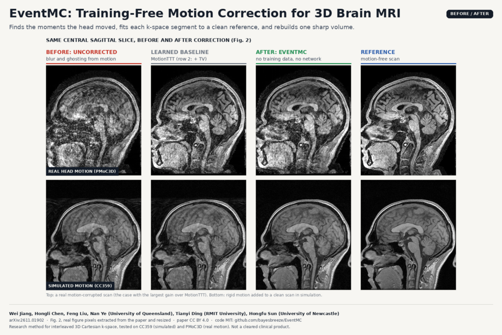
Head motion during a 3D brain MRI does more than spoil the picture. A patient who shifts a few millimetres during a T1 scan can blur the cortex enough to bias cortical thickness and grey-matter volume, and those numbers end up in research cohorts and clinical comparisons. In arXiv:2611.01902, Wei Jiang, Hongli Chen, Feng Liu and Nan Ye from the University of Queensland, Tianyi Ding from RMIT University and Hongfu Sun from the University of Newcastle describe EventMC, a retrospective motion correction method that needs no training data and no pretrained network.
EventMC works on raw multi-coil k-space, so it runs at reconstruction time and needs data that a DICOM viewer never sees. I come back to what that means for a workflow below.
What the method does
EventMC targets interleaved 3D Cartesian acquisitions. The scan is a sequence of shots, each acquiring one phase-encoding line, and consecutive shots are grouped into segments (13 in the paper). Because the ordering is interleaved, every segment samples both the centre and the edges of k-space. The method assumes the head holds one rigid pose per segment and estimates that pose, plus one motion-corrected image.
- Find the motion events. Head motion is usually slow drift with a few sudden jumps. EventMC fits a pose for each segment, then flags a jump wherever the displacement between neighbouring segments exceeds three times the local noise level and at least 0.6 mm. These jumps split the scan into stable intervals.
- Pick an anchor. A pre-run places the anchor segment in the middle of the longest stable interval. Its pose is fixed at zero, and the final image comes out in that head position.
- Stage A: fit each segment to a clean reference. Each segment’s pose is fitted against a total-variation reconstruction built only from the stable interval, leaving out segments next to an event and leaving out the target segment itself. Leaving the target out stops a bad pose estimate from reinforcing itself through its own data. This runs for five rounds, working outward from the anchor.
- Stage B: coarse-to-fine refinement. The image and all poses are refined together on a growing central band of k-space (a quarter, a half, then all of it), because low frequencies are less sensitive to small pose errors. A final reconstruction uses all samples with the poses fixed.
- Multi-anchor safety net. A single run can fail with a badly placed anchor, so the full method runs up to three candidates and keeps the one most consistent with the measured data.
Rotations are applied with Fourier shears and translations with phase ramps, so there is no interpolation kernel in the forward model, and coil sensitivities stay fixed to the scanner.
What comes out of it
The output is a complex 3D volume in the anchor’s head position plus a six-parameter pose (rotation in degrees, translation in mm) for every segment. The featured image shows Fig. 2 of the paper: the same central sagittal slice before correction, after a learned baseline, after EventMC, and the motion-free reference. On the real scan in the top row, the uncorrected image has ghosting across the whole head and a smeared cerebellum. EventMC’s version brings back the cerebellar folds and a clean corpus callosum outline, though it is noisier than the reference.
A QA step could also use the pose trace to log how much a patient moved and when, and flag studies where the motion was too frequent for the method to trust.
Where it would sit in a workflow
Because EventMC needs the multi-coil k-space, the segment ordering and the sampling mask, it belongs on the reconstruction side: a research reconstruction server or an offline reprocessing job that has access to raw data. A PACS or DICOM viewer that only holds magnitude images has nothing for it to work on. For a team that already stores raw data for its 3D T1 protocol, the practical path is to rerun reconstruction on flagged scans and push the corrected volume back as a new DICOM series next to the original, so the radiologist can compare the two.
Cost is reasonable for offline use. A single run takes a median of under 7 minutes per scan (12 coils, 170 x 218 x 256) on one NVIDIA H100, and the full multi-anchor version takes under 12 minutes end to end.
How they tested it
The authors added realistic rigid motion (slow drift plus one to three abrupt events) to the raw 12-coil k-space of nine CC359 subjects, at full, two-fold and four-fold sampling, and they ran 26 real motion-corrupted scans from PMoC3D, each with the same subject’s motion-free scan as reference. The main baseline is MotionTTT, a learned test-time-training method; to separate the motion estimate from the image prior, they also fed MotionTTT’s motion into EventMC’s own final reconstruction.
At full sampling in simulation, EventMC reaches 34.8 dB mean PSNR against 30.8 dB for MotionTTT with the same final reconstruction, so the gap comes from better motion estimates rather than a better prior. On the real scans, it lands at 32.7 dB against 32.6 dB for MotionTTT and 30.7 dB uncorrected. That is a tie with a learned method, without any training data.
Where it falls short
- Heavy undersampling. At four-fold acceleration, the TV prior cannot build reliable references and EventMC no longer beats MotionTTT with the same reconstruction. The real PMoC3D scans run at about 4.9-fold, which is why the real-motion result is a tie rather than a win.
- Motion within a segment. One pose per segment means a short movement inside a segment gets a compromise pose and stays partly uncorrected.
- Restless patients. Frequent movements leave no long stable interval for the anchor. The worst real-motion cases were exactly these.
- Acquisition requirements. Every segment must cover low and high frequencies, so linear or centric orderings are out, and so is any setup without raw k-space.
- Small evaluation. Nine simulated subjects and eight real subjects. Three test subjects also informed the multi-anchor rule, which the authors flag openly.
Code, weights and license
The code is public at github.com/bayesbreeze/EventMC under the MIT license, with a Python API (run_eventmc) and a synthetic phantom demo. There are no weights because there is nothing to train. The repository contains the method only: the motion simulation, baselines and evaluation scripts used for the paper are not included. The preprint is released under CC BY 4.0. For related reading, see our posts on faster follow-up brain MRI using a prior scan and cortical surfaces from any brain MRI.
My take
A careful physical model matched a learned method on real motion here, with no training set to collect and no domain shift to worry about. I like that EventMC is MIT-licensed, readable and open about where it loses. For a clinic or research site that keeps raw data for its 3D T1 scans, it is a cheap thing to try on the studies that would otherwise be repeated. It will not help anyone who only has the DICOM images, and it needs interleaved ordering, so check the protocol before planning around it.