
Most follow-up brain MRI is a repeat of a scan the hospital already has. A tumor patient comes back every few months for the same T1 protocol, and the previous exam sits in the archive, but the scanner reconstructs today’s data as if it had never seen this head before. Nikola Janjušević, Hersh Chandarana, Yao Wang and Li Feng, from NYU Grossman School of Medicine and NYU Tandon, posted arXiv:2610.22643 on 15 October 2026 with a reconstruction network called LGGS (Longitudinal Guided Group Sparsity). It takes a heavily undersampled follow-up acquisition plus the patient’s prior exam and uses the prior to decide which faint structures in the new image are real anatomy and which are undersampling noise. The payoff is a shorter follow-up scan that keeps detail an unguided reconstruction loses.
Why a viewer or PACS team should care
Reconstruction sounds like scanner-vendor territory, and today it mostly is. But this method also needs the right prior study for the same patient, and finding that study is a lookup a PACS or viewer platform already does every day when it hangs comparison studies. If longitudinally guided reconstruction reaches clinical use, someone has to find the matching prior series, ship it to the reconstruction server and then show the reader which prior was used. The second reason is display. A follow-up series that was rebuilt with help from an older exam should be labelled and shown next to that exam, so the reader can check that a new finding was not smoothed toward the old anatomy.
What goes in and what comes out
LGGS works on raw data, not on DICOM images. The inputs are the undersampled multi-coil k-space of today’s scan and the prior exam’s images, three neighbouring slices centred on the slice being reconstructed. The prior is first aligned to today’s scan with a whole-voxel 3D shift estimated from the fully sampled k-space centre of both exams, so no rotation is corrected at this step. Coil sensitivity maps come from ESPIRiT. The output is one reconstructed 2D slice at a time, complex-valued, which a scanner or reconstruction server would then turn into the usual magnitude series.
How it works
LGGS is an unrolled network. It alternates between a data-consistency step that keeps the image faithful to the measured k-space and a learned clean-up step, for a fixed number of iterations. It is built on two earlier models from the same group. LPDSNet supplies the backbone, which represents the image as feature codes and suppresses weak codes with a learned threshold, the way classical sparse reconstruction removes noise. GroupCDL supplies the idea of grouping similar codes so they share one threshold decision.
The new part is where the groups come from. For every pixel of today’s image, LGGS looks at a small 3 x 3 window in each of the three prior slices and measures how similar the feature codes are. Where a feature in today’s image matches a feature in the prior, the threshold is relaxed and the faint feature survives. Where nothing in the prior matches, the threshold stays as it would be without a guide. The prior never gets copied into the output; it only changes how hard the network cleans up each spot. With no guide at all, the network reduces exactly to the unguided LPDSNet, which makes the comparison between the two a clean test of what the prior adds.
Two details deal with real-world mess. MRI phase drifts between visits because of field inhomogeneity, coil loading and coil map estimation, even when the anatomy has not changed. LGGS compares codes with a similarity measure that ignores a shared phase offset, so the phase drift does not break the matching. And because it searches a small window across three slices, it tolerates about a pixel of leftover in-plane shift and a slightly different slice position.
What the numbers say
The data are in-house: 70 brain tumor patients from NYU, each with three T1-weighted exams and fully sampled raw k-space, 210 volumes in total. Twelve patients were held out for testing. Undersampling was retrospective, with nominal acceleration from 6x to 16x. LGGS beat its unguided twin LPDSNet, the unguided E2EVarNet and the longitudinal L-TGVN on NRMSE, SSIM and LPIPS at every rate, and the gap to the unguided models widened as acceleration went up. Read at equal error, the curves show LGGS reaching 10.32% NRMSE at 10x nominal, where L-TGVN needed 8x for the same error, which the authors describe as 25% more acceleration for the same image quality.
For a real archive, the accelerated-prior test is the one to read. Most prior exams were themselves acquired with acceleration, so the authors replaced the fully sampled prior with a 4x or 6x reconstruction. At 16x, a 4x prior cost only 0.09 percentage points of NRMSE compared with a fully sampled prior, while dropping the prior altogether cost 6.89 points. The model is also small, 1.08 million parameters against 43.6 million for L-TGVN, at a similar wall-clock cost per pass on an A100.
The figure
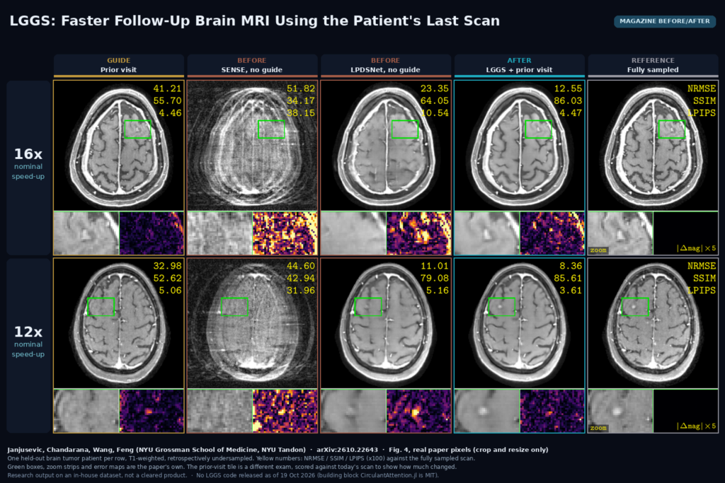
The featured image uses two rows of Fig. 4, one held-out patient per row. From left to right: the prior visit used as the guide, a classical SENSE reconstruction of today’s undersampled data, the unguided LPDSNet, LGGS and the fully sampled scan as reference. Under each image is a magnified region and an error map. At 16x, SENSE is buried in aliasing. LPDSNet looks clean at first glance, but the sulci are smeared and its error map in the zoom is bright and structured. LGGS keeps the sulcal detail and its error map is much flatter. In the 12x row, the magnified region contains a bright focal spot that is clear in today’s fully sampled scan and barely visible in the prior visit. LPDSNet shows it as a soft blur and its error map lights up on it. LGGS shows it sharply, so the difference between the two visits survived the guided reconstruction. The prior tile’s numbers are scored against today’s scan, so they also show how much changed between exams.
Limits
Everything was tested on T1-weighted brain MRI, where the head barely deforms between visits. The authors say plainly that large rotations and large anatomical deformation are not handled, so abdomen, pelvis or a patient repositioned at an angle are open questions. Undersampling was simulated from fully sampled data rather than acquired prospectively, and the authors call for prospective validation. The dataset includes tumor patients and the figures show lesions being preserved, but there is no systematic lesion-level study or reader study yet, and the authors list that as future work. The method needs raw k-space and coil data for today’s scan and, for the alignment step, the k-space centre of the prior as well, so it lives on the scanner or a reconstruction server with raw-data access, not in a PACS that only holds DICOM. The guide in the main experiments is complex-valued; a magnitude-only guide, which is what a DICOM archive usually holds, still beat the unguided model in the ablation, with a smaller gain.
Code, data and license
As of 19 October 2026 there is no public LGGS code or weights. The paper links one building block, CirculantAttention.jl, which is MIT-licensed, and all models were written in Julia with Lux.jl, so a Python shop would be porting rather than installing. The NYU dataset is in-house under an IRB protocol and is not released. If you want to try the idea, the realistic path today is to wait for the code or reimplement the guided threshold on top of an existing unrolled reconstruction, trained on your own raw data.
How we would wire it into a viewer
We would treat the prior lookup as part of the reconstruction request. When a follow-up exam is scheduled, query the archive (QIDO-RS or C-FIND on patient ID, body part and series description) for the most recent matching T1 series, and send its reference to the reconstruction server along with the raw data. After reconstruction, the server writes the series with a clear description such as “T1 recon guided by prior exam (LGGS)” and a reference to the prior’s Study and Series Instance UIDs. In the viewer, the hanging protocol should place the prior next to the guided follow-up with linked scrolling, which is how a reader would compare them anyway, and a small banner on the viewport should say the reconstruction used a prior exam. For any site evaluating this, we would also keep an unguided reconstruction of a sample of cases, so radiologists can check new or growing lesions in both before trusting the guided one. For background, see what a DICOM viewer does, our post on few-step reconstruction of undersampled brain MRI, and our post on registering follow-up PET/CT to the baseline.
Sources
- Janjušević, N., Chandarana, H., Wang, Y., Feng, L. LGGS: Longitudinal MRI Reconstruction via Guided Group Sparsity. arXiv:2610.22643, 2026. https://arxiv.org/abs/2610.22643. PDF: https://arxiv.org/pdf/2610.22643.
- Building block linked in the paper (MIT): CirculantAttention.jl, https://github.com/nikopj/CirculantAttention.jl.
- Featured image: Fig. 4 of arXiv:2610.22643, real paper pixels, cropped and resized only.