
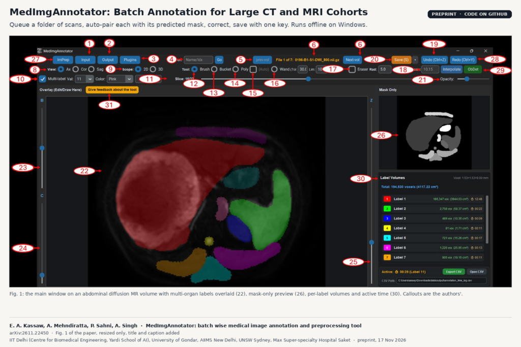
Anyone who has built a segmentation dataset knows that most of the hours go on opening each volume, finding the matching prediction file, loading it as an overlay, fixing the orientation, saving, and doing it all again for the next case. The team behind MedImgAnnotator hit this on a liver project with more than 14,270 abdominal MR and CT volumes to label. In their pilot, annotating 50 liver CT volumes in 3D Slicer took around 24 hours, mostly because of per-file loading, manual image and mask alignment, and menu navigation.
MedImgAnnotator, from the Indian Institute of Technology Delhi and partner institutions (arXiv:2611.22450, preprint), is the tool they built for it: a desktop application that loads a whole folder of scans as a queue, pairs each scan with its mask automatically, and puts the editing tools a few clicks away. The source code is on GitHub and a Windows installer is on Google Drive.
What it does
You point the tool at an image folder and, optionally, a folder of existing masks, such as nnU-Net predictions you want to correct. It sorts the files, remembers the last case you opened so you can resume later, and matches masks to images in three passes: exact file name first, then numeric tokens such as patient IDs, then approximate string matching (the Ratcliff-Obershelp algorithm, with a similarity cut-off of 0.6). If there is no mask, it creates an empty one so you can start from scratch. Prev and Next buttons step through the queue, and a single key press saves the mask as NIfTI.
The editing tools are the ones annotators expect, with a few extras:
- Brush and eraser in 2D (circle) or 3D (sphere), with gap filling between mouse samples so fast strokes stay continuous.
- Polygon with Chaikin smoothing and draggable vertices, plus an optional watershed snap that pulls the contour onto nearby intensity edges.
- Bucket and magic wand. The bucket fills closed regions; the wand grows a region from a click within an intensity tolerance and a radius limit. The wand runs on a 32-bit copy of the raw intensities, while the screen shows an 8-bit version for speed.
- Slice interpolation. You label a few key slices and the tool fills the ones between. When the key masks barely change shape, it blends their signed distance maps. When the structure shifts or deforms between slices, it registers neighbouring slices with Demons deformable registration and warps the distance maps along the motion before blending.
- Bounding boxes for detection datasets, exported as YOLO text files plus JSON.
- Label volumes and a time log. A side panel shows each label’s volume in cm³, and an idle-aware timer writes per-label active annotation time to a CSV file.
The preprocessing side
A built-in toolbox called ImPrep handles the jobs that usually need a separate script before labelling starts. The current repository lists 16 modules, including recursive DICOM to NIfTI conversion with in-phase and out-of-phase echo separation for MRI, reorientation to RAS, resampling to a target spacing, Hounsfield unit windowing, intensity normalisation, label merging and largest-component extraction, and a dataset curator that writes an nnU-Net folder layout. Volumes and masks can be passed between ImPrep and the annotation canvas without saving to disk.
Plugins and pre-labels
The plugin manager installs a Python script, a folder or a zip into its own directory. On first run it tries the plugin’s imports, records what is missing and installs those packages in a subprocess. The authors used this to ship an nnU-Net v2 liver segmentation plugin: you give it input and output folders, it installs PyTorch and nnU-Net on first launch, then writes liver masks that you open in the editor and correct. This gives the usual loop where the model predicts, a person corrects and the model retrains.
The authors ran this loop at scale. The paper reports that the tool was used to annotate and refine pseudo-labels for 6,412 unlabelled MR volumes, out of 7,093 abdominal MR scans, for the authors’ entry to the MICCAI CARE Liver 2026 challenge, which ranked first on the liver segmentation leaderboard.
What they measured
The main metric is active annotation time: opening a case, checking or aligning the initial mask, editing boundaries and saving. In the developer’s own test on one liver volume, the full cycle took 59 seconds in MedImgAnnotator, against 153 seconds in MRIcron, 205 in ITK-SNAP, 231 in 3D Slicer and 237 in ImageJ. Most of the gap came from creating, erasing and saving masks, and from batch opening: queuing a folder took 3 seconds, while only 3D Slicer offered batch opening at all (90 seconds).
Twelve researchers with one to five years of image processing experience then annotated their own regions of interest, from liver and lungs to brain tumours and coronary arteries, in MedImgAnnotator and in the tool they normally use. Across the 11 users with paired timings, the mean time fell from 420 to about 267 seconds, and nine of the 11 were faster (Wilcoxon p = 0.032). Mean ease-of-use ratings on a five-point scale rose from 3.67 to 4.50 (p = 0.008).
The median liver took 95.4 seconds on abdominal CT and 22.4 seconds on abdominal MR (20 volumes each). CT takes longer because it has more slices. The two slowest CT cases, at about 230 and 340 seconds, had severe cirrhosis, ascites and multiple metastases, which forced slice-by-slice editing.
Why a viewer or clinic team should care
For a team that delivers segmentation models to hospitals, the expensive part is the correction loop on the client’s own scans: run the model on a few hundred studies, have a radiographer or junior radiologist fix the masks, retrain. MedImgAnnotator is built for that loop. A practical setup looks like this:
- Export the studies from PACS and convert them to NIfTI with ImPrep’s DICOM module.
- Run your model, either outside the tool or as a plugin, and write the masks to a folder with matching names.
- Open both folders. The tool pairs every case and overlays the prediction.
- Annotators step through the queue, fix boundaries with the wand, polygon snap or interpolation, and save with one key.
- Use the time log CSV to see which cases and labels cost the most effort, and the dataset curator to write the nnU-Net layout for retraining.
It runs offline on an ordinary Windows laptop. The authors give 8 GB of RAM, a 2 GHz multi-core CPU and 5 GB of disk as the minimum, and the installed version needs no Python or CUDA. That makes it easier to place inside a hospital network than a web-based labelling server.
What it fails on and limits
- Windows only for the installer. The Python source should run elsewhere in principle, but the README only documents Windows 10 and 11.
- No multi-sequence view. You cannot show multiparametric or multiphase scans side by side, which matters for liver MRI, prostate MRI or anything read across phases. There is no 3D rendering either.
- NIfTI-centred. The paper describes NIfTI as the annotation input, with DICOM handled through conversion; the current README also lists .dcm files as input. The mask goes out as NIfTI, not as a DICOM SEG or RT Struct, so getting labels back into a PACS or a viewer that expects DICOM objects needs another step.
- Time, not quality. The study measures speed and user ratings. It does not compare the resulting masks with a reference or between annotators, so it says nothing about whether faster labels are as accurate.
- Small, friendly user study. Twelve researchers, all trained on MedImgAnnotator beforehand on five datasets, and two of the 11 with paired timings were slower with it. The paper reports the time reduction as both 32.0% and 36.5% in different places. The headline single-volume comparison was done by the developer.
- Not a medical device. The project page describes it as clinically uncertified research and training software.
Code and licence
As of 19 November 2026, the source code is public at github.com/EwunateAssaye/MedImgAnnotator (Python, CustomTkinter interface, version 2.2.0 in pyproject.toml). The repository has no licence file, so by default all rights are reserved: you can read the code and try it, but you should ask the authors before shipping it inside a product. The paper calls the compiled release a free trial version; the Windows installer is linked from the project page, and a video guide covers plugin installation. I did not find the weights for the nnU-Net liver plugin in the repository. The paper is an arXiv preprint with no venue or peer review stated.
If you are working on the correction side of the loop, see also our posts on GRACE, which fixes segmentation masks using retrieved verified cases, and Taurus, an LLM reviewer for aortic CT annotation.
Source
Paper: Ewunate Assaye Kassaw, Amit Mehndiratta, Peush Sahni and Anup Singh, “MedImgAnnotator: batch wise medical image annotation and preprocessing tool”, arXiv:2611.22450 (17 November 2026), preprint. Affiliations: Centre for Biomedical Engineering and Yardi School of Artificial Intelligence, Indian Institute of Technology Delhi; University of Gondar, Ethiopia; All India Institute of Medical Sciences, New Delhi; University of New South Wales, Sydney; Max Centre for Liver and Biliary Sciences, Max Super-specialty Hospital, Saket, New Delhi. Featured image: Fig. 1 of the paper (the main window with multi-organ labels on an abdominal diffusion MR volume), resized only, with labels added.