
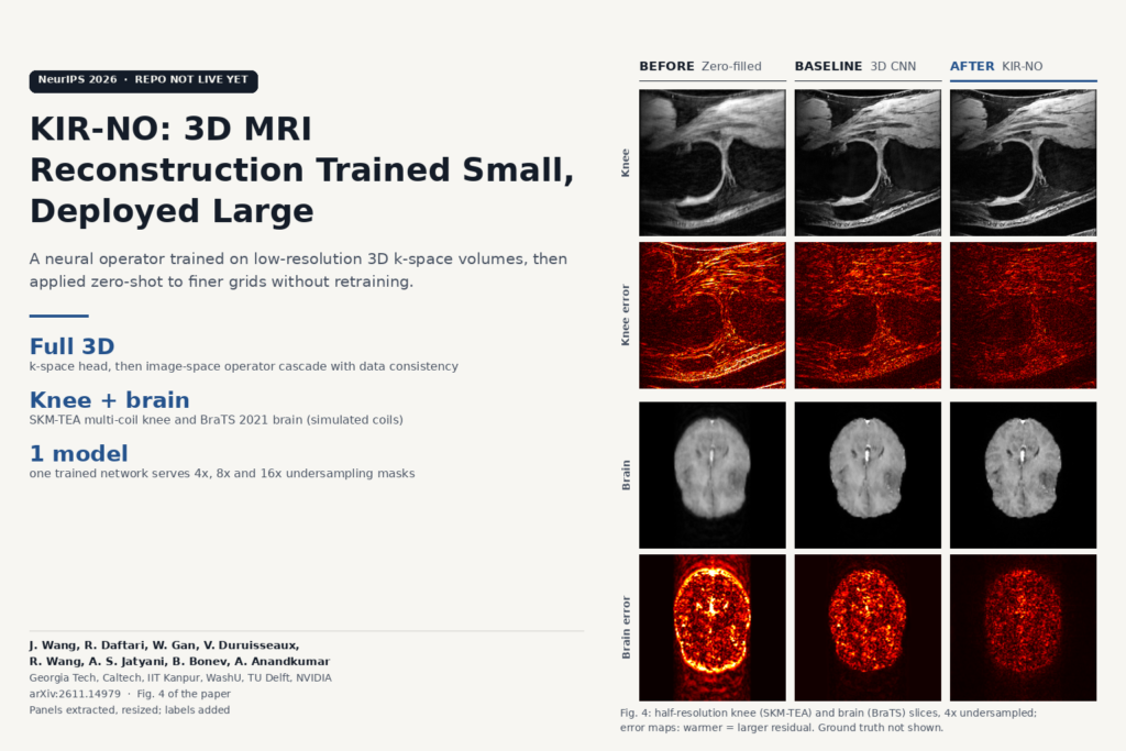
Anyone who has tried to train a 3D MRI reconstruction network on full-size volumes knows the wall: the backward pass needs every layer’s activations for the whole volume, and GPU memory runs out long before the dataset does. So most teams fall back to 2D models run slice by slice and stacked, and the through-plane direction gets ignored. Jiayun Wang, Ram Daftari and colleagues from Georgia Tech, Caltech, IIT Kanpur, Washington University in St. Louis, TU Delft and NVIDIA, with Anima Anandkumar as senior author (arXiv:2611.14979, 11 November 2026, NeurIPS 2026), propose a way around it: train a full 3D model on downsampled volumes, then run the same weights on the finer grid. They call it KIR-NO, the k-space-to-Image Reconstruction Neural Operator.
In their setup a single training step at 512 x 512 x 320 did not fit on a GH200 GPU for either their model or a 3D E2E-VarNet, and KIR-NO’s full-resolution inference alone needed 72.2 GiB.
Why a normal 3D CNN can’t do this
A convolution kernel in a CNN is a fixed grid of weights, say 3 x 3 x 3 voxels. Train it on 1 mm voxels and run it on 0.5 mm voxels and the same kernel now covers half the physical distance. The receptive field shrinks, the learned filters no longer mean what they meant in training, and output quality drops. The paper shows this directly: when the CNN baselines trained at quarter resolution on knee data were run at full resolution, the 2D and 3D E2E-VarNet scored below the zero-filled reconstruction, which involves no learning at all.
KIR-NO replaces those kernels with discrete-continuous (DISCO) convolutions. Each kernel is a continuous function defined over a fixed physical radius, written as a weighted sum of basis functions. At inference the kernel is sampled at whatever voxel spacing the volume has, so on a finer grid it keeps the same physical support and gains more taps. The authors designed two 3D bases for this: piecewise-linear hat functions in spherical coordinates and 3D Morlet wavelets. The piecewise-linear one won in their ablation, even though the Morlet kernel has more learnable coefficients.
The pipeline, step by step
The input is undersampled multi-coil 3D k-space plus coil sensitivity maps, which every method in the paper receives as a known part of the forward model (from scanner calibration for the knee data, simulated for the brain data). A k-space neural-operator head first refines the measured frequencies. After an inverse FFT, a cascade of image-space DISCO operators alternates with data-consistency steps that push the image back toward the acquired k-space samples. The default is a single image-space cascade; deeper cascades with gradient checkpointing improve results for both KIR-NO and the CNN baseline at extra compute.
When the model runs at a finer grid, the k-space head and data consistency still use the measurements at the training resolution. Only the image-space operators are resampled. The model gets no extra acquired frequencies, so anything it adds at full resolution comes from what it learned. In the ablation, removing the k-space head made PSNR fall as evaluation resolution went up, while the full model improved.
What the results show
The main test is SKM-TEA, a multi-coil qDESS knee dataset, with BraTS 2021 T1Gd brain volumes and simulated 8-coil k-space as the second anatomy. Models were trained at quarter or half resolution and evaluated at all three resolutions without fine-tuning.
Trained at half resolution on the knee data at 4x acceleration, KIR-NO reached 36.54 dB PSNR in-domain, 2.23 dB above the 3D E2E-VarNet. Run zero-shot at full resolution, its SSIM rose to 0.940 while the 3D E2E-VarNet’s was 0.870. One trained model also handled 4x, 8x and 16x undersampling masks with no retraining, and led the CNNs at each rate.
Fig. 4 of the paper, which we used for the featured image, shows a knee slice and a brain slice from half-resolution models. Zero-filled and l1-wavelet reconstructions carry obvious aliasing, and the error maps get visibly cooler from the 2D and 3D CNNs to KIR-NO on both anatomies.
Brain results are mixed. Trained at quarter resolution on BraTS, KIR-NO led in-domain, but the CNN baselines transferred upward better than it did on that simulated 8-coil data. Trained at half resolution, it led at full resolution by a small margin. The appendix also includes a recurrent transformer, ReconFormer, which scored higher PSNR than KIR-NO at all three resolutions in a slice-wise comparison.
Where it holds up and where it doesn’t
The appendix tests the model against harder conditions:
- Cross-scanner transfer. Trained on SKM-TEA and applied to fastMRI knee volumes from a different scanner, coil array and protocol, KIR-NO beat the 3D E2E-VarNet both zero-shot and after a 15-epoch fine-tune on 100 fastMRI volumes, and the gap widened after fine-tuning.
- Noise. With complex Gaussian noise added to k-space at test time, KIR-NO’s PSNR lead shrank as SNR fell, and it degraded faster than the CNN. At 14 dB SNR the CNN already had the better SSIM.
- Diffusion models. At one or ten steps KIR-NO was mostly ahead, but given their full sampling budgets (200 and 1200 steps) both diffusion baselines were more accurate. The authors state that KIR-NO’s advantage over them is inference time.
- Speed. A single-cascade KIR-NO reconstructed a quarter-resolution BraTS volume in 0.157 s on a B200 and a half-resolution one in 3.22 s. Cost grows with grid size, because the kernel’s fixed physical radius covers more voxels on a finer grid.
- Evaluation limits. On SKM-TEA the through-plane axis was interpolated from 160 to 320 slices for the full-resolution set, which the authors say may inflate metrics in that direction. Runs are single-seed, on two anatomies, and the “high-resolution” data are resized versions of the same acquisitions, so real noise, motion and coil differences between separate high-resolution scans are not tested.
Training cost is modest by 3D standards: about 43 GPU-hours on one NVIDIA B200 for the half-resolution knee model and about 5.4 GPU-hours for the quarter-resolution one.
Where it fits for a viewer or platform team
KIR-NO works on raw multi-coil k-space with coil maps, so it belongs on the reconstruction side: a scanner-adjacent recon server, an offline research pipeline that stores raw data, or a platform that reconstructs from vendor raw files before writing DICOM. A DICOM viewer only ever sees its output, and it can’t run on images that have already been reconstructed and exported.
For anyone building 3D imaging models, the training recipe carries over. If your bottleneck is fitting full-resolution volumes through backprop, a resolution-agnostic layer lets you train on coarse grids and still deploy at native spacing. For MRI recon, keep the caveats in view: it loses accuracy under k-space noise faster than the CNN, diffusion samplers win on accuracy when you can afford their runtime, and full-resolution inference still needs a high-memory GPU.
Code, weights and licence
The paper says code is available at github.com/neuraloperator/KIR-NO. When we checked on 21 November 2026 that repository returned a 404 and did not appear in the neuraloperator GitHub organisation’s public repositories, so there is no public code, no released weights and no stated licence yet. SKM-TEA, BraTS 2021 and fastMRI each come with their own data-use terms, which you would need to check before training anything commercial on them. The authors write that KIR-NO is a research contribution and is not validated for clinical deployment.
Source
Jiayun Wang, Ram Daftari, Weijie Gan, Valentin Duruisseaux, Ruibo Wang, Armeet Singh Jatyani, Boris Bonev and Anima Anandkumar, “Coarse-to-Fine 3D MRI Reconstruction via Resolution-Agnostic Neural Operators”, arXiv:2611.14979 (v1 11 November 2026), NeurIPS 2026. Affiliations: Georgia Tech, Caltech, IIT Kanpur, Washington University in St. Louis, TU Delft and NVIDIA. Featured image: Fig. 4 of the paper (knee and brain slices with error maps: zero-filled, 3D CNN and KIR-NO), panels extracted from the PDF, resized, with labels added.