
Anyone who reads dental or maxillofacial CBCT knows what the temporomandibular joint looks like on it: crisp condyle, crisp glenoid fossa, and a grey, featureless joint space in between. CBCT is built for bone. The articular disc, the synovial cavity and any fluid inside it are not something a reader can judge, so a patient with a painful jaw who already has a CBCT on file gets referred to MRI to find out whether the joint is inflamed. Evgeny Alves Limarenko of the Moscow Institute of Physics and Technology (arXiv:2611.23042, 17 November 2026, preprint) asks whether the CBCT already holds that answer below what the eye can see, and builds a pipeline to pull it out.
The target is MRI-confirmed synovial effusion in the TMJ, with articular disc displacement as a second, harder task. The method is a frozen 3D encoder called BertsWin plus a small classifier that flags effusion on CBCT well above chance, on a cohort of only 212 paired joints.
Why a standard 3D model struggles here
The paper names three problems. The first is data. Effusion comes and goes, so a CBCT can only be labelled with an MRI report if the two scans were taken close together. The author required 30 days or less between scans, against the roughly 90-day gap common in practice, and that left 99 patients and 212 joints. A high-capacity 3D network trained end to end on that many cases memorises each patient’s bone shape.
The second is the pre-training loss. Masked autoencoders usually learn by reconstructing hidden patches under a plain mean squared error. CBCT values span about 3,000 HU, and soft tissue sits in a thin slice of that range, so the error is dominated by mistakes at bright cortical edges. The encoder learns bone geometry very well and soft-tissue texture barely at all.
The third is the field of view. Even a good encoder sees a whole jaw region, and the joint space is a small part of it. Feed the full feature map to a classifier trained on 212 examples and the surrounding bone drowns out whatever signal sits in the joint.
How the pipeline works
Pre-training on unlabelled CBCT. The encoder is BertsWin, from the author’s earlier work with Anastasiia Studenikina (arXiv:2512.21769). It is a 3D masked autoencoder that keeps the full 14 x 14 x 14 token grid during encoding, with mask tokens in the hidden positions, instead of throwing 75% of patches away as sparse MAEs do. A 12-layer Swin transformer with local 3D windows processes that grid. It was pre-trained on a single-centre archive of 3,853 maxillofacial CBCT scans at 0.2 mm, cut into 6,550 training joints and 1,156 validation joints.
A loss aimed at soft tissue. The reconstruction error is split into brightness, contrast and structure terms, and the new objective, PhysLoss, weights those terms inside three masks: a soft-tissue mask covering voxels between -300 and 300 HU (half the weight), a thin shell around the bone surface, and the whole patch as a regulariser. Because CBCT intensities sit on the Hounsfield scale, that window picks out fluid and non-mineralised tissue in every scan. Training used GCond, the author’s optimiser, which combines sign-based LION-style updates with LARS-style layer-wise scaling.
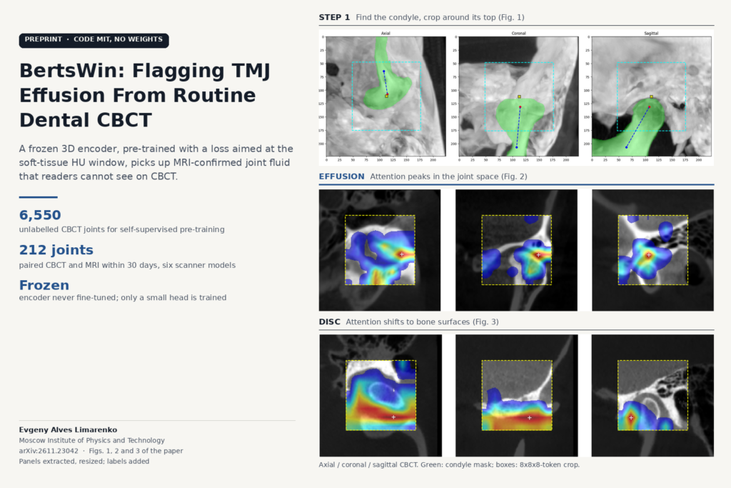
Find the joint, crop the features. A segmentation head on the frozen encoder outlines the mandibular condyle (Dice 0.947 against reference masks). From that mask the pipeline computes the condyle’s axis, finds its top point, and crops an 8 x 8 x 8 block of tokens around it, a 128-voxel cube. That throws away 81% of the feature grid, mostly bone that has nothing to do with the question.
A small head on frozen features. The encoder weights never change. A light attention projector and a two-layer classifier are the only trained parts for effusion. For disc displacement, the author adds a second input stream built from the raw CBCT crop: the vertical intensity gradient, normalised intensities and a coordinate grid.
The featured image above shows the three pieces on real scans from the paper: the condyle mask, axis and crop box in three planes (Fig. 1), then Grad-CAM maps for effusion (Fig. 2) and for disc displacement (Fig. 3).
What the results show
Evaluation is patient-isolated 10-fold cross-validation repeated three times, 30 runs in total. The 212 joints came from six CBCT scanner models with voxel sizes from 0.2 to 0.4 mm. Labels were pulled from MRI reports by clinical residents and, in parallel, by a large language model, with a senior gnathologist settling every disagreement.
For effusion, the PhysLoss and GCond encoder reached a ROC AUC of 0.793, with sensitivity 0.697 and specificity 0.762. The same BertsWin architecture pre-trained with plain mean squared error, or with the unmasked version of the split loss, scored 0.465 to 0.513, and a standard sparse MONAI MAE-ViT scored 0.499. None of those encoders found a usable signal, even with the same crop. Grad-CAM for the working model peaks in the dark joint space between condyle and fossa, which is where fluid collects.
Freezing the encoder mattered. Partial fine-tuning dropped AUC to 0.758, LoRA to 0.720, and full fine-tuning of all 86 million parameters collapsed to about 0.50 within a few epochs.
Disc displacement was harder. From soft-tissue features alone the classifier did not reach significance, because the disc has nearly the same attenuation as the muscle and ligament around it. With the vertical-gradient stream added, AUC was 0.679 with sensitivity 0.840 and specificity 0.585. In the Grad-CAM maps, attention moves off the joint space and onto the cortical surfaces of the condyle and articular eminence. The model is reading bony remodelling that follows long-standing disc problems, so it fires on joints whose bone stayed flattened after the disc returned to place, which explains the false positives.
Where it falls short
- Small cohort, no external test. 212 joints from 99 patients, about 21 joints per validation fold. The 30 runs reuse the same patients, and there is no held-out site. The author gives a conservative lower confidence bound of 0.724 for the effusion AUC.
- Unstable training. Even the best configuration had 2 of 30 folds collapse, and the paper reports that the order in which patients enter early mini-batches can decide whether a run finds the signal at all.
- Single-centre pre-training. All 3,853 pre-training scans came from one archive. The paired cohort spans six scanners, but how the encoder handles a new vendor’s noise and scatter has not been tested.
- Tasks left out. Osseous destruction was dropped because positives were too rare. The crop also excludes the lateral pterygoid muscle and other tissue outside the joint.
- Label noise. Effusion can change within 30 days, and labels come from report text rather than image annotation.
Where it fits for a dental imaging platform
The input is a scan the clinic already has. CBCT of the TMJ region is routine in dental and maxillofacial work, and the pipeline needs nothing beyond the volume: no extra sequence, no contrast, no manual landmark. In a CBCT viewer or a dental imaging platform, this kind of model fits as a background check on studies that include the joints, returning a per-joint flag such as “possible effusion, consider MRI” alongside the condyle segmentation it already computes.
At an AUC around 0.79 with specificity near 0.76, it works as a triage aid. About one in four effusion-free joints would still be flagged, so the output should prompt a look at the clinical picture and stay out of the report. The disc-displacement output would need even more care, since it partly re-describes bone changes a reader can already see.
For teams building on CT or CBCT more generally, the paper makes a practical case. When the signal lives in a known HU window, pointing the self-supervised loss at that window can matter more than the architecture, and on small labelled cohorts a frozen encoder with a tight anatomical crop did better than any fine-tuning the author tried.
Code, weights and licence
The paper links two repositories. AlevLab-dev/BertsWinMAE exists and is MIT-licensed; it contains the model code, pre-training scripts with GCond, and a stem-tuning script. AlevLab-dev/GCond exists but has no licence file, so reuse rights are unclear. When we checked on 22 November 2026, neither repository offered pretrained weights or releases, and the TMJ classifiers, localisation head and paired CBCT and MRI cohort are not public. To reproduce the result you would need your own CBCT archive for pre-training and your own paired cohort.
Source
Evgeny Alves Limarenko, “Uncovering the Invisible in Routine Radiology: Cross-Modal Detection of MRI-Verified Soft-Tissue Pathologies from CBCT via Physics-Guided 3D Self-Supervised Learning”, arXiv:2611.23042 (v1 17 November 2026), preprint. Affiliation: Moscow Institute of Physics and Technology. Featured image: Figs. 1, 2 and 3 of the paper (condyle localisation and crop, Grad-CAM for synovial effusion, Grad-CAM for disc displacement), panels extracted from the PDF, resized, with labels added.