
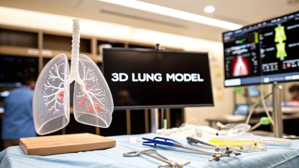
A 3D lung model is a patient-specific mesh of airways, parenchyma, and vessels, built from that person’s CT. You can rotate it, cut it, print it. It is not a picture of “the lungs.” It is not a bronchopulmonary-segment label on a slice.
If you meant S1–S10 / how to read lung segments on CT → lung segments on CT. If you meant how you draw the mask → medical image segmentation. If you meant turning slices into a volume you can spin → 3D reconstruction from CT. If you meant the printer job after the mesh → 3D printing anatomical models. If you meant MPR / MIP / volume-rendering software → 3D imaging software.
PYCAD builds the imaging piece (viewer / model) when a lung mesh has to live in a clinic app. It does not ship a thoracic-surgery platform or a 3D-print bureau.
What the model actually is
A chest CT is a stack of slices. A 3D lung model is the surfaces you extract from that stack so a person can see how a tumor sits against an artery and an airway in this chest, not a textbook one.
Three labels usually matter:
- Parenchyma. The lung tissue itself — left, right, sometimes lobes.
- Airway tree. Trachea down the generations you can resolve. The cut-plane for a segmentectomy lives here.
- Vessels. Pulmonary arteries and veins. A “safe” cut that nicks a vessel is not a safe cut.
Bronchopulmonary segment names (RB1–RB10 / LB1–LB10) are the language of that cut. They live on lung segments on CT. This page is the mesh you build once those labels exist.
Scan → mask → mesh
| Stage | What you do | What you have when it works |
|---|---|---|
| Acquisition | Thin-slice chest CT (sub-millimeter if you want distal airways). DICOM, not a JPEG of a slice | A volume you can reconstruct. How that scan was made is acquisition of images |
| Segmentation | Label parenchyma, airways, vessels — threshold, region-grow, or a U-Net. A person still checks the mask | A labelmap on the same grid as the CT. Methods sit on medical image segmentation |
| Reconstruction | Isosurface / marching cubes, or a volume render if you only need to look | A mesh (STL / OBJ) or a render you can spin. Reconstruction as its own job is 3D reconstruction from CT |
| Print (optional) | Watertight mesh, pick a material, slice, finish | A physical replica for a briefing or a box. Production is 3D printing anatomical models |
Garbage in, garbage mesh. Thick slices hide the bronchioles. Contrast timing that misses the pulmonary arteries gives you a hole where a vessel should be. A mask that leaks into the mediastinum prints a blob.
Where the mesh is used
- Thoracic surgery. Walk the tumor, the artery, and the airway before the table. A segmentectomy plan is a named segment plus a mesh you trust, not a slide that says “3D.”
- Inhalable drugs. A patient-specific airway tree is an input to an airflow / deposition sim. The paper is the sim. The mesh is the geometry. Do not sell the mesh as a drug.
- The briefing. A printed or spun model is how you show a patient the cut without a 200-slice stack. Communication, not a diagnosis.
AI that draws the mask faster is still a mask. It does not sign the operative plan. A Dice on someone else’s hospital is not a number you can paste onto the next scanner.
FAQ
Is this the same as lung segments on CT?
No. That page is the anatomy (which segment, which bronchus). This page is the mesh you build from a labeled volume.
Do I have to print it?
No. Most planning stays on a screen. Print when a physical replica changes the briefing. The printer job is 3D printing anatomical models.
How long does a model take?
A checked mask is the bottleneck, not the isosurface. Automated labels help. A person still has to say the airway tree is that patient’s airway tree.
Does PYCAD sell 3D lung models?
No. If the mesh or the viewer has to live in a clinic app, that is the imaging piece. Case studies.