
This is how to run 3D Slicer in a browser: pick a cloud host, get a volume in, run MONAI Label / DeepEdit, clean the mask in Segment Editor, export an STL. It is not the desktop nnUNet spine tutorial — that is automatic spine segmentation in 3D Slicer.
Desktop Slicer is still the same app (BWH / MIT, 1998; Qt rebuild as Slicer 4). The cloud path moves the GPU and the volume off your laptop. Your tab is a thin client into that session.
Pick a host
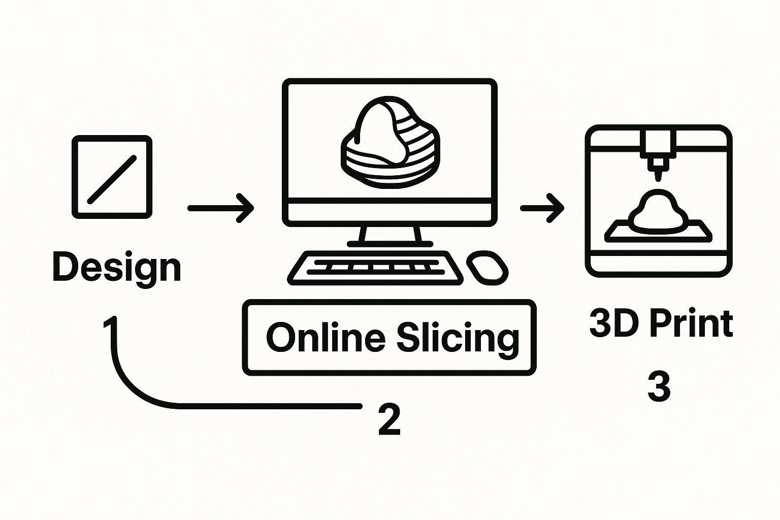
There is no single “3D Slicer online” product. You attach a Slicer session to a server that already has the volume and, usually, an AI labeler.
- NVIDIA Clara — hospital / vendor stack. Auth, audit, IT integration. You pay for the platform and the instance.
- Self-hosted MONAI Label — research default. You run the label server; Slicer in the browser is the client. You pick the models and the GPU.
A university box you SSH into and then open Slicer in a remote desktop is the same idea with worse UX. Same rule: the GPU lives next to the data.
CPU or GPU
- CPU — windowing, four-pane review, paint / erase. Cheap. Fine for a look.
- GPU — MONAI inference, DeepEdit, heavy 3D. Without it,
segmentation_liver_and_tumorsits in a queue or crawls.
Start small. Scale the instance when inference is the bottleneck, not when you are still clicking around an empty scene.
Desktop Slicer vs this
| Desktop 3D Slicer | Cloud / browser Slicer | |
|---|---|---|
| Where it runs | Your machine. Free download from slicer.org. | Remote session. Browser is the display. |
| Install | Local app + extensions (see the nnUNet spine guide). | Account or a URL your lab already hosts. |
| GPU | Whatever is in the desk. | Rented or the lab’s server GPU. |
| Data | Local disk. You carry the volume. | Bucket or PACS. Session can die; the bucket should not. |
| Share | Export a scene or a mask. | A session link, if the host supports it. |
| Cost | Software is free. Hardware is yours. | Instance hours + storage. Desktop Slicer itself is still free. |
A generic DICOMweb viewer (OHIF and friends) is a different product: view / measure / hang a study from PACS. This page is Slicer — Segment Editor, MONAI Label, STL out. For the wire format, see DICOM standards.
Get a volume in

Two ways:
- Browser upload. One series or a NIfTI. Same idea as attaching a file. Fine for a single case. Painful for a study with hundreds of series, and a dropped connection restarts it.
- Bucket. Put the data on S3, Azure Blob, or GCS. Point the session at that prefix. Cloud-to-cloud is faster than your office uplink, and the volume survives when you kill the instance.
If the host talks DICOMweb / WADO-RS to the site PACS, use that instead of a zip of slices. Do not copy PHI onto a laptop “just for Slicer” if the bucket or PACS path exists.
Four panes and windowing
Once the volume is loaded you get the same layout as desktop: axial, sagittal, coronal, 3D. Scroll, pan, zoom. Rendering is on the server; a slow tab is usually the network, not your CPU.
Window / level first. A chest CT is two different pictures: a lung window for parenchyma, a bone window for ribs and vertebrae. Wrong window, wrong impression, and a later AI overlay looks “wrong” when the tissue was just clipped.
The Data module is the layer list. Load a CT, an MRI, and a mask together. Toggle each in each pane. Fusion (PET on CT) is the same module, not a separate product.
MONAI Label
Open the MONAI Label module. Paste the label-server URL (lab box or vendor). On connect, Slicer lists the models that server actually has — not a global catalog.
Liver on an abdominal CT is the usual first run:
- Pick the model. The common bundled name is
segmentation_liver_and_tumor. Use whatever your server registered. - Run inference. The GPU on the server does the work. Your laptop does not need a card.
- A 3D segment appears on the volume. Treat it as a draft.
There is no honest single accuracy number. Contrast, protocol, and whether that checkpoint ever saw your scanner decide how much you will have to fix.
DeepEdit
DeepEdit is click-to-correct, not a second full auto-run from scratch.
- Foreground clicks inside tissue the model missed.
- Background clicks on tissue it stole (spleen on a liver model, for example).
Re-run. The model updates the boundary from those points. A few well-placed clicks beat painting 200 slices. If the checkpoint is a bad match for the series, stop and go to Segment Editor instead of fighting DeepEdit.
Segment Editor, then STL

Paint and Erase are slice-wise, same as desktop. Use them for the last millimeters the AI will not get: a vessel origin, a resection margin, a leak into bowel.
Logical Operators when two segments have to become one object:
- Add — kidney + tumor as one mesh for a print.
- Subtract — cut a cavity or remove an overlap.
- Intersect — keep only the overlap.
Color and opacity in the 3D view are for the meeting, not for the file. Export STL when someone will print or drop the surface into CAD. Check scale (STL is unitless) before a printer sees it. How to open or repair that mesh is how to open an STL file and how to edit an STL file.
A session link (if the host has it) is how a colleague sees the same view without a 2 GB mail. That is collaboration. It is not a substitute for an audit trail on clinical data.
Security, cost, speed
- PHI. The host has to be in a BAA / HIPAA (or local equivalent) posture: encryption in transit and at rest, auth, logs. A random GPU rental with a public URL is not that. Desktop-on-a-clinic-PC is not automatically safer; an unencrypted USB is worse than a locked bucket.
- Cost. You rent hours and disks. A weekly inference job is usually cheaper than a second workstation. A always-on GPU for one person who window-levels is a waste. Slicer itself is still free.
- Speed. Compare like to like: a cloud A100 vs the laptop, not vs a local 4090. The browser is a stream of frames. A bad VPN makes it feel slow even when the GPU is idle.
PYCAD builds custom web DICOM viewers when the clinical app needs a viewer, not a full Slicer session. Case studies.