Skip to content
  • Home
  • Services
    • All Services
    • Custom DICOM Viewer
    • Case Studies
  • Products
    • All Products
    • PYCAD DICOM Viewer
    • PYCAD Dental Viewer
    • PYCAD SSR Engine
  • Case Studies
  • Blog
  • About us
  • Home
  • Services
    • All Services
    • Custom DICOM Viewer
    • Case Studies
  • Products
    • All Products
    • PYCAD DICOM Viewer
    • PYCAD Dental Viewer
    • PYCAD SSR Engine
  • Case Studies
  • Blog
  • About us
Get in Touch

Category: Segmentation & AI

VoxForge: One Frozen 3D Encoder for MRI, CT and PET
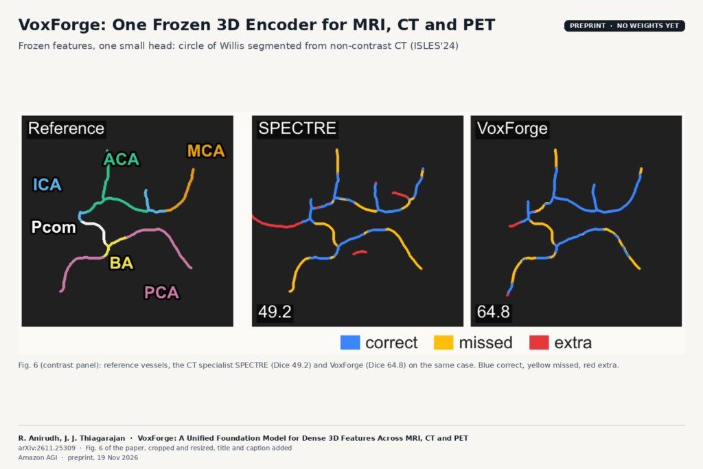
Fig. 6 of arXiv:2611.25309 by Anirudh and Thiagarajan: reference circle of Willis vessels, the SPECTRE result (Dice 49.2) and the VoxForge result (Dice 64.8) segmented from non-contrast CT, with correct, missed and extra vessel segments colour coded.

Amazon AGI’s 388M-parameter ViT, trained on 235K commercial-use volumes, segments, classifies and registers scans without fine-tuning.

MedImgAnnotator: Batch Annotation for Large CT and MRI Cohorts
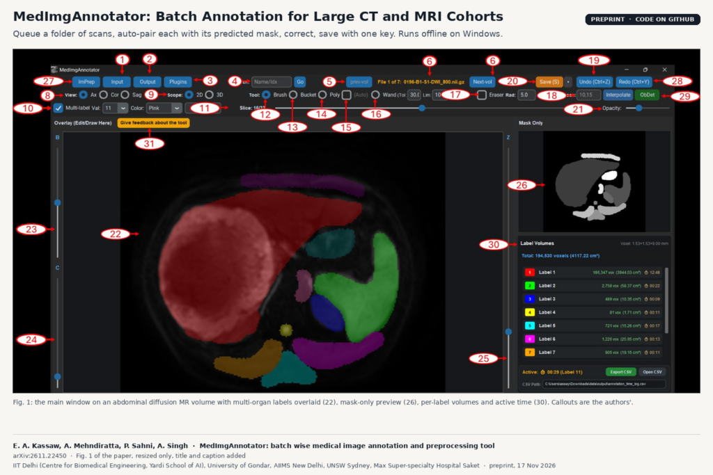
Fig. 1 of arXiv:2611.22450 by Kassaw et al.: the MedImgAnnotator main window showing an abdominal diffusion MR slice with multi-organ labels overlaid, a mask-only preview and a per-label volume and annotation time panel.

A Windows tool that queues folders of CT and MRI, pairs each scan with its predicted mask, and cuts the time spent per case.

SWIMSS: An Open MS Lesion Segmenter Trained on Seven Swiss Centres
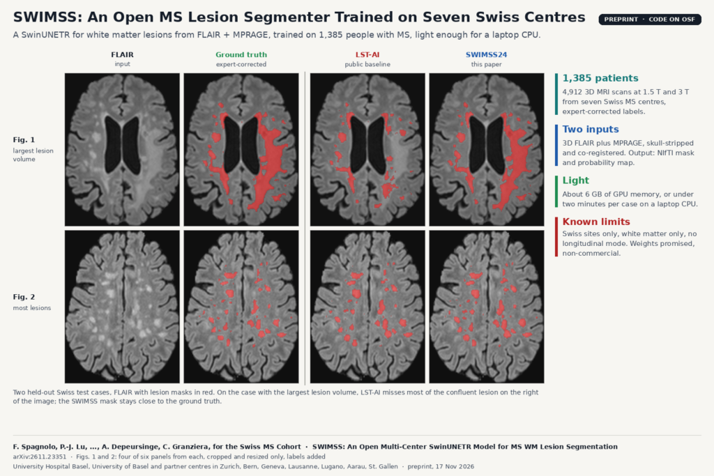
Figs. 1 and 2 of arXiv:2611.23351 by Spagnolo et al.: two axial FLAIR brain MRI test cases with multiple sclerosis, showing the input, the expert-corrected ground-truth lesion mask, the LST-AI mask and the SWIMSS24 mask in red.

A SwinUNETR trained on 1,385 people with MS from seven Swiss centres segments white matter lesions from FLAIR and MPRAGE, even on a CPU.

GRACE: Fixing Segmentation Masks With Retrieved Verified Cases
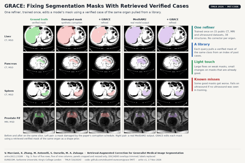
Fig. 5 of arXiv:2611.23189 by Marcianò et al.: liver, pancreas and spleen CT and prostate peripheral zone MRI slices showing the ground-truth mask, a synthetically damaged mask and its GRACE correction, and a MedSAM2 mask and its GRACE correction.

One refiner, trained once on CT, MRI and ultrasound, corrects a model’s mask using a verified case of the same organ from a library.

PathoDraw: One Zero-Shot Lesion Detector for Any Brain MRI
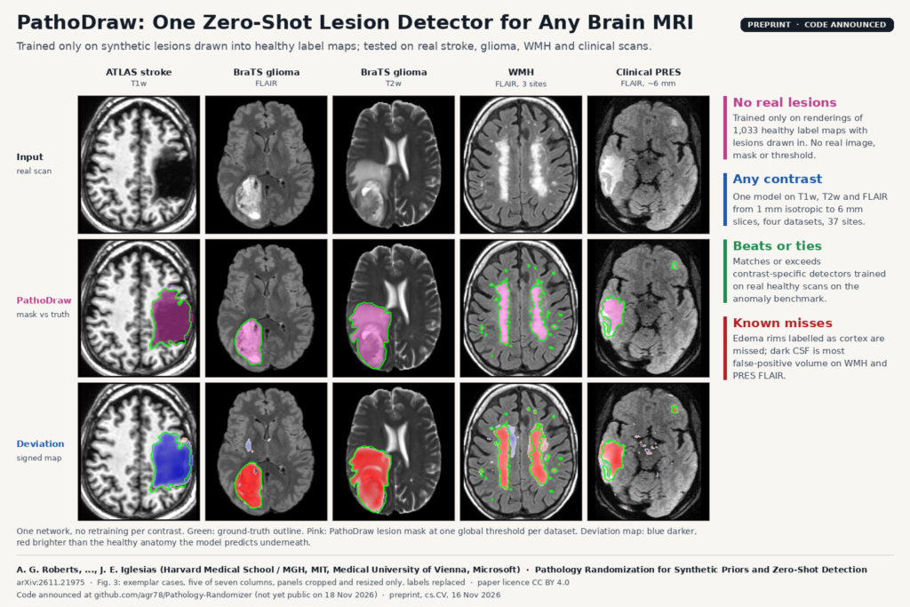
Fig. 3 of arXiv:2611.21975 by Roberts et al.: axial brain MRI from ATLAS T1w, BraTS FLAIR and T2w, WMH FLAIR and a clinical PRES FLAIR scan, with the input image, ground-truth outline in green with the PathoDraw lesion mask in pink, and the signed deviation map in blue and red.

Trained only on synthetic lesions, one network flags stroke, glioma and white matter lesions on T1w, T2w or FLAIR brain MRI.

Scribble Once: Echo Cine Segmentation From One Scribbled Frame
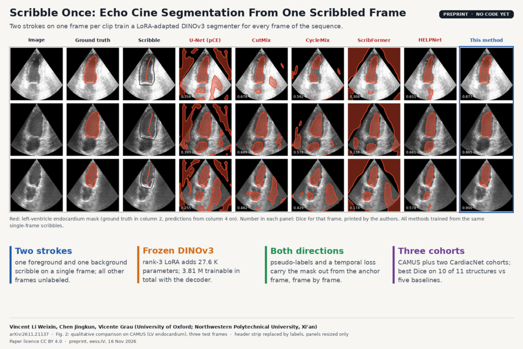
Fig. 2 of arXiv:2611.21137 by Vincent Li Weixin, Chen Jingkun and Vicente Grau (University of Oxford, Northwestern Polytechnical University): CAMUS two-chamber echo frames with left-ventricle endocardium masks from U-Net, CutMix, CycleMix, ScribFormer, HELPNet and the single-scribble method, per-frame Dice printed in each panel.

Two scribbles on one frame per clip train a LoRA-adapted DINOv3 model to segment heart chambers across the whole echo sequence.

X-Strata: Separating Aneurysm and Vessel Layers in 2D Angiography
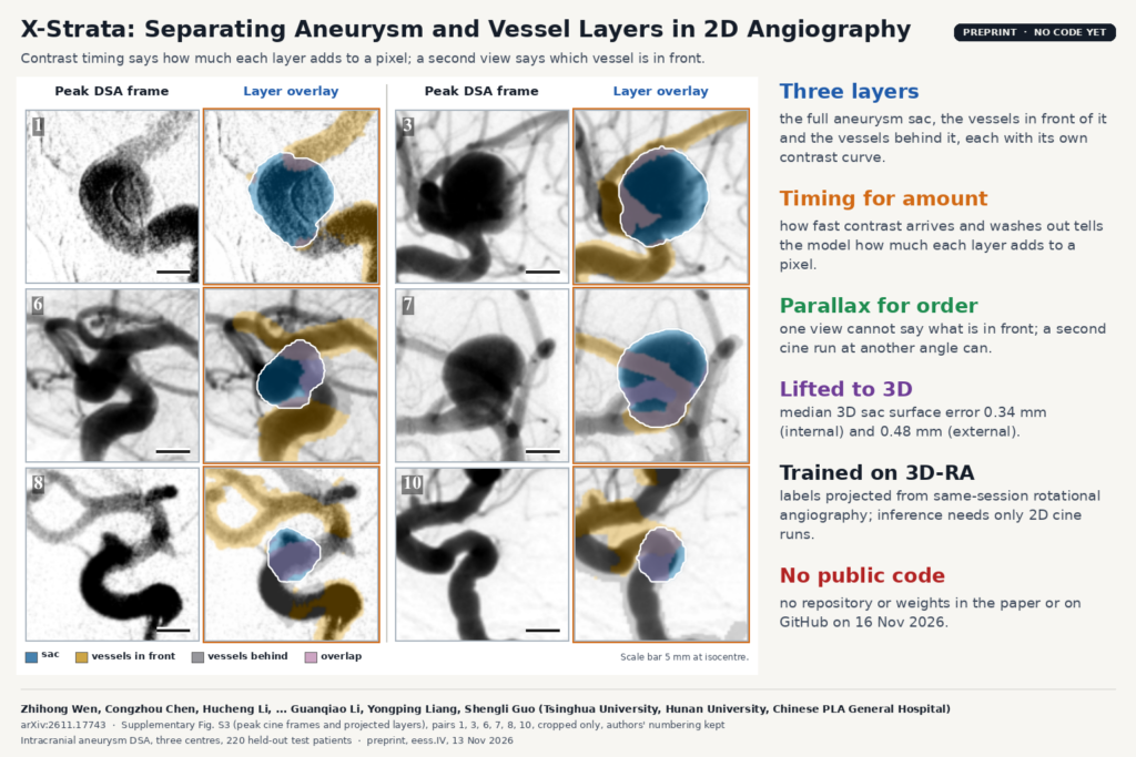
Supplementary Fig. S3 of arXiv:2611.17743 by Wen, Chen, Li and co-authors (Tsinghua University, Hunan University, Chinese PLA General Hospital): six pairs of peak cine DSA frames of intracranial aneurysms next to layer overlays with the sac in blue, vessels in front in gold, vessels behind in gray and overlap in pink.

From two routine DSA cine runs, X-Strata recovers the full aneurysm sac, sorts crossing vessels in front or behind, and lifts both to 3D.

TinyUNet3D: 3D Segmentation in Under a Million Parameters
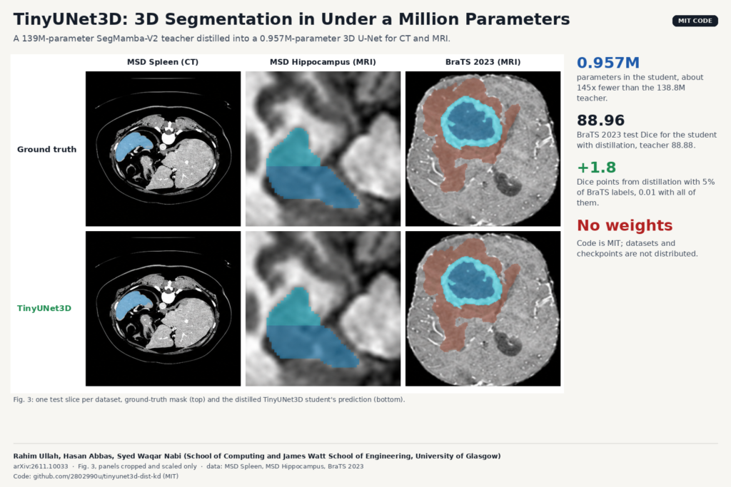
Fig. 3 of arXiv:2611.10033 by Ullah, Abbas and Nabi, University of Glasgow: one test slice each from MSD Spleen CT, MSD Hippocampus MRI and BraTS 2023 MRI, ground-truth mask on top and the distilled TinyUNet3D prediction below.

Glasgow distilled a 139M-parameter SegMamba-V2 into a 0.957M-parameter 3D U-Net. Distillation pays off mainly when labels are scarce.

Random Wavelet nnU-Net: Prostate Lesion Masks Across bpMRI Sites
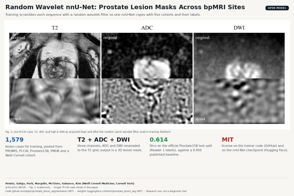
Fig. 1 of arXiv:2611.08535 by Heintz and colleagues, Weill Cornell Medicine and Cornell Tech: T2, ADC and DWI of a PI-CAI prostate MRI case as acquired and after the random sym4 wavelet filter used to train the open nnU-Net lesion model.

An open nnU-Net trained on 1,579 bpMRI cases from five cohorts, with a wavelet augmentation that scrambles each sequence.

Awakening ImageCAS: Open Coronary Ostia and Centerlines for CCTA
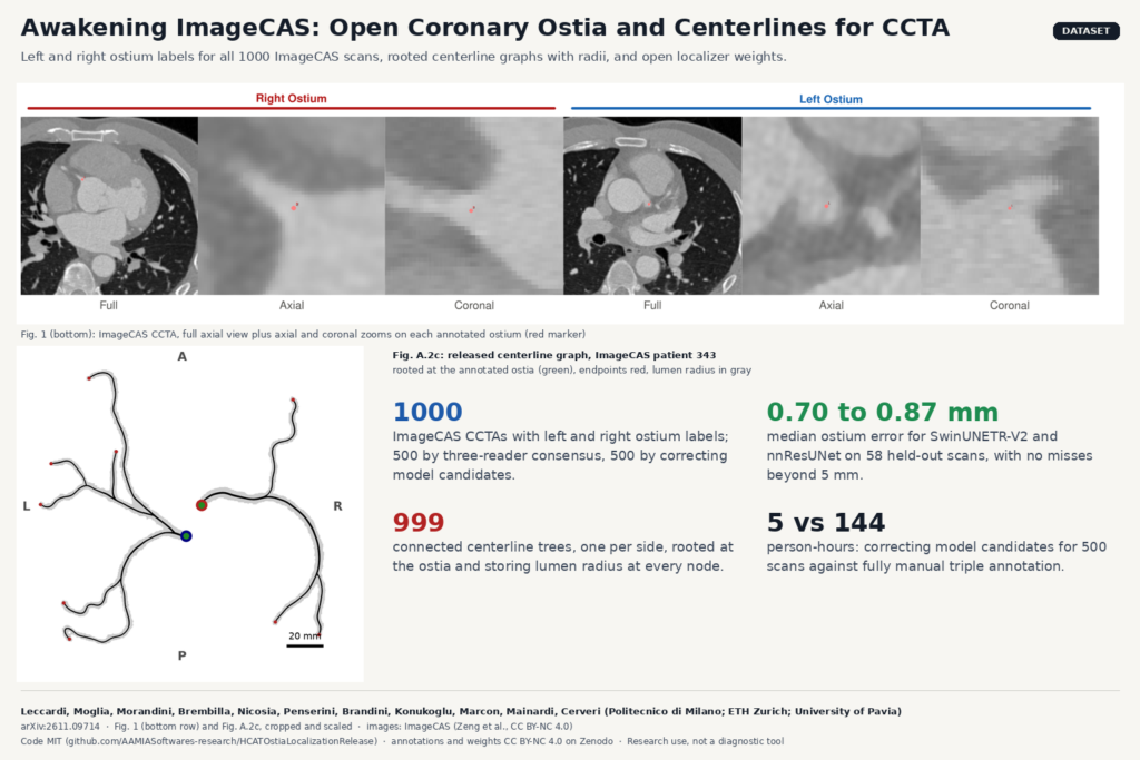
From arXiv:2611.09714 by Leccardi et al., Politecnico di Milano, ETH Zurich and University of Pavia: ImageCAS coronary CT angiography slices with the annotated right and left coronary ostium markers (Fig. 1), and the released centerline tree of ImageCAS patient 343 rooted at both ostia (Fig. A.2c).

Ostium labels for all 1000 ImageCAS scans, rooted centerline graphs with radii, and open localizers with sub-millimeter median error.

Next →