
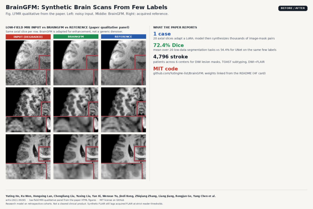
If you build brain tools for a DICOM viewer or a research PACS, the bottleneck is rarely the architecture. It is getting enough labeled T1, FLAIR, DWI, or head CT from the sites you actually serve. Protocols differ, lesions look different across scanners, and voxel masks take hours. In arXiv:2611.00265, Yuting He, Xu Wen, Hongxing Lan, Rongjun Ge, Yang Chen and colleagues (Case Western Reserve University, Southeast University, Jinling Hospital, Nanjing First Hospital and partners) present BrainGFM, a generative foundation model pretrained on about 1.3 million axial MRI and CT slices. Instead of shipping only frozen features, it turns that prior into synthetic training data or into a LoRA-adapted reconstructor you can hang on a real workflow.
On 20 low-data segmentation tasks, a UNet trained on BrainGFM-synthesized image-mask pairs reached a mean Dice of 72.4%, against 54.4% for the same UNet trained only on the few real labels. Across eight classification tasks the mean AUC was 94.0% under a one-case-per-class setup. A multicenter stroke study with 4,796 patients across six hospitals then tested lesion masks on DWI, TOAST subtyping, and DWI-to-FLAIR transforms. Code is MIT at github.com/YutingHe-list/BrainGFM, with pretrained weights linked from the README.
What hangs on a scan
BrainGFM is not a single-click segmenter you drop on a study. It is a controllable generator conditioned on modality or sequence (T1, T2, T1ce, FLAIR, DWI, CT, or mask). For segmentation you adapt with LoRA on about 20 labeled axial slices from one case, generate thousands of synthetic image-mask pairs, then train a plain UNet on those pairs. For classification you swap in class embeddings and synthesize class-conditioned images for a ResNet. For reconstruction you post-train on paired source and target images so the same prior maps low-field MRI to higher-quality MRI, low-dose CT to fuller-dose CT, or non-contrast T1 to a T1ce-like appearance.
That split matters for product design. The synthetic-data path keeps your production UNet or ResNet small and auditable. The reconstruction path is closer to a hanging-protocol helper: fill a missing contrast or clean a noisy acquisition before a reader opens the series. Both paths freeze the pretrained tokenizer and base transformer and only train LoRA (and light conditioning) on the target task.
How the prior is built
Pretraining uses BrainGFM-1.3M: 64,754 volumes from public resources spanning tumours, haemorrhage, neurodevelopmental and neurodegenerative cohorts. Each volume contributes 20 axial slices after skull stripping (HD-BET for MRI, SynthStrip for CT) and canvas placement at 256×256. A multi-scale VQ tokenizer shares a codebook between images and masks. An autoregressive transformer then predicts tokens coarse to fine under categorical modality conditions, following a ControlVAR-style next-scale setup.
Because masks live in the same discrete vocabulary, the model can emit a mask first and then an image conditioned on that mask. That is the mechanism behind few-shot segmentation synthesis: invent plausible lesion geometry, then invent a matching appearance for the requested sequence.
Where the numbers land
On the low-data segmentation suite (tumour, tissue, haemorrhage and vessel CT tasks), BrainGFM ranked first among the paper’s competing methods on 18 of 20 tasks and beat the full-data UNet reference on 14 tasks despite adapting from one case. Tissue tasks were especially strong (mean Dice 92.7%). Haemorrhage and vessel CT tasks averaged 73.4% Dice with the synthesis recipe.
Enhancement covers 20 tasks across low-field MRI, low-dose CT, and non-contrast-to-contrast MRI. Mean PSNR was 37.8 dB over those tasks. Low-dose CT saw the largest lift over the CAR baseline (about 12 dB PSNR on average). Qualitative panels show grainy low-field inputs cleaned toward the reference anatomy, which is the comparison used in the featured image for this post.
In the stroke cohorts, BrainGFM lesion Dice was 78.1% internal and 76.9% external under the 60-case low-data protocol. TOAST AUC was 87.4% internal and 84.5% external. DWI-to-FLAIR PSNR sat near 26.6 to 26.9 dB across centers. Blinded readers scored BrainGFM FLAIR overall diagnostic quality at 3.51 versus 3.57 for acquired FLAIR on a five-point scale, but at a strict threshold of 4 on all five criteria, synthetic FLAIR still trailed acquired FLAIR (15.6% versus 51.1%). In a 300-case human-AI subtyping study, mean radiologist accuracy rose from 55.6% to 63.4% with model suggestions (14% relative), with large reader-to-reader differences in how often wrong advice was accepted.
What breaks and what to watch
The model is mostly 2D axial. Volumetric continuity across slices is not the native unit, so 3D hanging protocols and multiplanar reformats still need your own stacking and QC. Pretraining follows the public corpus mix; rare protocols and populations outside that mix can fail quietly. Synthetic FLAIR is opportunistic when FLAIR is missing, not a drop-in replacement for an acquired series at high reader thresholds. Human-AI subtyping gains depend on how aggressively each reader trusts the suggestion, including when it is wrong.
Weights are advertised on Hugging Face via the README. As of this writing the HF model card for wenxuyyds/BrainGFM looks thin, so verify the checkpoint download before you promise a demo. License on the GitHub tree is MIT.
Why a viewer shop should care
BrainGFM is a practical answer to “we only have a handful of labeled brains from this hospital.” You can amplify those labels into a trainable set for a local segmenter, or adapt the same prior to clean low-field or low-dose series and to propose a missing FLAIR from DWI. Keep the synthetic path for training offline models, keep reconstruction behind a clear “generated” badge in the UI, and treat stroke subtyping outputs as decision support with an audit trail. That is the product shape that matches what the paper actually delivers.
Paper: arXiv:2611.00265 · Code: YutingHe-list/BrainGFM (MIT) · Weights: linked from the repo README (Hugging Face)