
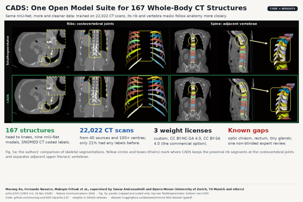
Anyone who has shipped organ segmentation inside a viewer knows the pattern. You start with one liver model, then a kidney model, then a vertebrae model a client asked for, and a year later you maintain six networks with six label conventions and nobody agrees where the “aorta” stops. A team led by the University of Zurich and the Technical University of Munich went the other way. CADS is one curated dataset and one model suite that segments 167 anatomical structures on CT, from the head to the knees. The paper is arXiv:2507.22953, revised on 10 November 2026 and published in Nature Communications, and the code, weights, dataset and a 3D Slicer extension are all public.
What CADS is
CADS has two parts. The CADS-dataset gathers 22,022 CT volumes from 40 sources: public archives such as TCIA, NLST and CT-RATE, challenge datasets, TotalSegmentator, and two new cohorts the authors contributed (484 head CTs and 586 triphasic contrast-enhanced abdominal CTs). The scans come from more than 100 centres in 16 countries, acquired between 2007 and 2024, with and without contrast, healthy and diseased. Every volume carries a label for all 167 targets.
The CADS-model is the network trained on that data. It is a standard nnU-Net, the residual-encoder ResEnc-L configuration from nnU-Net v2.5.1, split into nine region-specific models (abdominal organs and lungs, vertebrae, ribs, brain tissues, head-and-neck organs at risk, appendicular bones and muscles, and so on). The authors call the network standard on purpose and put the contribution in the data.
How they labelled 22,022 scans
Only 4,695 of the 22,022 volumes (21%) came with any annotation, and most of those covered one or two structures. To fill the rest, the team trained one nnU-Net per anatomical group on the labelled sources and used those models to pseudo-label everything.
Pseudo-labels at this scale go wrong in ugly ways: wrong spacing in the header, a cropped field of view, a vessel that leaks into the bowel. CADS screens them with a shape prior. An auto-decoder based on implicit neural representations is trained only on clean ground-truth masks. For each new pseudo-label it tries to reconstruct the mask from its learned shape space, and the 90th-percentile Hausdorff distance between the pseudo-label and the reconstruction becomes a plausibility score. Masks the prior cannot explain score badly. The worst 10% of images are withheld from one of the training runs.
That gives three “flavours” of model per group: one trained only on original ground truth, one on all pseudo-labels, and one on the shape-filtered pseudo-labels. For each of the 167 structures the team picked the flavour that scored best on held-out validation, preferring data from other sources than the training set. The final labels use original annotations wherever they exist and the chosen flavour everywhere else. All 167 targets map to SNOMED CT codes, with documented rules for where structures overlap or nest.
They also fixed labels by hand where public conventions were anatomically wrong. The example in the paper is the ribs: public rib masks often stop short of the spine, and CADS keeps the costovertebral joints.
What they found
The main baseline is TotalSegmentator v2.4.0, run in its strongest configuration, because it uses the same nnU-Net framework. Any gap then comes mostly from the training data. On the 117 structures both models segment, CADS reached a mean Dice of 90.52% against 88.12% for TotalSegmentator, and it was better on 15 of the 18 public test datasets. The gains were largest on structures that earlier datasets covered poorly or that have messy boundaries. Large, high-contrast organs moved less.
The second test used hospital data: 2,864 oncology CTs from the Radiation Oncology Department at University Hospital Zurich, contoured during routine care and never used for training. Here the biggest jumps were in radiotherapy organs at risk, with the brainstem 23.08 Dice points above TotalSegmentator. A radiation oncologist reviewed cases near the median score for each structure and judged most of them acceptable for treatment-planning delineation, singling out the parotid deep lobe, mandible and lungs.
While evaluating, the team also found many label errors in the TotalSegmentator test set, mostly ribs and vertebrae. Of 4,597 case-structure pairs reviewed, 3,002 were kept as reliable. If you benchmark a whole-body model on that test set, expect some of your “errors” to be label errors.
Why a viewer or clinic team should care
For a DICOM viewer, one model suite with one label map is easier to ship than a stack of single-organ networks. The 167 labels arrive grouped by region and tied to SNOMED CT codes, which is what you need to write DICOM SEG objects with proper coded concepts rather than free-text segment names.
The CADS 3D Slicer extension runs all nine tasks in one click and shows the segments grouped by model, each linked to its SNOMED code. For a research site, that removes most of the setup. For a product team, the command-line package is the more useful part. It runs in three stages: CPU preprocessing (reorient to RAS, resample to 1.5 mm), GPU inference, then CPU restoration back to the original geometry. That split maps well onto a queue-based pipeline where a PACS router drops studies and a GPU worker picks them up. The input is NIfTI, so you still need your own DICOM-to-NIfTI step and a SEG or RTSTRUCT writer on the way out.
Practical uses follow from the label list: organ-at-risk pre-contouring for radiotherapy, organ and muscle volumes for body-composition work, vertebra and rib numbering in a viewer, and anatomy-aware routing of findings (“this nodule sits in the right lower lobe”).
Licenses, read before you ship
The code is Apache-2.0. The weights come in three variants with different terms. The “reference” model used in the paper is under a custom CADS license that also carries the terms of four challenge datasets (EXACT09, LOLA11, SLIVER07, EMPIRE10). The “research” variant is CC BY-NC-SA 4.0, which rules out commercial use, and the README says it performs nearly identically to the reference model. The “open” variant is CC BY-SA 4.0 and allows commercial use under share-alike terms, but it was trained on fewer scans (13,269 at most per task, against 21,277 for the reference) and may be weaker on detailed head-and-neck structures. The dataset on Hugging Face is gated, and each scan stays under its source license, several of which are non-commercial.
For a commercial product, the “open” weights are the only clean path, and you should validate them on your own head-and-neck cases before relying on them.
Where it falls short
- Coverage is head to knees. Hands and feet are not included, and fine structures in the nervous, musculoskeletal and vascular systems are thin.
- Some labels follow conventions you may not want. The brain mask includes everything in the skull, the trachea covers the lumen but not the cartilage, the optic chiasm tends to be under-segmented, and the rectum is shorter than its anatomical extent.
- Very small structures are hard at 1.5 mm. Across the full label set the mean Dice drops to 85.87%, pulled down by structures such as the arytenoid cartilage (under 0.5 mL) and the buccal mucosa, which has only 30 ground-truth annotations.
- The shape filter can drop exactly the cases you care about. A tumour-distorted organ looks implausible to a prior trained on normal shapes, and the authors note it may filter clinically important atypical anatomy.
- The clinical review is one non-blinded radiation oncologist looking at three cases per structure. The authors are clear that it does not prove clinical utility.
- Faces in the new head CTs were blurred for anonymisation, so the dataset is not suited to detailed facial work.
Paper: Murong Xu, Fernando Navarro, Maksym Fritsak and 30 co-authors, with Tamaz Amiranashvili and Bjoern Menze as joint supervisors, “CADS: A Comprehensive Anatomical Dataset and Segmentation for Whole-Body Anatomy in Computed Tomography”, arXiv:2507.22953 (v2, 10 November 2026), published in Nature Communications (2026). Lead affiliation: Department of Quantitative Biomedicine, University of Zurich, with the Technical University of Munich, University Hospital Zurich, University Hospital Essen, Istanbul Medipol University and others. Code: github.com/murong-xu/CADS (Apache-2.0). Weights: GitHub releases, three license variants. Dataset: Hugging Face, gated, source licenses apply. Slicer extension: SlicerCADSWholeBodyCTSeg. Featured image: Fig. 5a of the paper, panels cropped and scaled.