Skip to content
  • Home
  • Services
    • All Services
    • Custom DICOM Viewer
    • Case Studies
  • Products
    • All Products
    • PYCAD DICOM Viewer
    • PYCAD Dental Viewer
    • PYCAD SSR Engine
  • Case Studies
  • Blog
  • About us
  • Home
  • Services
    • All Services
    • Custom DICOM Viewer
    • Case Studies
  • Products
    • All Products
    • PYCAD DICOM Viewer
    • PYCAD Dental Viewer
    • PYCAD SSR Engine
  • Case Studies
  • Blog
  • About us
Get in Touch

Category: Segmentation & AI

OraRefer: Oral Cancer Referral With a Guaranteed Sensitivity
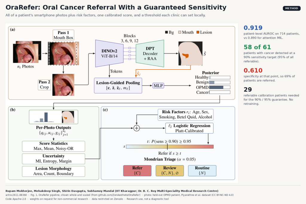
Fig. 1 of arXiv:2611.08360 by Mukherjee, Singh, Dasgupta and Mandal, IIT Kharagpur: an oral photograph is cropped to the mouth, segmented for lesions by a DINOv2 and DPT model, fused per patient with risk factors, and referred through a calibrated threshold or triage.

Fuses every smartphone photo of a patient with risk factors, then refers through a sensitivity threshold each clinic can calibrate.

EchoAppearance: Rebuilding the Vendor Echo Look From Raw Beams
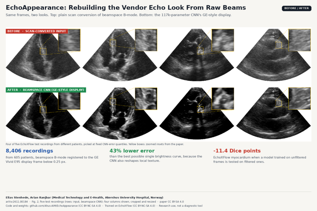
Comparison built from Fig. 2 of arXiv:2611.00184 by Elias Stenhede and Arian Ranjbar, Akershus University Hospital: four echocardiography test frames shown as plain scan conversion (top) and after the EchoAppearance beamspace CNN (bottom), with zoomed insets.

A 117k-parameter CNN turns native beamspace B-mode into GE-style display frames, and shows how a processing mismatch breaks segmentation.

OpenBrain: Brain MRI Parcellations Shipped With a Risk Score
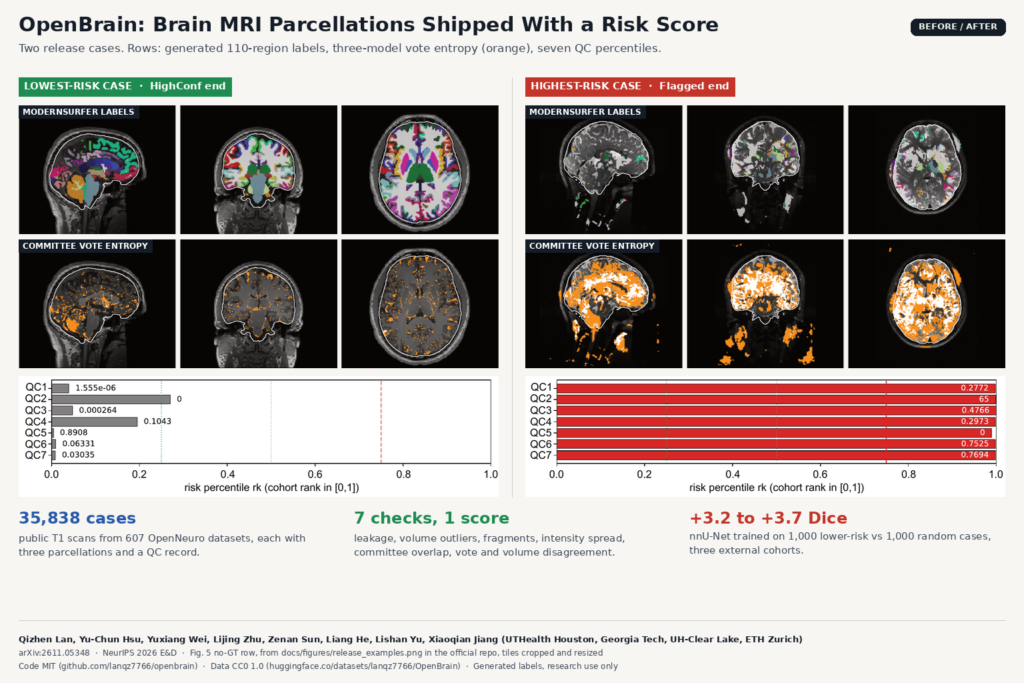
Comparison built from Fig. 5 of arXiv:2611.05348 by Qizhen Lan and colleagues, UTHealth Houston, Georgia Tech, UH-Clear Lake and ETH Zurich: sagittal, coronal and axial T1 views of the lowest-risk and highest-risk OpenBrain cases with ModernSurfer labels, committee vote entropy, and seven QC percentile bars.

Generated labels for 35,838 public T1 scans, each with seven quality checks and a tier, so you can drop bad cases before training.

Taurus: An LLM Reviewer for Aortic CT Annotation
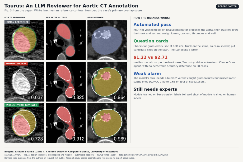
Comparison built from Fig. 3 of arXiv:2611.00247 by Bing Hu and Rishabh Sharma, University of Waterloo: axial CT angiography tiles from three public aortic datasets showing the human reference label, the Taurus automated pass, and the Taurus-hybrid result after LLM review.

An automated pass labels the aorta. A language model then picks among candidate fixes drawn on the CT, at under half the agent cost.

A-Fuse: Airway Tree Segmentation That Holds Up on Pediatric CT
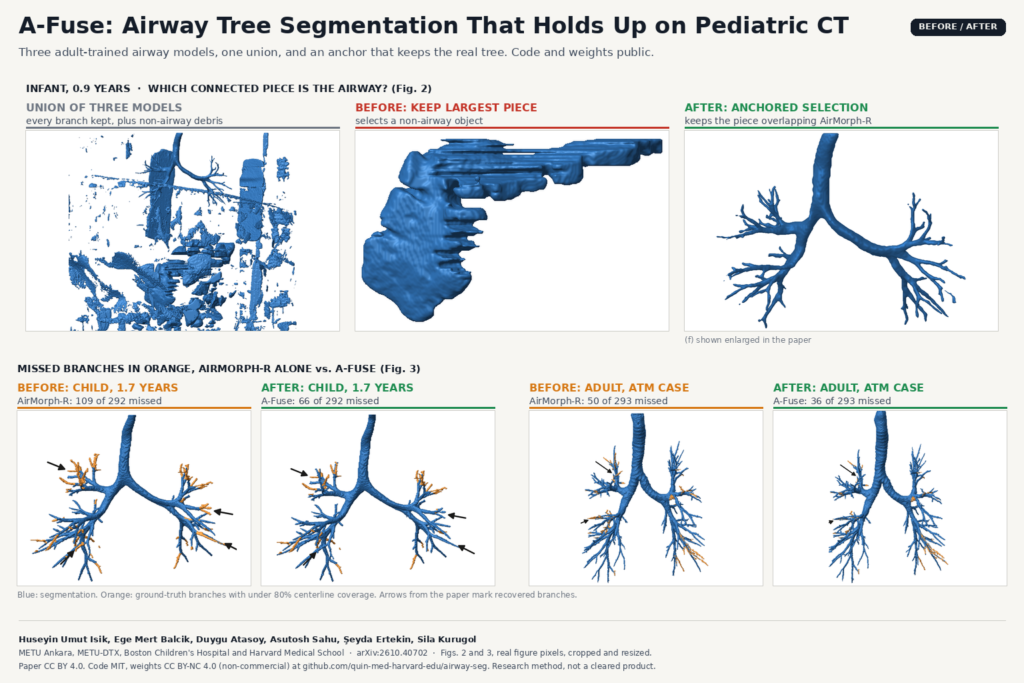
Before and after figure built from Figures 2 and 3 of arXiv:2610.40702 by Huseyin Umut Isik, Sila Kurugol and colleagues (METU and Boston Children's Hospital): for a 0.9-year-old infant, the union of three airway models full of non-airway debris, the largest connected piece that is not the airway, and the anchored selection that keeps the airway tree; below, 3D airway renders for a child and an adult comparing AirMorph-R alone with A-Fuse, with missed ground-truth branches in orange.

Three adult-trained airway models, one union, and an anchor rule that keeps the real tree on child and adult chest CT.

LoRe-MIM: Pretraining Whole-Body PET/CT on Unlabeled Scans
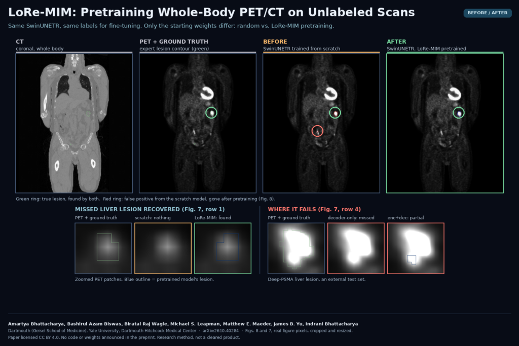
Before and after panel from Figures 8 and 7 of arXiv:2610.40284 by Amartya Bhattacharya, Bashirul Azam Biswas and colleagues (Dartmouth, Yale and Dartmouth Hitchcock Medical Center): coronal whole-body CT and PET with the ground-truth lesion, a SwinUNETR trained from scratch that adds a false positive, and the LoRe-MIM pretrained model that keeps the lesion and drops the false positive, plus zoomed PET patches of a recovered liver lesion and a Deep-PSMA liver lesion that the pretrained models miss or only partly segment.

A masked-image pretext that swaps PET and CT blocks helps a SwinUNETR find liver and bone lesions and drop false calls.

Self-Training Coronary Vessel Masks With Pre-Contrast Frames
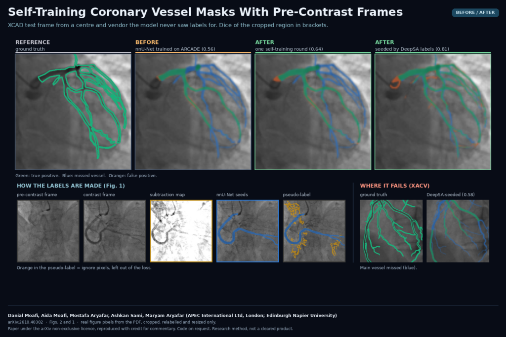
Before and after panel from Figures 2 and 1 of arXiv:2610.40302 by Danial Moafi, Aida Moafi, Mostafa Aryafar, Ashkan Sami and Maryam Aryafar (APEC International Ltd and Edinburgh Napier University): an XCAD coronary angiogram with ground truth, the ARCADE-trained nnU-Net, the self-trained model and the DeepSA-seeded model, a strip showing the pre-contrast frame, contrast frame, subtraction map, seed mask and pseudo-label, and an XACV frame where the main vessel is missed.

An nnU-Net labels a new hospital’s angiograms, and the frame before contrast arrives flags the branches it missed.

RepConTUR: Report-Prompted 3D Tumor Contours for Abdominal CT
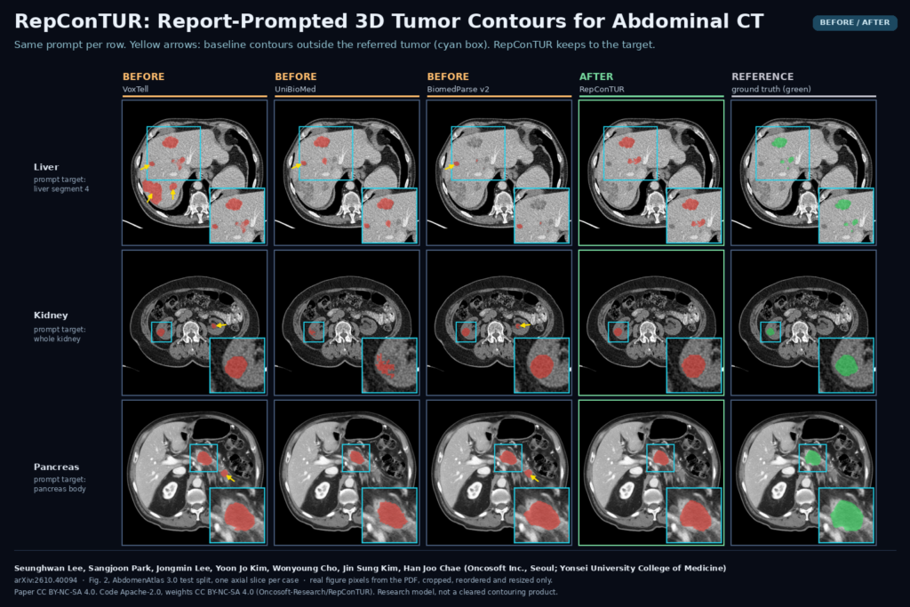
Before and after panel from Figure 2 of arXiv:2610.40094 by Seunghwan Lee, Sangjoon Park, Jongmin Lee, Yoon Jo Kim, Wonyoung Cho, Jin Sung Kim and Han Joo Chae (Oncosoft Inc. and Yonsei University College of Medicine): axial abdominal CT slices for liver segment 4, kidney and pancreas body tumor prompts, comparing VoxTell, UniBioMed and BiomedParse v2 masks with yellow arrows on off-target contours, the RepConTUR mask and the ground truth.

The report names the liver segment or pancreas region, and a 3D model contours that tumor with fewer stray masks elsewhere.

Pancreatic Duct Width From 3D MRCP, With a Review Flag
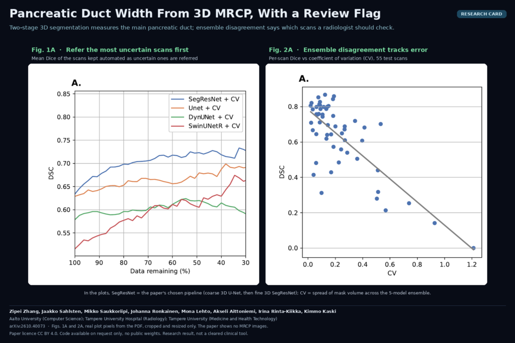
Research card with Figures 1A and 2A of arXiv:2610.40073 by Zipei Zhang, Jaakko Sahlsten, Mikko Saukkoriipi, Johanna Ronkainen, Mona Lehto, Akseli Aittoniemi, Irina Rinta-Kiikka and Kimmo Kaski (Aalto University, Tampere University Hospital, Tampere University): mean Dice of automated scans as uncertain cases are referred for four pipelines, and per-scan Dice against ensemble coefficient of variation.

A two-stage 3D model segments the main pancreatic duct on MRCP, and ensemble disagreement marks the scans a radiologist should check.

Plausible but Unimplantable: Foundation Models vs Cranial Implants
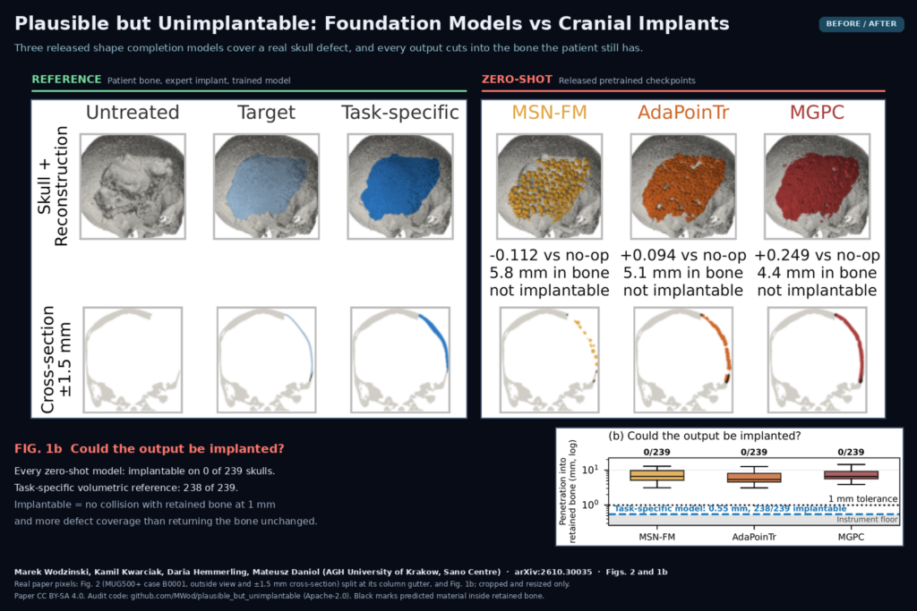
Figure 2 of arXiv:2610.30035 by Wodzinski, Kwarciak, Hemmerling and Daniol (AGH University of Krakow): a real craniectomy skull from MUG500+ with the expert target implant and a task-specific model beside the outputs of MSN-FM, AdaPoinTr and MGPC, each marked not implantable, plus Fig. 1b showing 0 of 239 implantable outputs per model.

An audit of three released shape completion models on 239 skull defects found no output that could be placed in a patient.

← Previous
Next →